
Thrombotic Disorders Janna M Journeycake MD MSCS UT
Thrombotic Disorders Janna M. Journeycake, MD, MSCS UT Southwestern Medical Center Dallas, Children’s Health
Disclosures • No relevant disclosures to this topic • Will discuss off label use of anticoagulant medications
Lecture Outline • • • Introduction Coagulation and Regulation of Coagulation Thrombophilia Diagnosis of thrombotic events Treatment of thrombotic events Outline of content specifications and corresponding slides – End of Slides and Handout
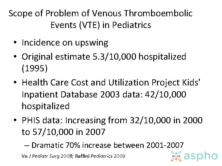
Scope of Problem of Venous Thromboembolic Events (VTE) in Pediatrics • Incidence on upswing • Original estimate 5. 3/10, 000 hospitalized (1995) • Health Care Cost and Utilization Project Kids' Inpatient Database 2003 data: 42/10, 000 hospitalized • PHIS data: Increasing from 32/10, 000 in 2000 to 57/10, 000 in 2007 – Dramatic 70% increase between 2001 -2007 Vu J Pediatr Surg 2008; Raffini Pediatrics 2009
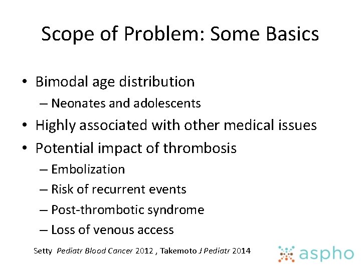
Scope of Problem: Some Basics • Bimodal age distribution – Neonates and adolescents • Highly associated with other medical issues • Potential impact of thrombosis – Embolization – Risk of recurrent events – Post-thrombotic syndrome – Loss of venous access Setty Pediatr Blood Cancer 2012 , Takemoto J Pediatr 2014
Review of Coagulation and Regulation of Coagulation • Dr. Young reviewed yesterday and I borrowed a few of his slides
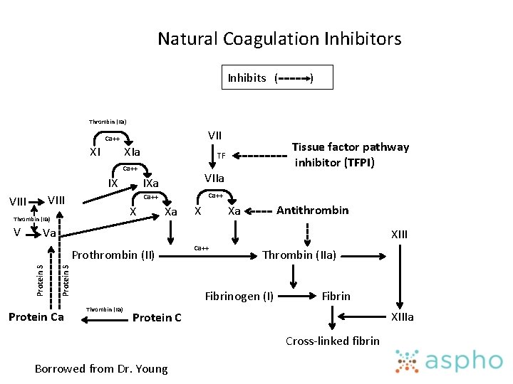
Natural Coagulation Inhibitors Inhibits ( ) Thrombin (IIa) VII Ca++ XI XIa Ca++ IX VIII V VIIa IXa Ca++ X Thrombin (IIa) Tissue factor pathway inhibitor (TFPI) TF Xa X Antithrombin Xa Va XIII Protein S Prothrombin (II) Protein Ca Ca++ Thrombin (IIa) Fibrinogen (I) Thrombin (IIa) Fibrin XIIIa Protein C Cross-linked fibrin Borrowed from Dr. Young
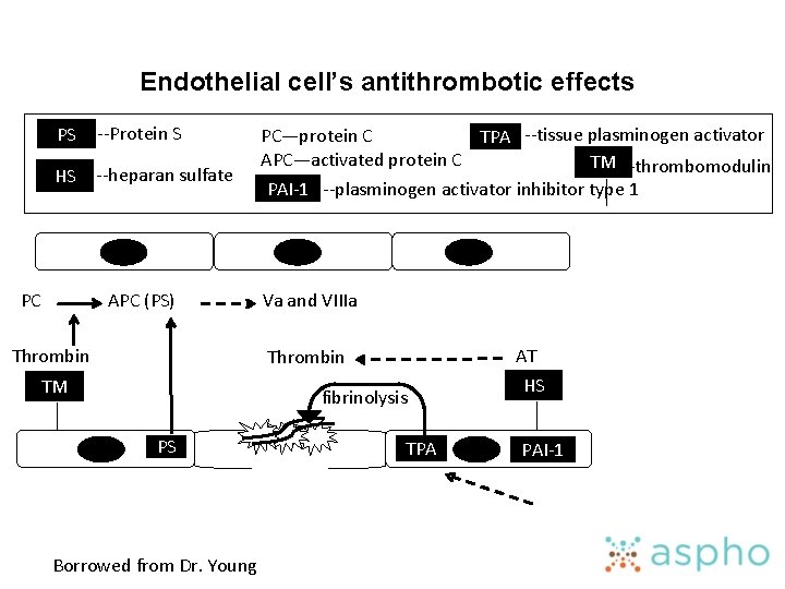
Endothelial cell’s antithrombotic effects PS --Protein S HS --heparan sulfate PC APC (PS) Thrombin PC—protein C TPA --tissue plasminogen activator APC—activated protein C TM --thrombomodulin PAI-1 --plasminogen activator inhibitor type 1 Va and VIIIa AT Thrombin TM fibrinolysis PS Borrowed from Dr. Young TPA HS PAI-1
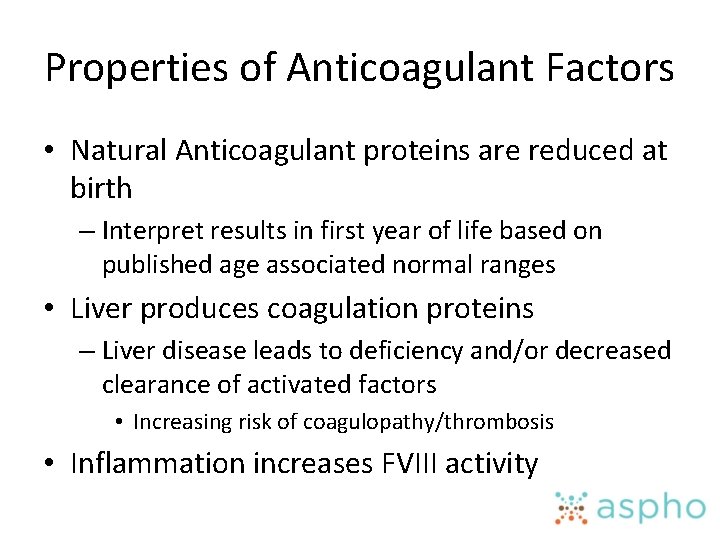
Properties of Anticoagulant Factors • Natural Anticoagulant proteins are reduced at birth – Interpret results in first year of life based on published age associated normal ranges • Liver produces coagulation proteins – Liver disease leads to deficiency and/or decreased clearance of activated factors • Increasing risk of coagulopathy/thrombosis • Inflammation increases FVIII activity
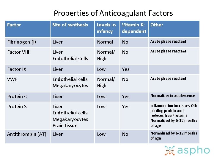
Properties of Anticoagulant Factors Factor Site of synthesis Levels in Vitamin K- Other infancy dependent Fibrinogen (I) Liver Normal No Acute phase reactant Factor VIII Liver Endothelial Cells Normal/ High No Acute phase reactant Factor IX Liver Low Yes VWF Endothelial cells Megakaryocytes Normal/ High No Acute phase reactant Protein C Liver Low Yes Normalizes in adolescence Protein S Liver Endothelial cells Megakaryocytes Brain tissue Low Yes Inflammation increases C 4 b binding protein and reduces free Protein S Normalized by 6 -12 months of age Antithrombin (AT) Liver Low No Normalized by 6 -12 months of age
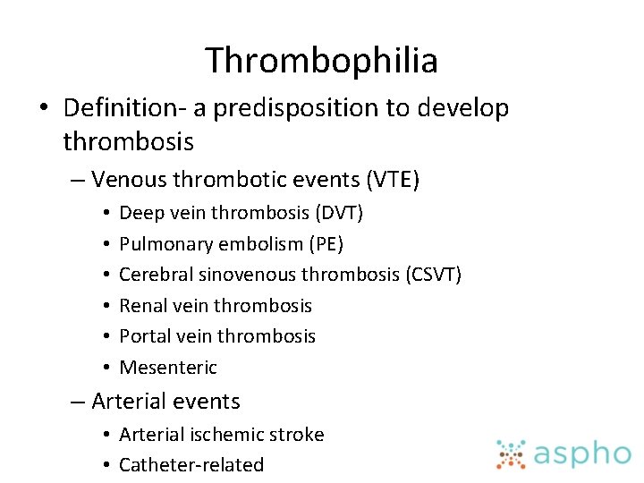
Thrombophilia • Definition- a predisposition to develop thrombosis – Venous thrombotic events (VTE) • • • Deep vein thrombosis (DVT) Pulmonary embolism (PE) Cerebral sinovenous thrombosis (CSVT) Renal vein thrombosis Portal vein thrombosis Mesenteric – Arterial events • Arterial ischemic stroke • Catheter-related
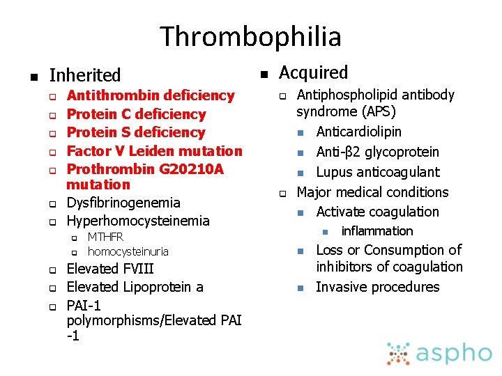
Thrombophilia n Inherited q q q q Antithrombin deficiency Protein C deficiency Protein S deficiency Factor V Leiden mutation Prothrombin G 20210 A mutation Dysfibrinogenemia Hyperhomocysteinemia q q q MTHFR homocysteinuria Elevated FVIII Elevated Lipoprotein a PAI-1 polymorphisms/Elevated PAI -1 n Acquired q q Antiphospholipid antibody syndrome (APS) n Anticardiolipin n Anti-β 2 glycoprotein n Lupus anticoagulant Major medical conditions n Activate coagulation n inflammation Loss or Consumption of inhibitors of coagulation Invasive procedures
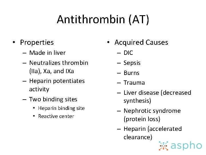
Antithrombin (AT) • Properties – Made in liver – Neutralizes thrombin (IIa), Xa, and IXa – Heparin potentiates activity – Two binding sites • Heparin binding site • Reactive center • Acquired Causes DIC Sepsis Burns Trauma Liver disease (decreased synthesis) – Nephrotic syndrome (protein loss) – Heparin (accelerated clearance) – – –
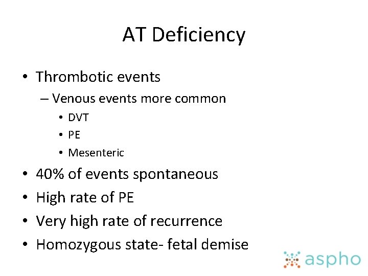
AT Deficiency • Thrombotic events – Venous events more common • DVT • PE • Mesenteric • • 40% of events spontaneous High rate of PE Very high rate of recurrence Homozygous state- fetal demise
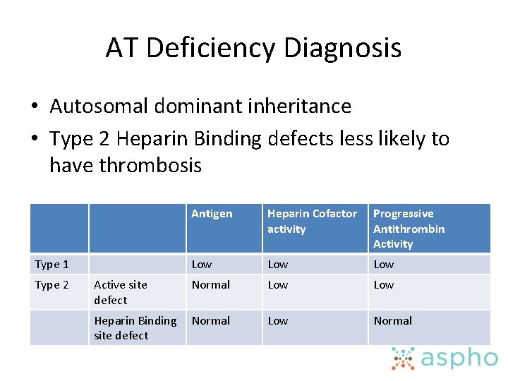
AT Deficiency Diagnosis • Autosomal dominant inheritance • Type 2 Heparin Binding defects less likely to have thrombosis Antigen Heparin Cofactor activity Progressive Antithrombin Activity Low Low Normal Low Heparin Binding Normal site defect Low Normal Type 1 Type 2 Active site defect
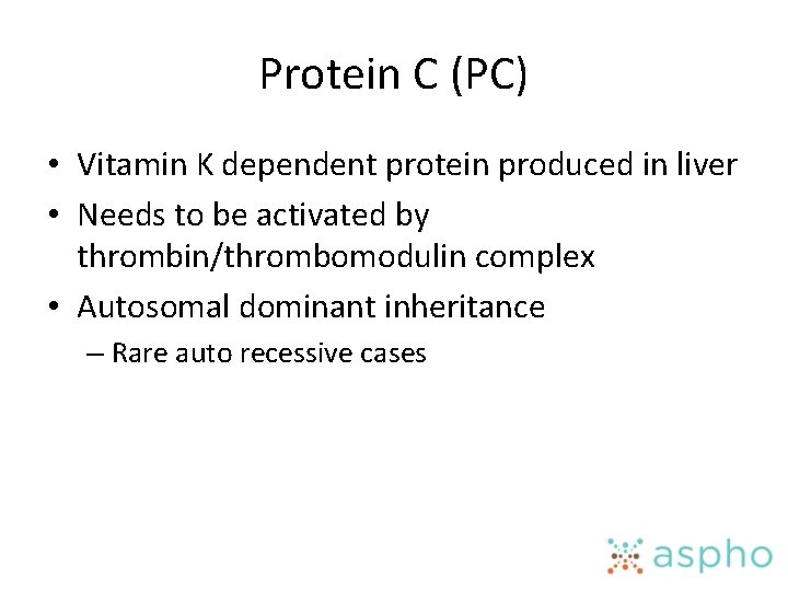
Protein C (PC) • Vitamin K dependent protein produced in liver • Needs to be activated by thrombin/thrombomodulin complex • Autosomal dominant inheritance – Rare auto recessive cases
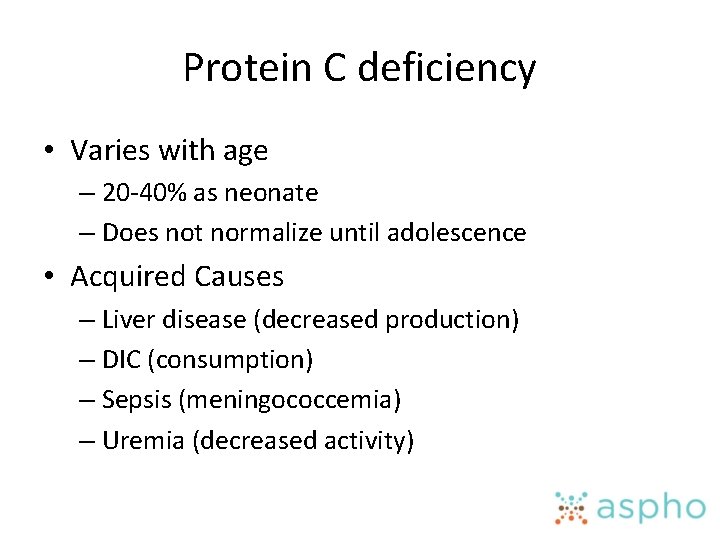
Protein C deficiency • Varies with age – 20 -40% as neonate – Does not normalize until adolescence • Acquired Causes – Liver disease (decreased production) – DIC (consumption) – Sepsis (meningococcemia) – Uremia (decreased activity)
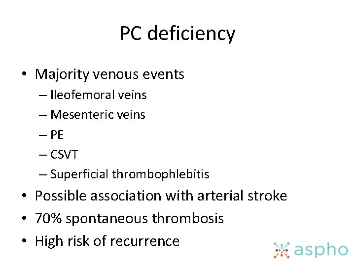
PC deficiency • Majority venous events – Ileofemoral veins – Mesenteric veins – PE – CSVT – Superficial thrombophlebitis • Possible association with arterial stroke • 70% spontaneous thrombosis • High risk of recurrence
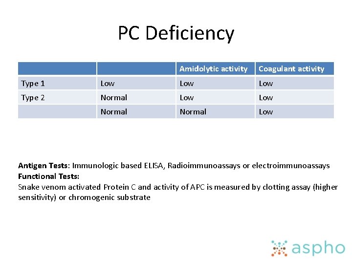
PC Deficiency Amidolytic activity Coagulant activity Type 1 Low Low Type 2 Normal Low Antigen Tests: Immunologic based ELISA, Radioimmunoassays or electroimmunoassays Functional Tests: Snake venom activated Protein C and activity of APC is measured by clotting assay (higher sensitivity) or chromogenic substrate
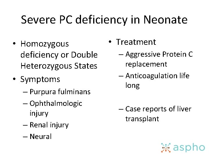
Severe PC deficiency in Neonate • Treatment • Homozygous – Aggressive Protein C deficiency or Double replacement Heterozygous States – Anticoagulation life • Symptoms – Purpura fulminans – Ophthalmologic injury – Renal injury – Neural long – Case reports of liver transplant
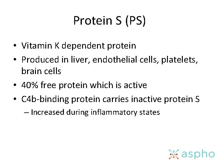
Protein S (PS) • Vitamin K dependent protein • Produced in liver, endothelial cells, platelets, brain cells • 40% free protein which is active • C 4 b-binding protein carries inactive protein S – Increased during inflammatory states
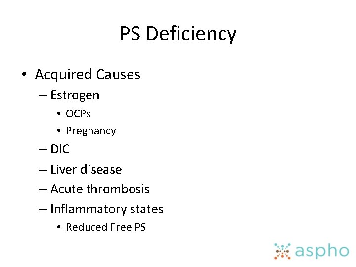
PS Deficiency • Acquired Causes – Estrogen • OCPs • Pregnancy – DIC – Liver disease – Acute thrombosis – Inflammatory states • Reduced Free PS
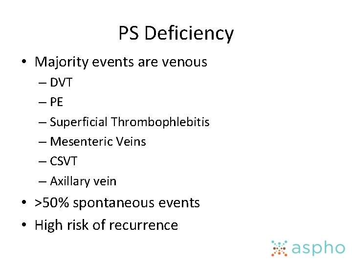
PS Deficiency • Majority events are venous – DVT – PE – Superficial Thrombophlebitis – Mesenteric Veins – CSVT – Axillary vein • >50% spontaneous events • High risk of recurrence
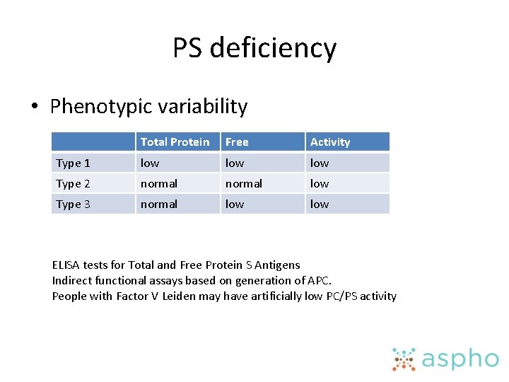
PS deficiency • Phenotypic variability Total Protein Free Activity Type 1 low low Type 2 normal low Type 3 normal low ELISA tests for Total and Free Protein S Antigens Indirect functional assays based on generation of APC. People with Factor V Leiden may have artificially low PC/PS activity
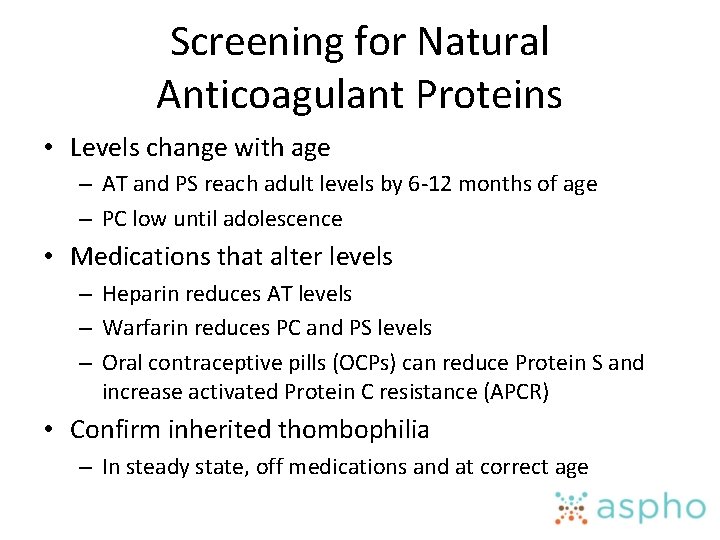
Screening for Natural Anticoagulant Proteins • Levels change with age – AT and PS reach adult levels by 6 -12 months of age – PC low until adolescence • Medications that alter levels – Heparin reduces AT levels – Warfarin reduces PC and PS levels – Oral contraceptive pills (OCPs) can reduce Protein S and increase activated Protein C resistance (APCR) • Confirm inherited thombophilia – In steady state, off medications and at correct age
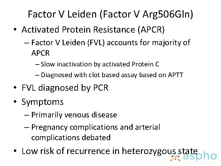
Factor V Leiden (Factor V Arg 506 Gln) • Activated Protein Resistance (APCR) – Factor V Leiden (FVL) accounts for majority of APCR – Slow inactivation by activated Protein C – Diagnosed with clot based assay based on APTT • FVL diagnosed by PCR • Symptoms – Primarily venous disease – Pregnancy complications and arterial complications debated • Low risk of recurrence in heterozygous state
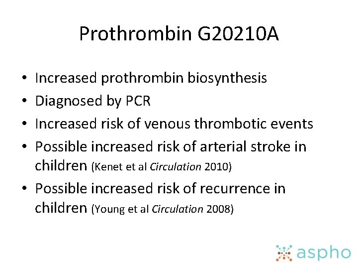
Prothrombin G 20210 A Increased prothrombin biosynthesis Diagnosed by PCR Increased risk of venous thrombotic events Possible increased risk of arterial stroke in children (Kenet et al Circulation 2010) • Possible increased risk of recurrence in children (Young et al Circulation 2008) • •
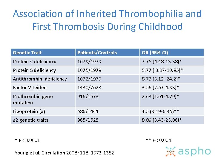
Association of Inherited Thrombophilia and First Thrombosis During Childhood Genetic Trait Patients/Controls OR (95% CI) Protein C deficiency 1079/1979 7. 75 (4. 48 -13. 38)* Protein S deficiency 1075/1979 5. 77 ( 3. 07 -10. 85)* Antithrombin deficiency 1072/1979 8. 73 (3. 12 - 24. 2)* Factor V Leiden 1430/2623 3. 56 (2. 57 -4. 93)* Prothrombin gene mutation 916/1673 2. 63 (1. 61 -4. 29)* Lipoprotein (a) 586/1441 4. 5 (3. 19 -6. 35)** ≥ 2 genetic traits 965/1625 8. 89 (3. 43 -23. 06)* * P< 0. 0001 ** P< 0. 001 Young et al. Circulation 2008; 118: 1373 -1382
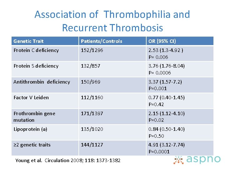
Association of Thrombophilia and Recurrent Thrombosis Genetic Trait Patients/Controls OR (95% CI) Protein C deficiency 152/1296 2. 53 (1. 3 -4. 92 ) P= 0. 006 Protein S deficiency 132/857 3. 76 (1. 76 -8. 04) P= 0. 0006 Antithrombin deficiency 150/969 3. 37 (1. 57 -7. 2) P=0. 001 Factor V Leiden 112/1160 0. 77 (0. 40 -1. 45) P=0. 42 Prothrombin gene mutation 171/1397 2. 15 (1. 12 -4. 10) P=0. 02 Lipoprotein (a) 135/1020 0. 84 (0. 50 -1. 40) P=0. 50 ≥ 2 genetic traits 144/1127 4. 91 (3. 12 -7. 74) P=0. 0001 Young et al. Circulation 2008; 118: 1373 -1382
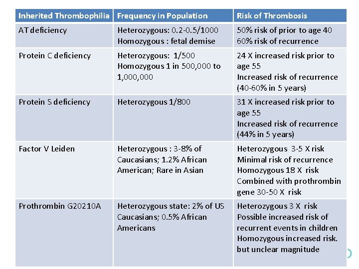
Inherited Thrombophilia Frequency in Population Risk of Thrombosis AT deficiency Heterozygous: 0. 2 -0. 5/1000 Homozygous : fetal demise 50% risk of prior to age 40 60% risk of recurrence Protein C deficiency Heterozygous: 1/500 Homozygous 1 in 500, 000 to 1, 000 24 X increased risk prior to age 55 Increased risk of recurrence (40 -60% in 5 years) Protein S deficiency Heterozygous 1/800 31 X increased risk prior to age 55 Increased risk of recurrence (44% in 5 years) Factor V Leiden Heterozygous : 3 -8% of Caucasians; 1. 2% African American; Rare in Asian Heterozygous 3 -5 X risk Minimal risk of recurrence Homozygous 18 X risk Combined with prothrombin gene 30 -50 X risk Prothrombin G 20210 A Heterozygous state: 2% of US Caucasians; 0. 5% African Americans Heterozygous 3 X risk Possible increased risk of recurrent events in children Homozygous increased risk. but unclear magnitude
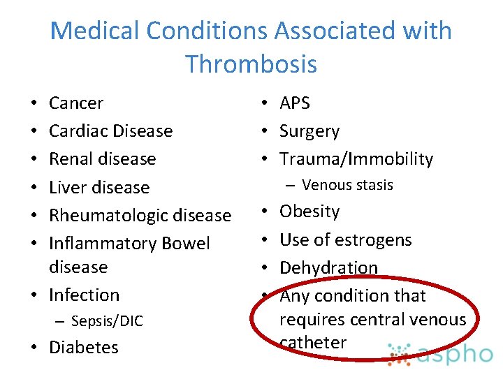
Medical Conditions Associated with Thrombosis Cancer Cardiac Disease Renal disease Liver disease Rheumatologic disease Inflammatory Bowel disease • Infection • • • – Sepsis/DIC • Diabetes • APS • Surgery • Trauma/Immobility – Venous stasis • • Obesity Use of estrogens Dehydration Any condition that requires central venous catheter
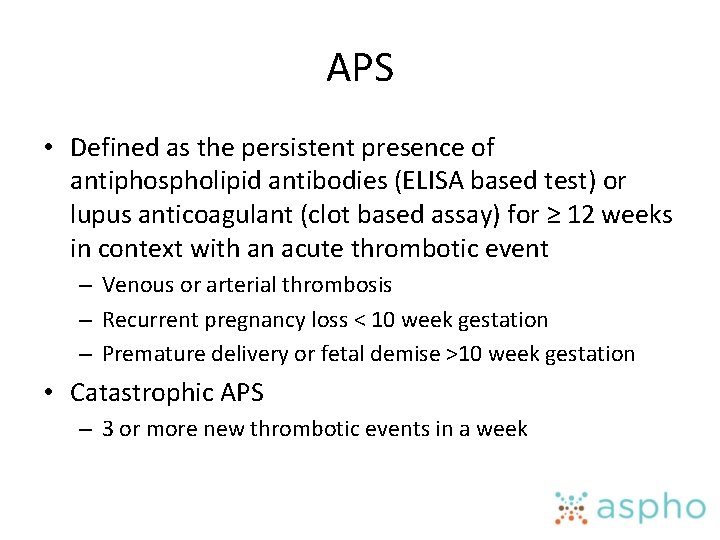
APS • Defined as the persistent presence of antiphospholipid antibodies (ELISA based test) or lupus anticoagulant (clot based assay) for ≥ 12 weeks in context with an acute thrombotic event – Venous or arterial thrombosis – Recurrent pregnancy loss < 10 week gestation – Premature delivery or fetal demise >10 week gestation • Catastrophic APS – 3 or more new thrombotic events in a week
Impact of Acquired Thrombophilia • Which has greater impact? Inherited or Acquired risk factors? – Prevalence of inherited thrombophilia in children with thrombosis is 13 -79% • Depends on which tests were performed and timing • Close to 90% of children with thrombosis have underlying medical condition • 75 -85% also have central venous catheters – Thrombophilia plays a stronger role in spontaneous thrombosis in neonates and adolescents Revel-Vilk Thromb Res 2006
Thrombophilia Testing • During acute event transient abnormalities? – Consumption of Protein C and S – Elevated FVIII and D-Dimers – Transient antiphospholipid antibodies/lupus anticoagulant (LA) • Any non-genetic abnormality should be confirmed by repeat testing in stable state – Off warfarin for Protein C and S – Off anticoagulation for clot based assays of LA
Thrombophilia Testing • Guidelines – screen patients with thrombosis <50 yrs, unusual sites, recurrent events, first event and family history • Factor V Leiden Prothrombin Gene Protein C Protein S AT Lipoprotein a Homocysteine (arterial) Antiphospholipids/LA
Presentation of DVT
Diagnosis • Symptoms • Imaging • Lab tests – No specific test that makes diagnosis – If suspicious for thrombosis, do the imaging
Venous Thrombosis • Superficial Thrombophlebitis – Painful – Palpable cord – Often from intravenous stick or PICC line – Varicose veins • Deep Vein Thrombosis (DVT) – Acute swelling of extremity – Pain – Discoloration (red to purple) – Heat
Specific Syndromes • Lower Extremity – Associated with Trauma, Immobilization, Bed rest, Post-operative state Long-haul travel Central venous catheter • Anatomic abnormalities • • Upper Extremity – Associated with • Central catheters • PICC • Anatomic abnormalities
Specific Syndromes • May Thurner – Left-sided ileofemoral thrombosis • Narrowed left iliac vein due to chronic compression from right iliac artery • More likely in adolescent girls • Axillary/Subclavian vein thrombosis – Paget-Schroetter, Thoracic Outlet Syndrome, Exertional or Effort-induced Thrombosis • More likely in adolescents • Associated with repetitive activity • Compression of veins by extra rib, muscle group
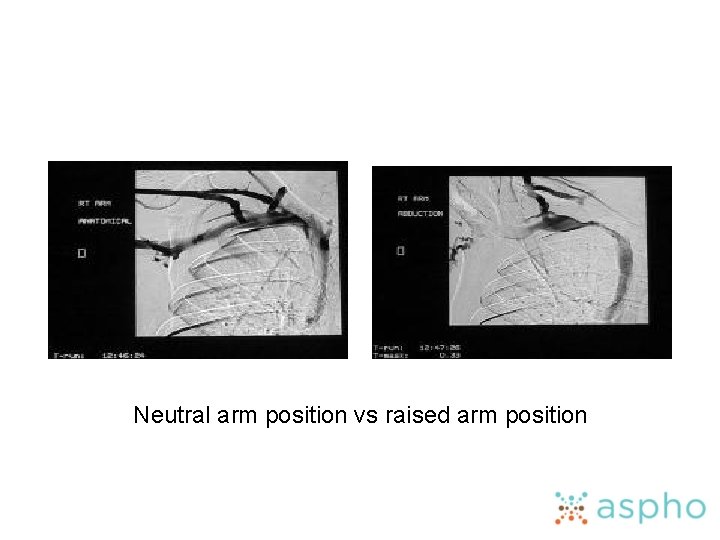
Neutral arm position vs raised arm position
Catheter-related DVT • Most common cause of DVT in children – 90% of DVT in neonates and 60% of DVT in children are catheter-related
Catheter-related DVT • Acute (10 -15%) – – Extremity Swelling Extremity Pain Discoloration Line not working well • Chronic ”asymptomatic” – Catheter occlusion – Prominent chest wall veins – Arm swelling – Arm pain – Recurrent bacteremia?
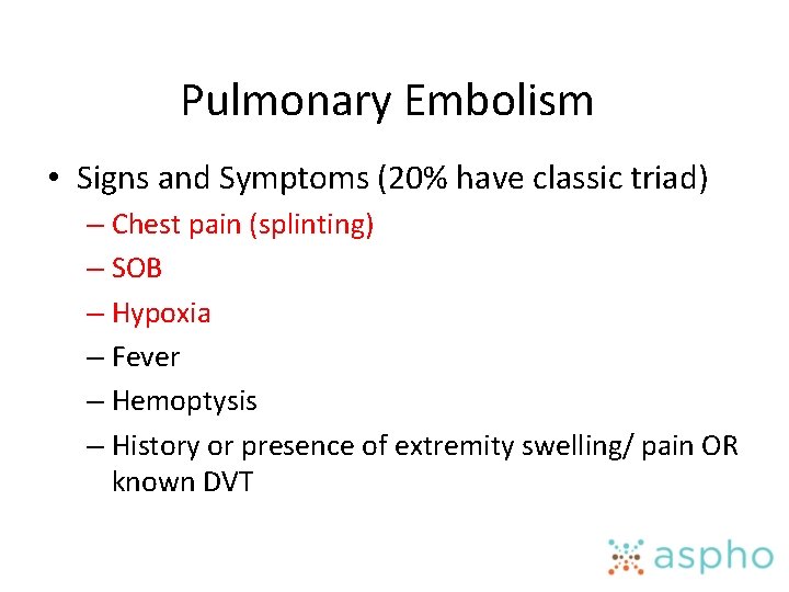
Imaging for Diagnosis for DVT • Contrast Venography – – Requires IV contrast “gold standard” Sensitivity lower up to 90% Sensitivity upper 79 to 94% • Improve images of contrast in central veins by elevating extremity and using tourniquet • Reduces “washout” phenomenon of influx of blood without contrast into jugular vein – Reduced ability to identify jugular vein thrombosis
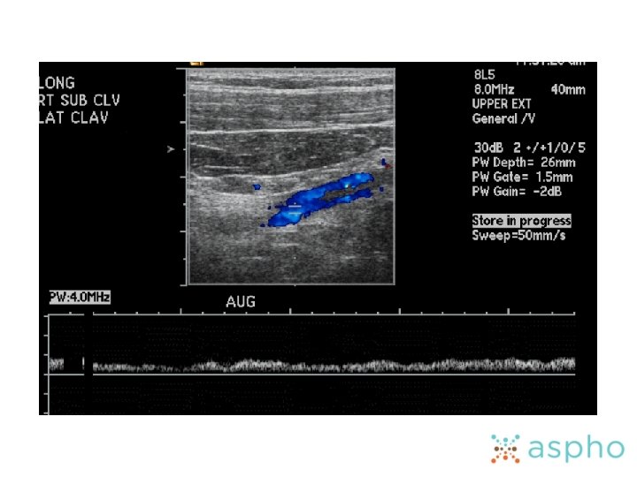
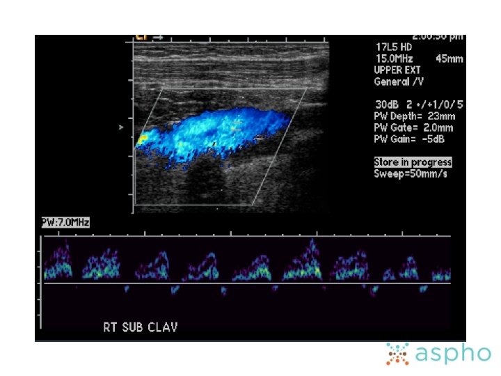
Ultrasonography • Non-invasive • Diagnosis – Lack of compressability of vessels, – Visualization of echogenecity in lumen, – Lack of wave form response with respirations • Lower extremity • 95% sensitive for proximal, 50% for isolated calf • Limited ability to see iliac veins • Upper extremity • Sensitivity 54 to 100%, Specificity 94 to 100% • Technically difficult in upper extremity- no compressibility through ribs and clavicles
Imaging for Diagnosis of DVT • CT venography – High dose radiation, risk of contrast – High rate of false positives and false negatives – 3 -D reconstruction may help increase reliability • Detected 100% of thrombi in central and jugular veins vs 82% by sono – Farinasso et al. Leukemia 2007; 21: 552 -556
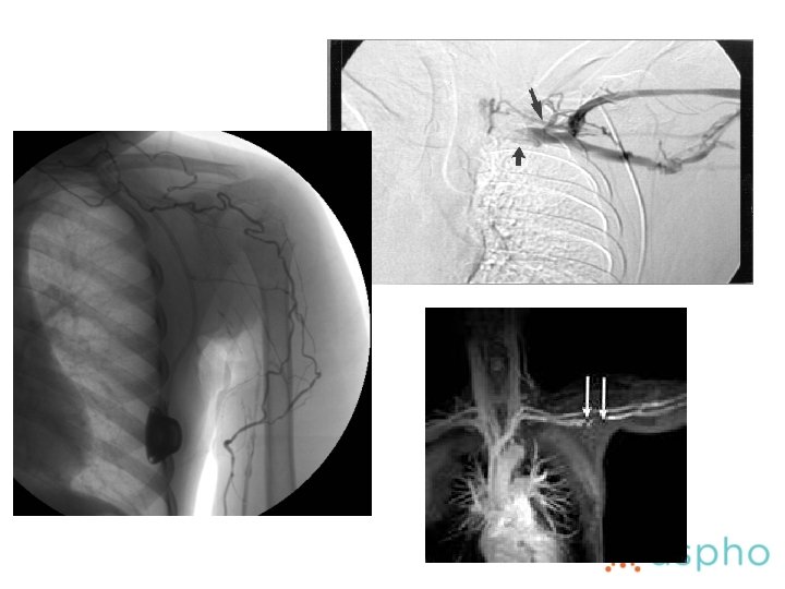
Imaging for Diagnosis of DVT • MR Venography – No radiation – Requires cooperation and prone to motion artifact – 100% sensitivity for proximal DVT, 92% for distal in adults • Not yet replicated in children – Baskin et al. Lancet 2009; 374: 159 -169
Pulmonary Embolism • Signs and Symptoms (20% have classic triad) – Chest pain (splinting) – SOB – Hypoxia – Fever – Hemoptysis – History or presence of extremity swelling/ pain OR known DVT
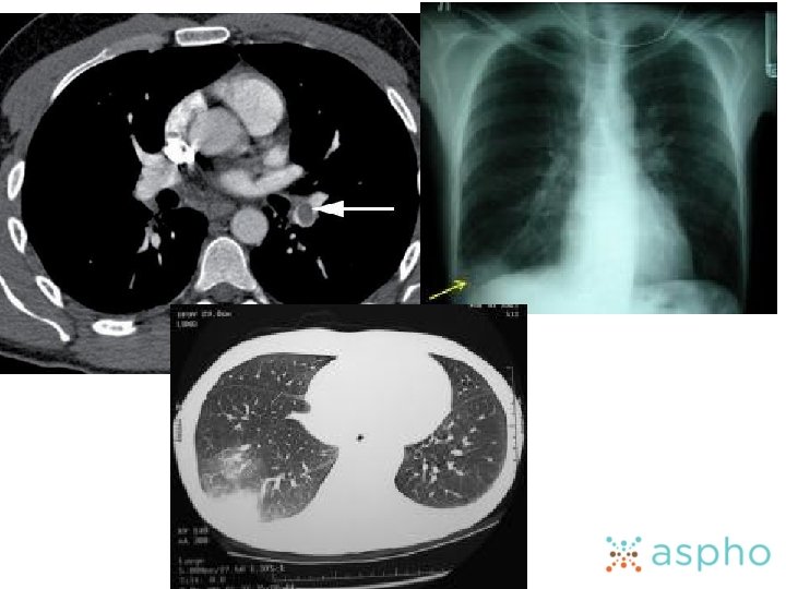
Imaging for Diagnosis of PE • CXR – May be initially normal in patient with PE and hypoxia – Knuckle sign and enlarged right heart • V/Q scan – Use as initial study if CT scan not available
Imaging for Diagnosis of PE • CT – Spiral CT/CT angiography • Central PE 100% sensitivity • Sometimes difficult to diagnose sub-segmental PE • 8 -10% of scans will be sub-optimal • Contrast pulmonary angiography – “gold standard” – Invasive
Additional Studies to Evaluate Severity of PE • Laboratory – Troponin- right ventricular overload – Brain Natriuretic Protein- evidence of heart stress • ECHO – Enlarged right ventricle and evidence of pulmonary hypertension • EKG – Tachycardia, atrial fibrillation, ST-T wave abnormality
Cerebral Sinovenous Thrombosis (CSVT) • Presentation – Headache – Evidence of increased intracranial pressure – Seizures – Cranial nerve palsies – Papilledema – Focal neurologic signs (cortical vein thrombosis) – Eyelid swelling, proptosis (cavernous thrombosis) • History – Ear infection progressing to mastoiditis – Lymphadenitis – Dehydration – Jugular vein catheterization – Neonate
Imaging for Diagnosis of CSVT • Conventional CT scan – Before and after contrast • Delta sign in non-contrast CT and reverse delta with contrast – Cerebral edema, venous infarction – May need CT venography in increase sensitivity • MRI/MR venography – Best non-invasive test for diagnosis – Direct visualization of thrombosis – 2 D Time of flight or phase contrast MRV • Occlusion of vessel
Portal Vein Thrombosis • Presentation – Often slowly evolves • Discovered with first GI bleed from varices • Asymptomatic hepatomegaly and splenomegaly • Ascites – Acute presentation • Abdominal pain • May be masked by underlying pathology (pancreatitis) • History – UVC – Intra-abdominal tumor – Intra-abdominal infection – Thrombophilia – OCP/Pregnancy
Portal Vein Thrombosis Imaging • Doppler Ultrasonography with Color Flow – Best initial method • CT – CT angiography with helical CT – Visualize collaterals • MRI – 99% sensitivity and 96% specificity for main portal vein thrombosis – False positive with noncontrast MRI
Renal Vein Thrombosis • Presentation – Generally in first month of life – Flank mass – Hematuria • History – Maternal gestational diabetes – Dehydration – Perinatal asphyxia – IVC catheters – Nephrotic syndrome
Imaging for Renal Vein Thrombosis • Doppler Ultrasound and MR Venography – Equal ability to diagnose obstructed flow and do not require radiation or contrast media of CT – If inconclusive, perform angiography/IVC imaging • Radionuclide renography – Identify poor perfusion to kidneys
Cautionary Interpretation of Laboratory Testing in Diagnosing Thrombosis • D-Dimer as a tool for diagnosis – Not well studied in children – Type of D-Dimer important • Latex agglutination test less sensitive than ELISA – Not specific • Can be positive in patients with cancer, infection, trauma • Can be normal with subacute thrombus (CVL thrombosis) – Negative test in person with low pretest probability is reliable – Le Gal, et al Ann Intern Med. Feb 7 2006; 144(3): 165 -71
Laboratory Testing AT Diagnosis (not FOR diagnosis) • CBC – Thrombocytopenia • Coagulation Studies – PT/PTT, fibrinogen, D-Dimer and FVIII • Baseline prior to initiating anticoagulation • Elevated D-Dimer and FVIII prognostic • Creatinine needed prior to initiating low molecular weight heparin • Thrombophilia evaluation – Not necessary urgently • False results in very acute phase
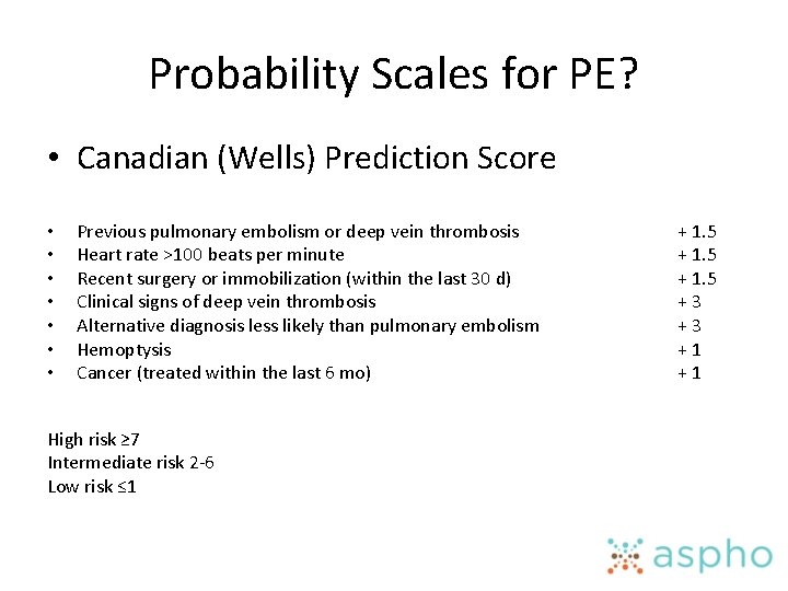
Probability Scales for PE? • Canadian (Wells) Prediction Score • • Previous pulmonary embolism or deep vein thrombosis Heart rate >100 beats per minute Recent surgery or immobilization (within the last 30 d) Clinical signs of deep vein thrombosis Alternative diagnosis less likely than pulmonary embolism Hemoptysis Cancer (treated within the last 6 mo) High risk ≥ 7 Intermediate risk 2 -6 Low risk ≤ 1 + 1. 5 + 3 + 1
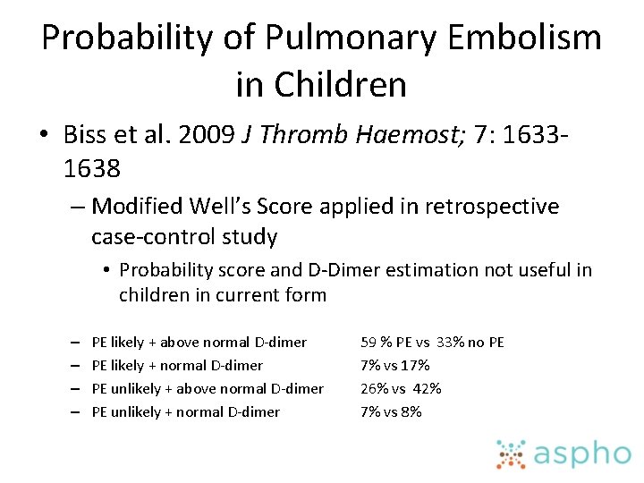
Probability of Pulmonary Embolism in Children • Biss et al. 2009 J Thromb Haemost; 7: 16331638 – Modified Well’s Score applied in retrospective case-control study • Probability score and D-Dimer estimation not useful in children in current form – – PE likely + above normal D-dimer PE likely + normal D-dimer PE unlikely + above normal D-dimer PE unlikely + normal D-dimer 59 % PE vs 33% no PE 7% vs 17% 26% vs 42% 7% vs 8%
Treatment
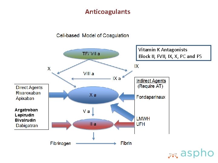
Anticoagulants Vitamin K Antagonists Block II, FVII, IX, X, PC and PS Argatroban Lepirudin Bivalrudin
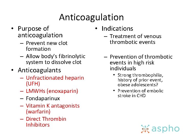
Anticoagulation • Purpose of anticoagulation – Prevent new clot formation – Allow body’s fibrinolytic system to dissolve clot • Anticoagulants – Unfractionated heparin (UFH) – LMWHs (enoxaparin) – Fondaparinux – Vitamin K antagonists (warfarin) – Direct Thrombin Inhibitors • Indications – Treatment of venous thrombotic events – Prevention of thrombotic events in high risk individuals • Strong thrombophilia, history of prior event, obese adolescents? • Prevention of embolic stroke in CHD
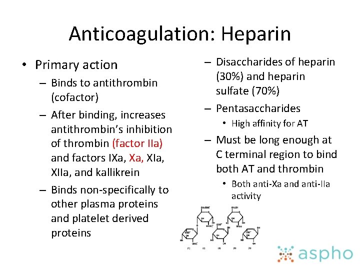
Anticoagulation: Heparin • Primary action – Binds to antithrombin (cofactor) – After binding, increases antithrombin’s inhibition of thrombin (factor IIa) and factors IXa, XIa, XIIa, and kallikrein – Binds non-specifically to other plasma proteins and platelet derived proteins – Disaccharides of heparin (30%) and heparin sulfate (70%) – Pentasaccharides • High affinity for AT – Must be long enough at C terminal region to bind both AT and thrombin • Both anti-Xa and anti-IIa activity
Heparin • Properties – Short half-life – Reversible with protamine sulfate – Excreted by kidney – Risk of heparin induced thrombocytopenia (HIT) – a. PTT very sensitive to heparin contamination • Blood drawn through catheters • Heparinase can detect presence of heparin
Monitoring Heparin • PTT (Target range 65 -80 seconds or 1. 5 -2. 5 X baseline) – Sensitive to improper collection, anemia, thrombocytopenia, factor deficiencies, lupus anticoagulant, age • ACT (target range depends on clinical use e. g. ECMO, cardiac catheterization) – Sensitive to platelet counts and function, factor deficiencies, ambient temperature, hypothermia, lupus anticoagulant • Anti-Xa most consistent result (Target 0. 3 -0. 7 units/ml) – Affected by hemolysis, high bilirubin, high triglycerides – Not affected by age or coagulopathy – Monitor every 4 hours after dose change and daily
Heparin Resistance • Defined as the need for greater than average doses to achieve a therapeutic goal • Possible reasons for heparin resistance: – AT deficiency • AT levels <60% - higher likelihood of heparin resistance, – Increased heparin clearance • Lasix , CVVH – Elevation in level of heparin-binding proteins • Transfusion of plasma products – High levels of factor VIII or fibrinogen – DIC
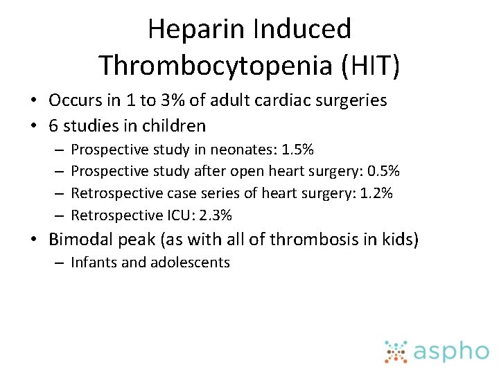
Heparin Induced Thrombocytopenia (HIT) • Occurs in 1 to 3% of adult cardiac surgeries • 6 studies in children – – Prospective study in neonates: 1. 5% Prospective study after open heart surgery: 0. 5% Retrospective case series of heart surgery: 1. 2% Retrospective ICU: 2. 3% • Bimodal peak (as with all of thrombosis in kids) – Infants and adolescents
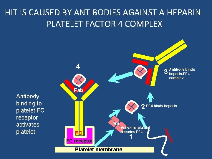
HIT IS CAUSED BY ANTIBODIES AGAINST A HEPARINPLATELET FACTOR 4 COMPLEX 4 binds 3 Antibody heparin-PF 4 complex 2 PF 4 binds heparin PF 4 Platelet membrane 1 rin FC receptor pa FC Activated platelet secretes PF 4 He Antibody binding to platelet FC receptor activates platelet Fab
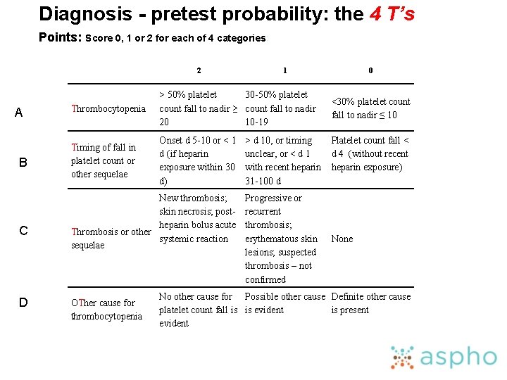
Diagnosis - pretest probability: the 4 T’s Points: Score 0, 1 or 2 for each of 4 categories: 2 A B C D 1 Thrombocytopenia > 50% platelet 30 -50% platelet count fall to nadir ≥ count fall to nadir 20 10 -19 Timing of fall in platelet count or other sequelae Onset d 5 -10 or < 1 d (if heparin exposure within 30 d) New thrombosis; skin necrosis; postheparin bolus acute Thrombosis or other systemic reaction sequelae OTher cause for thrombocytopenia > d 10, or timing unclear, or < d 1 with recent heparin 31 -100 d Progressive or recurrent thrombosis; erythematous skin lesions; suspected thrombosis – not confirmed 0 <30% platelet count fall to nadir ≤ 10 Platelet count fall < d 4 (without recent heparin exposure) None No other cause for Possible other cause Definite other cause platelet count fall is is evident is present evident
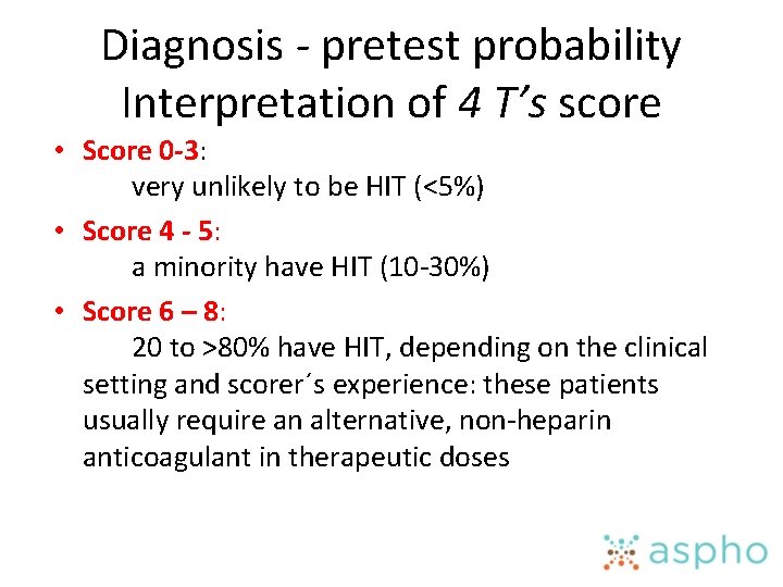
Diagnosis - pretest probability Interpretation of 4 T’s score • Score 0 -3: very unlikely to be HIT (<5%) • Score 4 - 5: a minority have HIT (10 -30%) • Score 6 – 8: 20 to >80% have HIT, depending on the clinical setting and scorer´s experience: these patients usually require an alternative, non-heparin anticoagulant in therapeutic doses
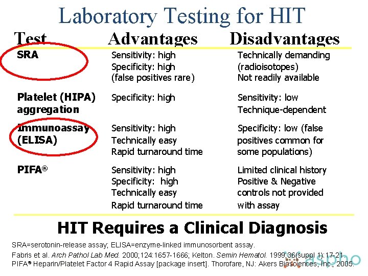
Laboratory Testing for HIT Test Advantages Disadvantages Sensitivity: high Specificity: high (false positives rare) Technically demanding (radioisotopes) Not readily available Platelet (HIPA) aggregation Specificity: high Sensitivity: low Technique-dependent Immunoassay (ELISA) Sensitivity: high Technically easy Rapid turnaround time Specificity: low (false positives common for some populations) PIFA® Sensitivity: high Specificity: high Technically easy Rapid turnaround time Limited clinical history Positive & Negative controls not provided with assay SRA HIT Requires a Clinical Diagnosis SRA=serotonin-release assay; ELISA=enzyme-linked immunosorbent assay. Fabris et al. Arch Pathol Lab Med. 2000; 124: 1657 -1666; Kelton. Semin Hematol. 1999; 36(suppl 1): 17 -21. PIFA® Heparin/Platelet Factor 4 Rapid Assay [package insert]. Thorofare, NJ: Akers Biosciences, Inc. ; 2005.
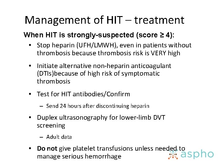
Management of HIT – treatment When HIT is strongly-suspected (score ≥ 4): • Stop heparin (UFH/LMWH), even in patients without thrombosis because thrombosis risk is VERY high • Initiate alternative non-heparin anticoagulant (DTIs)because of high risk of symptomatic thrombosis • Test for HIT antibodies/Confirm – Send 24 hours after discontinuing heparin • Duplex ultrasonography for lower-limb DVT screening – Adult data • Do not give platelet transfusions unless needed to manage serious hemorrhage

Direct Thrombin Inhibitors Argatroban Synthetic L-arginine derivative Half-life in healthy subjects Lepirudin Bivalirudin Recombinant Semi-synthetic hirudin hirulog 39 -51 min 1. 3 hours 25 minutes Hepatic Renal 80% Enzymatic 20% Renal Monitoring needed a. PTT, ACT Thrombin binding Reversible Irreversible Partially reversible None Elimination Antidote . Adapted from Chen JL. Heart Dis. 2001; 3: 189 -198. Warkentin, TE, Greinacher A. Heparin-Induced Thrombocytopenia. 3 rd ed. Revised and Expanded. 2004; : 339 -479. Warkentin TE, Greinacher A. Chest 2004; 126: 311 s– 337 s.
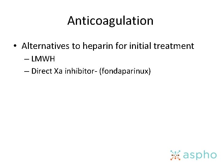
Anticoagulation • Alternatives to heparin for initial treatment – LMWH – Direct Xa inhibitor- (fondaparinux)
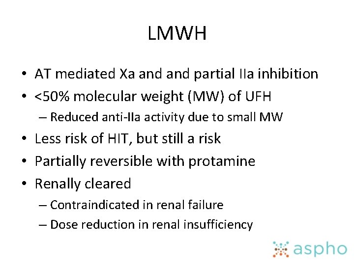
LMWH • AT mediated Xa and partial IIa inhibition • <50% molecular weight (MW) of UFH – Reduced anti-IIa activity due to small MW • Less risk of HIT, but still a risk • Partially reversible with protamine • Renally cleared – Contraindicated in renal failure – Dose reduction in renal insufficiency
Enoxaparin • Most clinical data for LMWH use in children is based on studies of enoxaparin • Peak anti-Xa level is 2 to 6 hours after administration – Obtain level 4 to 6 hours after – Goal 0. 5 to 1. 0 U/ml for treatment • 0. 1 -0. 3 for prophylaxis – Half life 4. 5 to 7 hours • Infants <3 months or 5 kg need higher doses – Larger volume of distribution
Enoxaparin • Monitoring in children recommended – Wide interpatient variability – Weight based; obesity and neonate concerns – Age; infants need higher dose – Decreased renal function increases risk of accumulation • Additional monitoring – Bleeding reported in 0 to 10% of patients • Primarily neonates
Anticoagulation • UFH – Continuous IV infusion – Monitored with PTT to correspond to an anti-factor Xa activity of 0. 3 to 0. 7 units – Good first choice in unstable patient or need procedure • Reversible with protamine and short half-life – Starting dose • 75 units/kg bolus*** • 20 units/kg/hr • 28 units/kg/hr- neonate • LMWH – Subcutaneous injection – Monitor with anti-factor Xa activity (goal 0. 5 to 1. 0 units) – Easier to use in stable patient – More predictable pharmacokinetics – Requires good renal function – Incomplete reversal with protamine – enoxaparin 1 mg/kg SQ BID – Neonates 1. 5 mg/kg
Fondaparinux • • AT mediated selected inhibition of Xa Renal clearance Peak level reached at 3 -4 hours Elimination half life 17 -21 hours Dose in kids 0. 1 mg/kg SQ daily Target therapeutic range 0. 5 - 1 mg/L Not reversible – Hemodialysis reduces level by 20% – r. FVIIa has treated bleeding
Anticoagulation • Hard to use heparin for all of therapy • May use LMWH for duration of therapy • Transition to warfarin or other Vitamin K antagonist – Must bridge with a heparin/LWMH/DTI – Acquired hypercoagulability for first 5 days of warfarin therapy • Protein C levels reduced before the Factor X levels • Potential for clot extension • Potential for warfarin skin necrosis
Vitamin K Antagonists (VKA) • Difficulty with VKA – Slow onset of action • HEPARIN BRIDGE – Multiple food and drug interactions • FREQUENT MONITORING of PT/INR – Narrow therapeutic index • FREQUENT MONITORING of PT/INR – Genetic polymorphisms • INCONSISTENT DOSING • Reversible – Vitamin K 2 -5 mg PO, SQ or IV • IV slow infusion due to anaphylaxis – FFP • Stop bleeding but not completely reverse – Prothrombin Complex Concentrates • Contain many Vit K dependent proteins at high concentration
Vitamin K antagaonists • Warfarin- most common – Monitor with PT/INR • Target range INR 2 -3 for most DVT/PE • 2. 5 -3. 5 for mechanical valves and some APS patients • INR is a calculation made to standardize prothrombin time. INR is based on the ratio of the patient's prothrombin time and the normal mean prothrombin time • Check pregnancy status prior to initiation in teen girls and change medication in pregnancy – Warfarin embryopathy
Monitoring Warfarin Therapy • INR can be altered by: – – – – Initiation and discontinuation of medications (antibiotics) Febrile illnesses Gastroenteritis- diarrhea Liver disease Growth Changes in nutrition Alcohol “Coumadin diet”- feeding tube issues • Alterations can place kids at risk for recurrent clot or for bleeding • Check at least once a month when stable, and when changes suspected
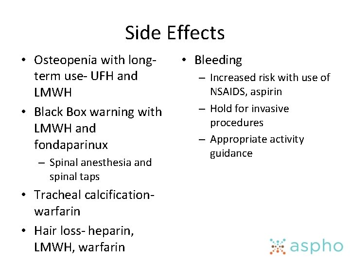
Side Effects • Osteopenia with longterm use- UFH and LMWH • Black Box warning with LMWH and fondaparinux – Spinal anesthesia and spinal taps • Tracheal calcificationwarfarin • Hair loss- heparin, LMWH, warfarin • Bleeding – Increased risk with use of NSAIDS, aspirin – Hold for invasive procedures – Appropriate activity guidance
Novel oral anticoagulant agents (NOAC)/Target Specific Oral Anticoagulant Agents (TSOAC) • Not currently indicated as first line therapy in children – Studies ongoing – Dosing and monitoring not established
Additional Treatment Considerations
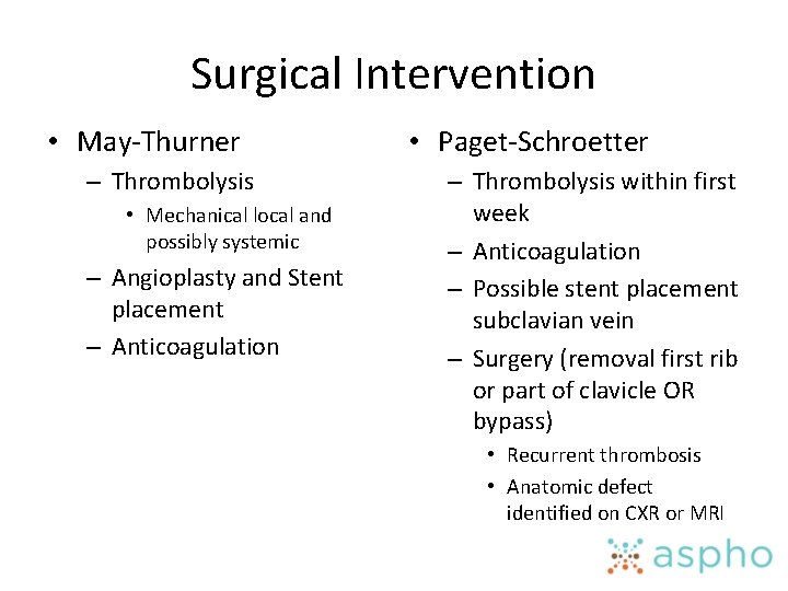
Surgical Intervention • May-Thurner – Thrombolysis • Mechanical local and possibly systemic – Angioplasty and Stent placement – Anticoagulation • Paget-Schroetter – Thrombolysis within first week – Anticoagulation – Possible stent placement subclavian vein – Surgery (removal first rib or part of clavicle OR bypass) • Recurrent thrombosis • Anatomic defect identified on CXR or MRI
IVC interruption • Indications – Large ileofemoral thrombosis when anticoagulation contraindicated • Temporary – Recurrent ileofemoral thrombosis and PE despite anticoagulation
Thrombolytic Agents • Indications – Arterial thrombosis- limb threatening – Intracardiac thrombus – Pulmonary embolus- massive and submassive – Extensive deep venous thrombosis • SVC syndrome • Occlusive IVC thrombosis • Bilateral renal vein thrombosis with renal compromise • Large iliofemoral clot • Contraindications – Active major bleeding including intracranial, retroperitoneal or gastrointestinal hemorrhage – Significant potential for uncontrolled local bleeding – Surgery within the preceding 10 days – Neurosurgery, including spinal surgery, within preceding 3 weeks – New stroke
Thrombolytic Therapy • Need to determine if systemic or catheter-directed – Catheter-directed less bleeding risk • Systemic dosing – Arterial Thrombosis: t. PA 0. 3 -0. 6 mg/kg/h for 6 -12 hours – Venous Thrombosis: t. PA 0. 1 -0. 5 mg/kg/h for 6 to 12 hours • Newer data suggests that lower doses for longer duration (12 to 24 hours) may also be effective for venous thrombosis. Consider 0. 03 -0. 06 mg/kg/h.
Thrombolytic Therapy • Monitoring/Therapeutic Goals – PTT/Fibrinogen/D-dimer every 6 hours – CBC every 12 hours, unless sudden drop in blood pressure or extensive active bleeding – Goals • Fibrinogen 100 -200 (expect to decrease by 20 -50% with adequate therapy) • D-dimers elevated • PTT 50 -70 (prophylactic heparin) • Hb stable (major bleeding if >2 g/dl drop) • Plt >100 K (transfuse to maintain)
Thrombolytic Therapy • Expect oozing from line/puncture sites, this is an indication that thrombolysis is occurring and NOT an indication to stop the TPA infusion. – Major bleeding • Drop of Hemoglobin by 2 g/dl • ICH, retroperitoneal, Intraspinal, intramuscular with compartment syndrome, intraarticular, etc • Neonates and infants may need FFP empirically prior to thrombolysis secondary to naturally low levels of plasminogen. • Continue Prophylactic heparin during t. PA infusion
Duration of Anticoagulant Therapy • Standard therapy – DVT/venous thrombosis at unusual sites: 3 -6 months – PE: 6 -12 months • Long term anticoagulaiton – Antiphospholipid antibody syndrome – Protein C, Protein S and AT deficiency- long term/indefinite – Combined inherited traits – Persistence of D-Dimer or FVIII elevation may lengthen course Goldeberg and Bernard Hematol Oncol Clin N Amer 2010; 151 -166
Hypercoagulable Thrombotic States ABP Content Specifications (1) GENERAL a. Know the important antithrombotic properties of vascular endothelium (SLIDE 8) b. Know the removal of the activated coagulation factors by the liver is important in preventing thrombosis (SLIDE 9) c. Know that estrogen containing contraceptives are associated with an increased risk of venous thromboembolism, stroke, MI (SLIDE 22, 31)
(1) Diagnosis a. Know how to evaluate a hypercoagulable state (SLIDE 15, 19, 24 -27) b. For each of the known hereditary thrombotic states, know how to diagnose the defect in infants and older children (SLIDE 10, 17, 25) c. Understand the clinical manifestations of antithrombin III and protein C and S deficiencies (heterozygous and homozygous) (SLIDE 14, 18, 20, 23) d. Know how to evaluate child with established deep vein thrombosis of unknown cause (laboratory tests, imaging) (SLIDE 37 -65) e. Know how the normal concentrations of hemostatic factors in neonate complicates the diagnosis of hypercoagulable states (SLIDE 20) f. Know the predisposing causes of DVT in infants children and adolescents (SLIDE 12, 30 -33) g. Know the acquired conditions that have been associated with venous and arterial thromboembolism (SLIDE 12, 13, 17, 22, 31 -33) h. Know the clinical signs and symptoms of venous thrombosis in children (SLIDE 38, 39, 43, 51, 56, 58, 60, )
a. Know the clinical signs and symptoms of pulmonary embolism in children (SLIDE 49) b. Know the laboratory measures important in evaluating children with venous thrombosis and pulmonary embolism (SLIDE 37, 62 -65) c. Know the clinical presentation, laboratory features and epidemiology of activated Protein C resistance (Factor V Leiden) (SLIDE 26) d. Know the problems inherent in diagnosing hypercoagulable states in patients receiving Coumadin therapy (SLIDE 25) e. Know the clinical presentation and lab diagnosis of patient with prothrombin mutation (SLIDE 27)
(1) Treatment a. General Considerations i. Know the treatment of a hypercoagulable state in newborn infant (SLIDE 20, 99) ii. Know the action of anticoagulant drugs used in thrombotic states or in patients with thrombophilia (SLIDE 67, 69, 79, 81, 85) iii. Know the indications for and treatment of acquired hypercoagulable states (SLIDE 20, 68, 93, 94, 99, ) b. Warfarin i. Recognize the embryopathic potential of warfarin therapy (SLIDE 88) ii. Know the mechanism of action of warfarin and other Vit K antagonists used as anticoagulant for therapeutic purposes (SLIDE 67) iii. Recognize the association of skin necrosis with warfarin therapy in patients with protein C or S deficiency (SLIDE 86) iv. Know the indications for use of Vit K antagonists (SLIDE 20, 68, 86) v. Know the importance of INR and its use in monitoring Vita K antagonists (SLIDE 87, 88) vi. The Recognize the many dietary, and drugs that can interact with VIt k antagonists (SLIDE 89)
a. Heparin i. Recognize the clinical and lab correlates or iatrogenic bleeding secondary to heparin administration (SLIDE 84, 90) ii. Know how to screen blood samples for presence of heparin (SLIDE 70) iii. Understand the structure and mechanism of action of heparin and heparin like substances (SLIDE 68, 69) iv. Know the causes of heparin resistance (SLIDE 72) v. Recognize the effect of heparin on coagulation assays in specimens from plasma, including those drawn from catheters (slide 69) vi. Know the syndrome and treatment of heparin induced thrombocytopenia (HIT) including association with thrombosis (SLIDE 73 -79) vii. Know the relative advantages and disadvantaged of standard heparin vs low molecular weight heparin (SLIDE 81, 84) b. Direct Thrombin Inhibitors i. Know the benefits , pharmacokinetics, monitoring of direct anti-thrombin inhibitors (SLIDE 79) ii. Know the various means to reverse Vit K antagonists (SLIDE 87) c. Fibrinolytic drugs i. Know the indications and risk for using fibrinolytic therapy (SLIDE 95) ii. Know how to monitor fibrinolytic therapy (SLIDE 96 -98)

Questions? • If your eyes are like mine and you need a word document of the tables, email me. • If you need the updated content specifications, email me • Janna. journeycake@childrens. com
- Slides: 105