THROMBOSIS Niazy B Hussam Defenition Thrombosis is the

THROMBOSIS Niazy B Hussam

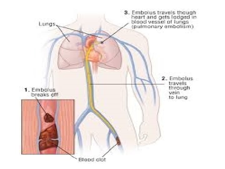
Defenition Thrombosis is the development of a ‘thrombus’ consisting • of platelets, fibrin, red cells and white cells in the arterial or • venous circulation. If part of this thrombus in the venous • circulation breaks off and enters the right heart, it may be • lodged in the pulmonary arterial circulation, causing • pulmonary embolism (PE). In the left-sided circulation, an embolus may result in peripheral arterial occlusion, either in the lower limbs or in the cerebral circulation (where it may cause thromboembolic stroke)
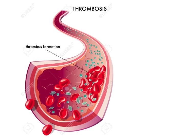

Hypercoagulation: more clotting activity than normal • Thrombosis: formation of platelet and/or fibrin mass in • a vessel Thrombus: stationary fibrin mass consisting of fibrin, • platelets and trapped cells Embolus: piece of thrombotic material that moves • Embolism: obstruction in circulatory system caused by • embolus Blood Clot: a mass that forms extravascularly, either in • vitro or in tissue

Plaque: consists of lipids, fibrous connective • tissue, macrophages and smooth muscle cells Thrombophlebitis: thrombus of superficial veins • of legs; self-limiting and benign Deep vein thrombosis: involvement of deep veins • of legs Thrombophilia: any disorder associated with an • increased tendency to cause venous thromboembolism Ischemia: Local obstruction of a blood vessel by a • thrombus

Venous thrombo embolism Epidemiology • VTE is common, with an incidence of 2– 5%. PE is • now the commonest cause of maternal death, and deep vein thrombosis may result in not only PE but also subsequent morbidity as a result of the post-phlebitic limb. Thromboembolism • appears to increase in prevalence over the age of • 50 years, and the diagnosis is more often missed in this age group


Aetiology VTE occurs primarily due to a combination of stagnation of • blood flow and hypercoagulability. Vascular injury is also a recognised causative factor but is not necessary for the development of venous thrombosis. In VTE, the structure of the thrombus is different from that in arterial thromboembolism. In the former, platelets seem to be uniformly distributed • through a mesh of fibrin and other blood cell components, • whereas in arterial thromboembolism the white platelet • ‘head’ is more prominent and it appears to play a much more important initiatory role in thrombus.

Protein C deficiency is inherited by an • autosomal dominant transmission. Such patients are at increased risk not only of VTE but also of warfarin skin necrosis. This occurs because protein C (and its closely related cofactor, protein S) is a vitamin K-dependent antithrombotic factor that can be further suppressed by the administration of warfarin. •

Protein S deficiency • Protein S deficiency is probably even rarer than protein C • deficiency, but the familial form, inherited in an autosomal • dominant fashion, is a high-risk state, accounting for • possibly 5– 8% of cases of thromboembolism in patients less than 45 years old. Factor V Leiden • The presence of factor V Leiden, a point mutation in the • factor V gene, causes the activated factor V molecule to • be resistant to deactivation by activated protein C (APC) • Fibrinogen gamma 10034 T Approximately 6% of individuals carry this variant gene, • which increases thrombotic risk approximately two fold. . •

Oestrogens • Oestrogens increase the circulating concentrations of clotting • factors I, II, VIII, IX and reduce fibrinolytic ctivity. They also depress the concentrations of antithrombin III which is protective against thrombosis. This effect is dose related, • and venous thrombosis was more often seen with the high (50 μcg) oestrogen-containing contraceptive Malignancy VTE is also commoner in malignancy (the risk may be up to fivefold • greater). Although first described in association with carcinoma of the pancreas, all solid tumors' seem to be associated Surgery The increased risk of VTE in surgery is related in part to • stagnation of venous blood in the calves during the operation and also to tissue trauma, since it appears to be more common in operations that involve marked tissue damage, such as orthopaedic surgery with this problem Other risk factor • There are several other patient-related risk factors for VTE Age over • 60 years is an important factor. Critical care Admission, dehydration, and one or more significant medical comorbidities such as heart disease, metabolic, endocrine or respiratory pathologies, acute infectious diseases and inflammatory conditions are all important risk factors for VTE

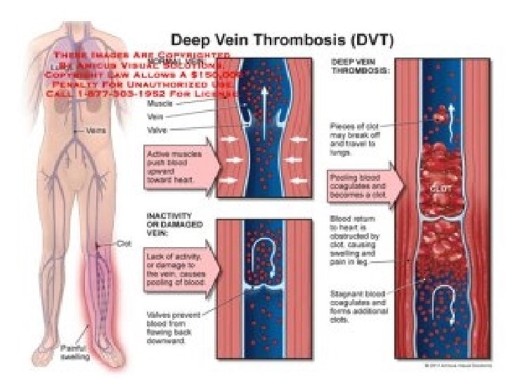
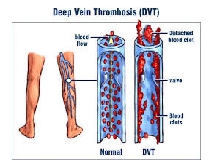
Clinical manifestations In 90% of patients, deep vein thrombosis occurs in the • veins of the lower limbs and pelvis. In up to half of cases, this may not result in local symptoms or signs, and the onset of PE may be the first evidence of the presence of VTE. In other cases, patients classically present with pain involving the calf or thigh associated with swelling, redness of the overlying skin and increased warmth. In a large deep venous thrombosis that prevents venous return, the leg may become discolored and oedematous. Massive venous thrombus can occasionally result in gangrene, although this occurs very rarely now that effective drug therapies are available


Investigations Deep vein thrombosis Venography involves injection of radio-opaque • contrast medium, normally into a vein on the top of the foot, • and subsequent radiography of the venous system. • Ultrasound is a non-invasive alternative to • venography that does not involve exposure to ionizing radiation • or potentially allergenic contrast media. It is now the initial • investigation of choice in clinically suspected deep vein thrombosis, although it is less sensitive for below-knee and isolated pelvic deep vein thrombosis. Magnetic resonance imaging (MRI). MRI is also non-invasive • and avoids radiation exposure. When used with direct • thrombus imaging (DTI), which detects methaemoglobin in • the clot, • •

Investagation -PE Pulmonary arteriography. The diagnosis of PE is most often • made using one of two techniques: pulmonary arteriography or • ventilation–perfusion scanning. Pulmonary arteriographyis the Ventilation–perfusion scanning involves the injection of a radiolabelled substance into the vein and measurement of perfusion via the pulmonary circulation , using a Spiral computed tomography. Computed tomography angiography (CT angiography) using helical or spiral CT (s. CT) is now being • increasingly used in some centers and has a high accuracy rate. •

Spiral computed tomography

Treatment The aim of treatment of venous thrombosis is to allow • normal circulation in the limbs and, wherever possible, to prevent damage to the valves of the veins, thus reducing the risk of the swollen post-phlebitic limb. Second, it is important to try to prevent associated PE and also recurrence of either venous thrombosis or PE in the risk period after the initial episode In acute massive PE, the initial priority is to correct the circulatory defect that has caused the haemodynamic upset, and in these circumstances, rapid removal of the obstruction using thrombolytic drugs or surgical removal of the embolus may be necessary. In acute submassive PE,

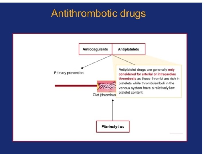
Treatment The treatment of VTE consists of the use of • anticoagulants and, in severe cases, thrombolytic drugs. Anticoagulant • therapy involves the use of immediate-acting agents (particularly heparin) and oral anticoagulants, the commonest of which is warfarin. Not only do these treat the acute event, but they also prevent recurrence and may be necessary for • some time after the initial event, depending on the persistence of risk factors for recurrent thromboembolism
 is a • heterogeneous mixture of large mucopolysaccharide")
Heparins Conventional or unfractionated heparin (UFH) is a • heterogeneous mixture of large mucopolysaccharide molecules ranging • widely in molecular weight between 3000 and 30, 000, with immediate anticoagulant properties. It acts by increasing the rate of the interaction of thrombin with antithrombin III by a factor of 1000. It, thus, prevents the production of fibrin (factor I) from fibrinogen. Heparin also has effects on the inhibition of production of activated clotting factors IX, X, XI and. XII, and these effects occur at concentrations lower than its effects on thrombin

Heparins The major adverse effect of all heparins is • haemorrhage which is commoner in patients with severe heart or liver disease, , renal disease, general debility and in women aged over 60 years. The risk of haemorrhage is increased in those with prolonged clotting times and in those given heparin by intermittent intravenous bolus rather than by continuous intravenous administration. UFH is monitored by derivatives of the activated partial thromboplastin time (APTT),
 • contain polysaccharide chains ranging in molecular • weight")
low molecular weight heparins (LMWHs) • contain polysaccharide chains ranging in molecular • weight between 4000 and 6000. Whereas UFH produces its anticoagulant effect by inhibiting both mbin and factor Xa LMWHs predominantly inactivate only factor Xa. In addition, unlike UFH, they inactivate platelet-bound factor Xa and resist inhibition by platelet factor 4 (PF 4), which is released during coagulation. Bemiparin, dalteparin, enoxaparin reviparin and tinzaparin are LMWHs with similar efficacy and adverse effects.

Heparinoids Danaparoid is a heparinoid that is licensed for • prophylaxis of deep vein thrombosis in patients undergoing general or orthopaedic surgery. It is a mixture of the low molecular weight sulphated glycosaminoglycuronans: heparin • sulphate, dermatan sulphate and a small amount of chondroitin sulphate. It acts by inhibiting factor Xa and, like LMWHs, is given by subcutaneous injection. It normally has a low cross-reactivity rate with heparin-associated antiplatelet antibodies and if this is not present can be used in the treatment of individuals who develop HIT but still need ongoing anticoagulation

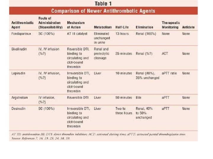
Hirudins Lepirudin, a recombinant hirudin, is licensed for • anticoagulation in patients with type II (immune) HIT who require • parenteral antithrombotic treatment. The dose of lepirudin is adjusted according to the APTT, and it is given intravenously by infusion. Haemorrhage is greater in those with poor • renal function. Severe anaphylaxis occurs rarely in association with lepirudin treatment and is more common posed patients Bivaluridin is an analogue of hirudin but acts as a direct • thrombin inhibitor. It is licensed for anticoagulation in patients undergoing percutaneous coronary intervention (PCI). It has to be administered parenterally and the activated clotting time (ACT) is used to assess its activity. Haemorrhage is also an important adverse effect of this agent.

Fondiparinux sodium is a synthetic • pentasaccharide that binds to antithrombin III, thus inhibiting factor Xa but without effect on factor IIa. Therefore, at doses normally used for treatment, it does not significantly affect coagulation tests and out line monitoring of these is not necessary. It has to be given parenterally. It is used for prophylaxis of VTE in high-risk situations and for treatment of acute deep vein thrombosis and treatment of acute PE, except in haemodynamically unstable patients or patients re thrombolysis or pulmonary embolectomy.

Oral anticoagulant Warfarin. Although not the only coumarin anticoagulant • available, warfarin is by far the most widely used drug in • this group because of its potency, duration of action and more • reliable bioavailability. Acenocoumarol (nicoumalone) has a much shorter duration of action and phenindione may be associated with a higher incidence of non-haemorrhagic adverse effects. When given by mouth, warfarin is completely and rapidly absorbed, although food decreases the rate (but not the extent) of absorption. It is extremely highly plasma protein-bound (99%) and, therefore, has a small volume of distribution (7– 14 L).

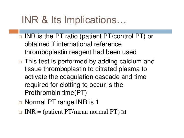
Warfarin The effect of warfarin is monitored using the one-stage • thrombin time, for example the international normalised ratio (INR). This test is sensitive chiefly to factors VII, II and X (and to a lesser extent factor V, which is not a vitamin K-dependent clotting factor). However, factor VII, to which the INR is sensitive, is the most important factor in the extrinsic pathway of clotting. The optimum therapeutic range for the INR differs for different clinical indications since the lowest INR consistent with therapeutic efficacy is the • best in reducing the risk of haemorrhage.

Dabigatran is an orally active inhibitor of both free and clot-bound • thrombin (Wittkowsky, 2010). It has a rapid onset of action and does not require laboratory monitoring. Dabigatran etexilate is a pro-drug which is hydrolysed to active dabigatran in the liver. Since 80% of activated dabigatran is excreted unchanged through the kidneys, it should be avoided in patients with severe renal impairment (creatinine clearance < 30 m. L/min) and the dose should be reduced in moderate renal impairment (creatinine clearance 30– 50 m. L/min). Dabigatran is a substrate for the transport protein p-glycoprotein (p-GP), which facilitates renal elimination • of certain drugs. Amiodarone, an inhibitor of p-GP reduces the • clearance of dabigatran and so doses should be reduced in patients who are on concurrent treatment with amiodarone.

Rivaroxaban is an orally active inhibitor of both the ‘free’ • and prothombinase complex-bound forms of activated factor X (Xa) (Wittkowsky, 2010). Two thirds of the dose is metabolised, principally by CYP 450 enzymes and the remaining third is excreted unchanged in the urine. Like dabigatran, Rivaroxaban also appears to be a p-GP substrate and it • should be used with caution when prescribed concomitantly with p-GP inhibitors and potent p-GP inducers. It should also be used with caution in patients with creatinine clearance less than 30 m. L/min (severe renal impairment) It is recommended as an option for prophylaxis of VTE in adults after hip or knee replacement surgery

Fibrinolytic drugs Thrombolytic therapy is used in life-threatening • acute massive pulmonary embolus. It has been used in deep vein thrombosis, particularly in those patients where a large amount of clot exists and venous valvular damage is likely. However, fibrinolytic drugs are potentially more dangerous • than anticoagulant drugs, and evidence is not available in situations other than acute massive embolism to show a sustained benefit from their use.

Streptokinase was the first agent available in • this class. It was produced from streptococci • and is a large protein that binds to and activates plasminogen, thus encouraging the breakdown of formed fibrin to fibrinogen degradation products. It also acts on the circulating fibrinogen to produce a degree of systemic anticoagulation
 or alteplase was • developed using recombinant DNA technology.")
Alteplase. Tissue plasminogen activator (rt-PA) or alteplase was • developed using recombinant DNA technology. Although this agent is much more expensive than streptokinase, it can be used in those situations where streptokinase may be less effective because of development of antibodies, for example within 1 year of previous streptokinase use or where allergy to streptokinase has previously occurred. Because it produces a • lesser degree of systemic anticoagulation (it is more active against plasminogen associated with the clot), immediate use of heparin uently is necessary to prevent recurrence of thrombosis. Reteplase and tenectaplase. Reteplase, and more recently tenecteplase, are also fibrin-specific agents and so heparin is • required to prevent rebound thrombosis. They are indicated for the treatment of acute myocardial infarction In this clinical situation, reteplase is administered as an intravenous bolus, followed by a second bolus 30 min later (double bolus), and tenecteplase is given as a single intravenous bolus.

Urokinase, like alteplase and streptokinase, can • be used for the treatment of deep vein thrombosis and PE. It • is also licensed to restore patency in intravenous catheters and • cannulas blocked by fibrin thrombi. • Patient care • The patient on oral anticoagulants should be given full information • on what to do in case of problems and what circumstances • and drugs to avoid. An anticoagulant card with previous INR values • and doses should also be provided. The patient should be told of the colour code for the different strengths of warfarin tablet and advised to carry their treatment card at all times. The likely duration of anticoagulant

Arterial thromboembolism Acute myocardial infarction is the commonest clinical • presentation of acute arterial thrombosis. Stroke is commonly • caused by atherothromboembolism from the great vessels or embolism arising from the heart (approximately 80% of strokes). These two conditions are discussed elsewhere Peripheral arterial thrombosis or thromboembolism may also occur, most often in the lower limb. Antiplatelet drugs are often used for prophylaxis, but surgical embolectomy and/ or fibrinolytic therapy may be needed for treatment of acute thrombotic or thromboembolic events to avoid consequent ischaemic damage.

Aetiology Arterial thromboembolism is normally associated with vascular • injury and hypercoagulability. Vascular injury is most often due to atheroma, itself aggravated by smoking, hypertension, • hyperlipidaemia or diabetes mellitus. Although the exact • mechanism is not clear, it is thought that platelet aggregation may be induced by the sheer stresses caused by stenosis of • anatherosclerotic vessel. This thrombotic material may embolise to cause occlusion further downstream. Hypercoagulability is also a risk factor. It may be associated with increased plasma fibrinogen levels and an increase in circulating cellular components for example polycythaemia or thrombocythaemia •
 is a potent inhibitor of the")
Treatment and prevention Aspirin • Aspirin (acetylsalicylic acid) is a potent inhibitor of the enzyme cyclo • -oxygenase, which catalyses the production of prostaglandins. It reduces the production of pro-aggregatory prostaglandin, thromboxane A 2 in the platelet, an effect that lasts for the life of the platelet Clopidogrel • Clopidogrel is a pro-drug that is metabolised in part to an active • thiol derivative. The latter inhibits platelet aggregation by rapidly and irreversibly inhibiting the binding of adenosine • diphosphate (ADP) to its platelet receptor, thus preventing the ADP -mediated activation of the glycoprotein IIb/IIIa receptor for the life of the platelet. It is an orally active pro- drug and is given once daily for the reduction of atherosclerotic events in those with preexisting atherosclerotic disease

Dipyridamole is used by mouth as an adjunct to oral anticoagulation • for prophylaxis for thromboembolism associated with prosthetic heart valves. Modified-release preparations are licensed (alone or preferably in combination with low dose • aspirin) for secondary prevention of ischaemic stroke and TIAs (see • treatment of stroke. Glycoprotein IIb/IIIa inhibitors prevent platelet aggregation by blocking the binding of fibrinogen to receptors on platelets. Abciximab is a monoclonal antibody which binds to • coronary glycoprotein IIb/IIIa receptors and to other related sites. It is licensed as an adjunct to heparin and aspirin for the prevention of ischaemic complications in high risk patients undergoing percutaneous transluminal coronary intervention.

Eptifibatide and tirofiban also • inhibit glycoprotein IIb/IIIa receptors; they are licensed for use with • heparin nd aspirin to prevent early myocardial infarction in patients with unstable angina or non-ST-segment • elevation myocardial infarction (NSTEMI). Abciximab, eptifibatide and tirofiban all have to be administered • parenterally and should be used by specialist clinicians only • Patient care • Aspirin is normally well tolerated at the doses used for stroke • prevention. However, it should not be given to patients with a history of gastro-intestinal ulceration. Since it may induce bronchospasm in susceptible individuals, it should be used cautiously in such circumstances. It is best tolerated if taken once daily as soluble aspirin after food. •
- Slides: 43