Thorax Region of the body between the neck


Thorax § Region of the body between the neck and abdomen § Flattened in front and behind, but rounded on the sides § The bony framework of the walls is called the thoracic cage, cage which is formed of: § Vertebral column posteriorly § Ribs & intercostal spaces on the sides § Sternum and costal cartilages anteriorly

• Superiorly: It communicates with the neck through an opening bounded: § Posteriorly by 1 st thoracic vertebra § Laterally by medial border of the 1 st ribs and their costal cartilages § Anteriorly by superior border of manubrium sterni • This opening is occupied: § In the midline, by the structures that pass between the neck and the thorax § On either sides, it is closed by a dense suprapleural membrane 1 st rib 1 Suprapleural membrane

Suprapleural Membrane § Tent shaped dense fascial sheet that covers the apex of each lung. § An extension of the endothoracic fascia § Extends approximately an inch superior to the superior thoracic aperture § It is attached: Laterally to the internal border of the first rib & costal cartilage At its apex to the transverse process of C 7 vertebra. Medially to the fascia covering the structures passing through the superior thoracic aperture

• Inferiorly: It communictes with the abdomen through a large opening bounded: § Posteriorly by the 12 th thoracic vertebra § Laterally by curving costal margin § Anteriorly by xiphisternal joint • This opening is closed by the diaphragm 12 Costal margin 2 th rib

• The thoracic cage: § Protects the lungs, heart and large vessels § Provides attachment to the muscles of thorax, upper limb, abdomen & back • The cavity of thorax is divided into: • A median partition, the mediastinum • Laterally placed pleurae & lungs

• Anterior wall: Cutaneous Nerves § Above the level of sternal angle: Supraclavicular nerves § Below the level of sternal angle: Segmental innervation by anterior and lateral cutaneous branches of the intercostal nerves • Posterior wall: § Segmental innervation by posterior rami of the thoracic spinal nerves

Thoracic Dermatomes

The Intercostal Space

Intercostal Space • It is the space between two ribs • Since there are 12 ribs on each side, there are 11 intercostal spaces. • Each space contains: § Intercostal muscles § Intercostal neurovascular bundle § Lymphatics

Intercostal muscles • Each intercostal space has three muscles: • External Intercostal • Innermost Intercostal • Supplied by corresponding intercostal nerves • Action: • Tend to pull the ribs nearer to each other § Strengthen the tissue of the space

External Intercostal Muscle • Most superficial • Fibers directed downward & forward • Origin: Origin from lower border of the rib above • Insertion: Insertion upper border of rib below • Extends from the rib tubercle behind to the costo-chondral junction in front • Deficient anteriorly & replaced by external (anterior) intercostal membrane Costo-chondral junction

Internal Intercostal Muscle • Intermediate layer • Fibers directed downward & backward • Origin: Origin from subcostal groove of the rib above • Insertion: Insertion upper border of rib below • Extends from the sternum in front to the angle of the rib behind • Deficient posteriorly & replaced by internal (posterior) intercostal membrane

Angle of the rib posterior intercostal membrane & internal intercostal muscle V CC S costochondral junction anterior intercostal membrane & external intercostal muscle

Innermost Intercostal Muscle • Deepest layer • Incomplete layer, divided into three portions • Fibers cross more than one intercostal space • Related externally to intercostal nerve and vessels, and internally to endothoracic fascia

Endothoracic Fascia • It is the extrapleural fascia that lines the wall of the chest • It is located between the muscles and bones of the thoracic wall and the parietal pleura, extends over the apex (cupola) of the pleura as the suprapleural membrane, and forms a thin layer between the diaphragm and the pleura.

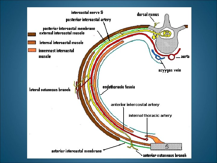
Intercostal Neurovascular Bundle • Lies between the innermost and the internal intercostal muscles • Runs high in the intercostal space, related to subcostal groove of the rib above • Has a strict order in arrangement: Vein-Artery-Nerve (VAN), from top to bottom • As the innermost intercostal muscle is not forming a complete layer, the bundle is generally covered on the inside by the endothoracic fascia

Intercostal Nerves • Twelve pairs • Are the anterior primary rami of the thoracic spinal nerves. • 1 -6 distributed in the intercostal spaces, 7 -11 th supply the anterior abdominal wall • Anterior ramus of 12 th nerve runs forward in the abdomen as the subcostal nerve

Intercostal Nerves cont’d • These are mixed nerves & supply the: • The skin • Muscles of the intercostal space & abdomen • The parietal pleura & parietal peritoneum • Branches: • • § § § Rami communicantes Collateral branches Lateral cutaneous Anterior cutaneous Muscular branches Pleural Peritoneal (7 th-11 th)

Atypical Intercostal Nerves • First thoracic nerve: • Has no anterior cutaneous branch • Is joined to the brachial plexus by a large branch that corresponds to the lateral cutaneous branch • Second thoracic nerve : • Is joined to the medial cutaneous nerve of the arm by brachial plexus by a branch called the intercostobrachial nerve that corresponds to the lateral cutaneous branch

Intercostal Arteries • Each intercostal space contains: § A single posterior & § Two anterior intercostal arteries • Each artery gives off branches to the muscles, skin, parietal pleura (& breast)

Posterior Intercostal Arteries • In the upper two spaces, arise from the superior intercostal artery (a branch of costocervical trunk of the subclavian artery) • In the lower nine spaces, arise from the branches of thoracic aorta • The course and branching of the intercostal arteries follow the intercostal nerves

Anterior Intercostal Arteries • In the upper six spaces, arise from the internal thoracic artery • In the lower five spaces arise from the musculophrenic artery (one of the terminal branch of internal thoracic) • Form anastomosis with the posterior intercostal arteries

Intercostal Veins • Accompany intercostal arteries and nerves • Each space has posterior & anterior intercostal veins • Eleven posterior intercostal and one subcostal vein • Lie deepest in the costal grooves • Contain valves which direct the blood posteriorly

Posterior Intercostal Veins • On right side: • The first space drains into the right brachiocephalic vein • Rest of the intercostal spaces drain into the azygos vein • On left side: • The upper three spaces drain into the left brachiocephalic vein. • Rest of the intercostal spaces drain into the hemiazygos and accessory hemiazygos veins, which drain into the azygos vein

Anterior Intercostal Veins • The lower five spaces drain into the musculophrenic vein (one of the tributary of internal thoracic vein) • The upper six spaces drain into the internal thoracic vein • The internal thoracic vein drains into the subclavian vein.

Lymphatics • Lymph vessels of the intercostal space conform to the general rule, that deep lymphatics follow arteries • Anteriorly drain into anterior intercostal nodes that lie along the internal thoracic artery • Posterioly drain into posterior intercostal nodes that lie in the posterior mediastinum

Applied Anatomy § Sternum: § used for marrow biopsy § May be split to make surgical access to heart, great vessels and thymus § Sternal angle as an important landmark for counting ribs, costal cartilages and intercostal spaces § Thoracic outlet syndrome: Compression of nerves /vessels at the superior aperture of thorax § Cervical rib § Referred pain: Disease in the thorax may reveal pain in the anterior abdominal wall… Why?

Applied Anatomy cont’d § Traumatic injuries to the thorax: § Fracture of rib is extremely painful condition as periosteum of the rib is supplied by the intercostal nerves above & below the rib § Fractured rib may penetrate the lung (and produce pneumothorax) or may damage the upper abdominal organs § Injuries involving multiple ribs result in flail chest. The flail segment is sucked in during inspiration and pushed out during expiration

Applied Anatomy cont’d § The intercostal spaces are important access points for: § Surgical procedures, e. g. resection of (part of) the lung § Percussion and auscultation of underlying structures e. g. heart & lung § To obtain a sample of pleural fluid or drain pus/blood from the pleural cavity. The needle/drain is passed through the intercostal space just above the upper border of the rib to avoid the neurovascular bundle.

- Slides: 33