Thorax and Abdomen Orthopedic Assessment III Head Spine

Thorax and Abdomen Orthopedic Assessment III – Head, Spine, and Trunk with Lab PET 5609 C

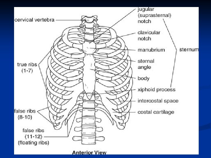
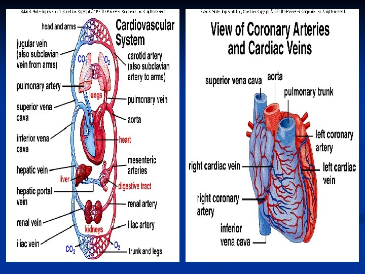
Clinical Anatomy n Thorax – bone cavity n n Formed by 12 pairs of ribs that join posteriorly with the thoracic spine and anteriorly with the sternum Thoracic Cavity: n n n Lined with a thin layer of tissue (pleura) One lung in each thoracic cavity Mediastinum is between the chest cavity n n Heart, Aorta, Superior and Inferior Vena Cava, Trachea, Major Bronchi, and Esophagus Spinal cord – protected by vertebral column

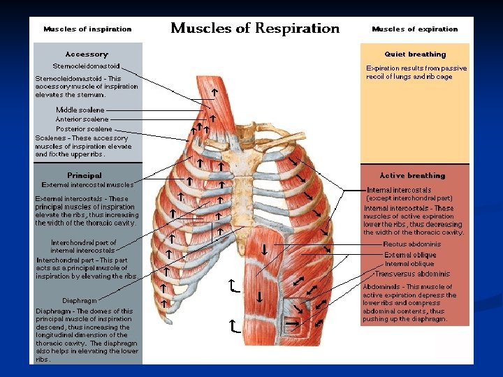
Clinical Anatomy n Muscles of Inspiration: n Diaphragm: n n Separates thoracic and abdominal activities Innervation: phrenic nerve Inhalation – diaphragm contracts enlarging the thoracic cavity and reducing intra-thoracic pressure (air drawn into lungs) Exhalation – diaphragm relaxes and air is exhaled by elastic recoil of the lungs

Clinical Anatomy

Clinical Anatomy n Muscles of Inspiration: n Intercostal muscles: n External intercostal muscles: (outside of the ribcage) n n Internal intercostal muscles: (inside the ribcage) n n n Depress the ribs decreasing the transverse dimensions of the thoracic cavity (aid in forced expiration) Scalene muscles: n n Elevate the ribs and expand the transverse dimensions of the thoracic cavity (aid in quiet and forced inhalation) Elevate the 1 st and 2 nd ribs SCM, trapezius, serratus anterior, pectoralis major/minor and latissimus dorsi (secondary muscles) Muscles of Expiration: n Abdominal muscles (rectus abdominis, internal/external obliques, transverse abdominis

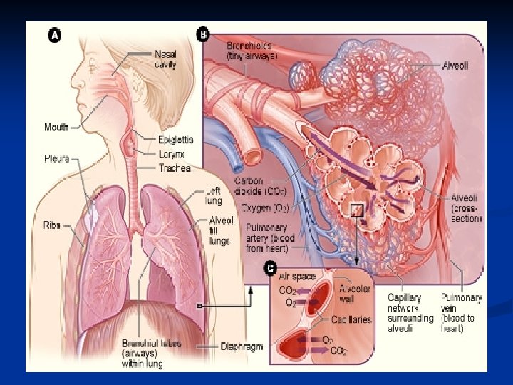
Clinical Anatomy n Respiratory Tract Anatomy: n Trachea: n n Pleura: n n n Connects larynx to 2 principle bronchi Left bronchus → 2 segmental bronchi (2 lobes) Right bronchus → 3 segmental bronchi (3 lobes) Parietal pleura – lines thoracic wall Visceral pleura – surrounds lungs Alveoli: n n n Terminal branches of bronchioles Gas exchange Capillary system → blood exchanged (pulmonary arteries and veins)

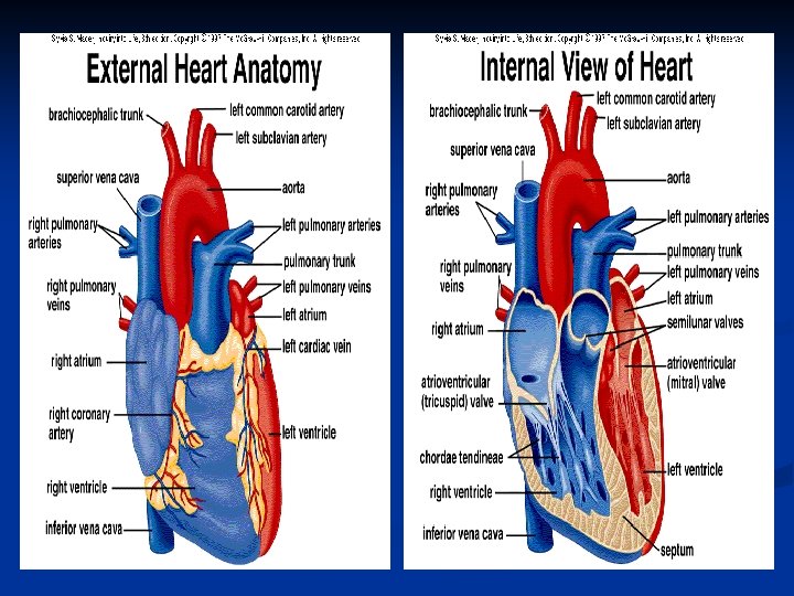
Heart Chamber Function Right Atrium Receives deoxygenated blood via: Superior vena cava (head, neck, upper extremities) Inferior vena cava (trunk and lower extremities) Role: Delivers blood to right ventricle Right Ventricle Receives deoxygenated blood from right atrium Role: Delivers blood to lungs via left and right pulmonary arteries Left Atrium Receives oxygenated blood from lungs via right and left pulmonary veins Role: Delivers blood to left ventricle Left Ventricle Delivers oxygenated blood through aortic valve to ascending aorta

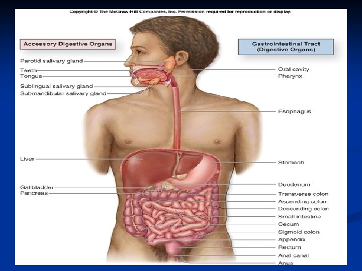
Clinical Anatomy n Digestive Tract Anatomy: n Esophagus: n Carries n food/liquid to stomach Small intestine: n Duodenum, n jejunum, ileum Large intestine: n Cecum, ascending colon, transverse colon, descending colon, sigmoid colon n Rectum and Anus

Clinical Anatomy n Lymphatic Organ Anatomy: n Spleen: n n n Left upper quadrant (level of 9 th-11 th ribs) Solid organ Function: n n n Produce and destroy red blood cells Blood reservoir Increased risk of injury → mononucleosis

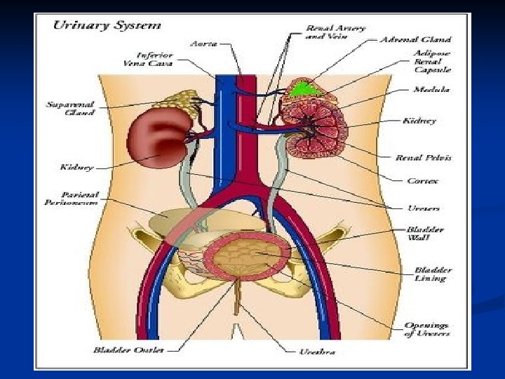
Clinical Anatomy n Urinary Tract Anatomy: n Kidneys: n n Filter blood Regulate electrolyte levels: n n Maintain balance of water, sodium, potassium Location: n Posterior part of the abdominal cavity: (level of T 12 – L 3 vertebrae) n Right kidney: sits below the diaphragm and posterior to the liver; sits slightly lower than left kidney n Left kidney: sits below the diaphragm and posterior to the spleen n Note: Lower portion of kidneys susceptible to trauma (unprotected by ribs)
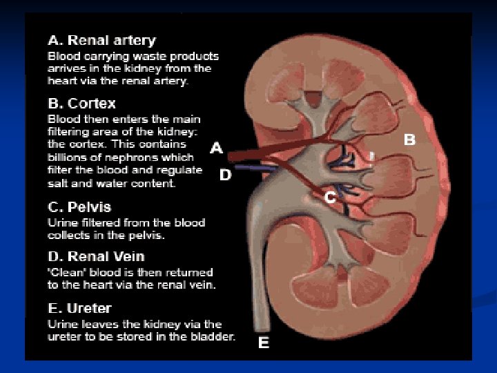

Clinical Anatomy n Urinary Tract Anatomy: n Ureters: n Muscular ducts that propel urine from the kidneys to the urinary bladder n n Urinary Bladder: n n Length: 10 -12 inches (adults) Solid, muscular, and elastic organ Collects urine excreted by the kidneys Urine enters the bladder via the ureters and exits by urethra Urethra: n n Tube connects urinary bladder to outside the body excretory function in both sexes (pass urine)

Clinical Evaluation n Anatomy: n n Abdominal cavity separated from the thorax by the diaphragm Lined with a membrane (Peritoneum) Lower portion of abdominal cavity: (Pelvic region) Surrounded by pelvis, vertebrae, and sacrum

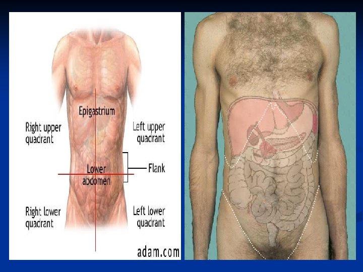
Clinical Evaluation Upper Right Quadrant Liver Kidney Pancreas Lung Upper Left Quadrant Heart, Lung Spleen Kidney Stomach Lower Right Quadrant Appendix Ureter Bladder Colon Gonads Lower Left Quadrant Ureter Bladder Colon Gonads

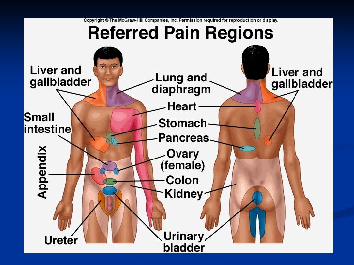
Clinical Evaluation n History: n Location of Pain: Musculoskeletal pain → ribs, costal cartilage, abdominal muscles (tender at injury site) n Injury to internal organs → diffuse pain; referred pain sites (Kehr’s sign) n n Onset of Symptoms: Gradual (internal bleeding can accumulate within cavity) n Pain ↑ with breathing (rib, abdominal injury) n n Mechanism of Injury: n Direct blow (thoracic, abdominal, pelvic injuries)

Clinical Evaluation n History: n Symptoms: n n n Medical History: n n Pain, difficulty breathing Diffuse abdominal pain Nausea, dizziness Vomiting of blood, blood in urine/stool Not common (acute injury) Exercise-induced asthma Illnesses (mononucleosis) General Medical Health: n Medications

Clinical Evaluation n Inspection: n n Start → observe patient’s posture Throat: n n Inspection: n n Rate, respiration rate, depth, quality Nail beds: n Capillary refill (cyanosis) Muscle tone Discoloration of skin: n n Breathing pattern: n n Position of trachea and larynx n Vomiting: n n Contusions, wounds, abrasion Presence of blood Hematuria

Clinical Evaluation n Inspection: n Auscultation: n Lungs: n Inhalation – smooth unobstructed sound n n n Absence: pneumothorax, collapsed lung Rales: pneumonia Abdomen: n Gurgling noises (peristalsis)

Clinical Evaluation n Palpation: n Sternum: n Manubrium, body, xiphoid process n Costal cartilage and ribs: n Palpate anterior to posterior n Pain, crepitus, deformity

Clinical Evaluation n Palpation: n Spleen: n n Palpate for enlarged spleen under left rib cage Have patient raise arms above head

Clinical Evaluation n Palpation: n Kidneys: n Location → under posterolateral portion of rib cage n Right kidney rests more inferior than left

Clinical Evaluation n Palpation: Liver n Method 1: Place your fingers just below the costal margin and press firmly n Ask the patient to take a deep breath n May feel the edge of the liver press against or slide under your hand n n Normal liver is not tender

Clinical Evaluation n Palpation: Liver n Method 2: n Hands "hooked" around the costal margin from above n Instruct patient to breath deeply to force the liver down toward your fingers

Clinical Evaluation n Palpation: Mc. Burney’s Point n n Location → one-third of way between right ASIS and naval Tenderness → may indicate acute appendicitis

Clinical Evaluation n Palpation: Abdomen n Rigidity: n n n Occurs secondary to muscle guarding or blood accumulation Indication of internal injury Rebound Tenderness: n Tests for peritoneal irritation. n n Palpate deeply and then quickly release pressure ↑ pain = peritoneal irritation

Clinical Evaluation n Palpation: Abdomen n Tissue density: Percussion n Patient position: hook-lying Examiner: Lightly places one hand over abdomen (palm down); Index/middle fingers of opposite hand tap the DIP joints Findings: (normal) n n n Solid organs have a dull thump Hollow organs more resonant sound Findings: (positive) n n Hard, solid sounding echo over areas that should sound hollow Internal bleeding

Clinical Evaluation Quadrant Pain: Upper Lower Right Left Liver: Pain associated with cholecystitis or liver laceration Gall bladder: Pain without trauma indicates gall bladder disease Spleen: Rigidity under the last several ribs Appendix: Rebound tenderness indicates appendicitis Colon: Colitis or diverticulitis may cause pain Pelvic inflammation: Diffuse tenderness

Clinical Evaluation n Vital Signs: n Heart Rate: n Pulse: n n n Normal pulse is 60 -100 beats per minute n n Athletes tend to have a slower pulse than non athletes (well-conditioned strong heart) Normal pulse is 60 -100 beats per minute n n Regular / Irregular Strong / Weak Athletes tend to have a slower pulse than non athletes (40 -60 bpm) Abnormal: n n Tacchycardia: > 100 bpm Bradycardia: < 60 bpm

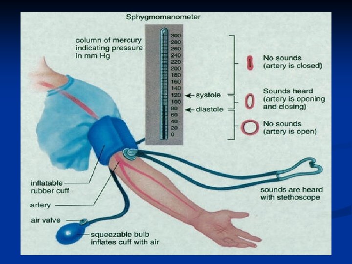
Clinical Evaluation n Vital Signs: Blood Pressure n Patient position: n n Seated or supine Procedure: n n n Cuff secured over upper arm Stethoscope placed over brachial artery Inflate cuff to 180 -200 mm Hg Air slowly released Note point at which 1 st pulse sound is heard Note point at which last pulse sound is heard

Clinical Evaluation n Vital Signs: Blood Pressure n Affected by: n Decrease in blood volume (severe bleeding or dehydration) – Hypovolemic shock n Decreased capacity of vessels (shock) n Rapid/weak pulse; ↓ BP n Decreased n ability of heart to pump blood ↓ nutrients/oxygen to organs of body (anoxia)

Clinical Evaluation n Vital Signs: Respiratory Rate n n Normal: 12 – 20 bpm Abnormal: n Rapid, shallow breaths: n n n Deep, quick breaths: n n n Internal injury Shock Pulmonary instruction Asthma Noisy, raspy breaths: n Airway obstruction

Clinical Evaluation n Rib Fractures: n Most common injured: n n 5 th-9 th ribs (anterior and lateral portions) History: n n Onset: acute (single traumatic blow) Pain: over fracture site n n ↑ pain with deep inspirations, coughing, sneezing, movement of torso MOI: n n Force (anteroposterior direction) – outward displacement Force (lateral side) – inward displacement n Internal injury (i. e. lungs)

Clinical Evaluation n Rib Fractures: n Inspection: n Splinting posture: n n Discoloration / swelling Shallow, rapid respirations (minimize chest movement) Palpation: n n Holding the painful area to limit chest wall movement during inspiration Point tenderness, crepitus, possible deformity Functional Tests: n n Movement of torso causes pain ↑ pain with deep respiration, coughing, sneezing

Clinical Evaluation n Rib Fractures: n Stress Fractures: n Rowing, swimming, golf n Posterolateral portion of 4 th-9 th ribs n Causes: n n n Overtraining, sudden increases in training Improper biomechanics Special Tests: n Rib n compression test: Contraindicated in presence of obvious fracture/lung trauma

Clinical Evaluation n Lateral Rib Compression Test: n Test position: n n Action: n n Subject supine Examiner compresses the lateral aspect of the rib cage then quickly releases Positive finding: n Pain with compression or release of pressure indicates possible rib fracture, contusion, or costochondral separation

Clinical Evaluation n Anterior/Posterior Rib Compression Test: n Test position: n n Action: n n Subject supine Compress rib cage anterior to posterior and quickly release Positive test: n Pain with compression or release of pressure indicates possible fracture, rib contusion, costochondral separation

Clinical Evaluation n Costochondral Injury: n MOI: n Overstretching the costochondral junction n n Hyperflexion Horizontal abduction “Snap” or “pop” at time of injury Symptoms: n n Anterior pain (cartilage junction) ↑ pain with deep breathing, coughing, sneezing

Clinical Evaluation n Pneumothorax: n n Accumulation of air in pleural activity Spontaneous pneumothorax: n Diagnosis dependent on signs/symptoms – rare condition n Contributing Factors: n n n Family history, tall and thin body build Sports-related spontaneous pneumothorax – documented in weight lifting, football, jogging Primary spontaneous pneumothorax: n n n Chest pain, dyspnea, diminished breath sounds Chest pain – usually localized to the side of the affected lung n Can radiate to shoulder, neck, back Primary cause: Bleb (imperfection in the lining of the lung) bursts causing lung to deflate Tall thin men (ages 20 -40) Secondary spontaneous pneumothorax:

Clinical Evaluation n Pneumothorax: n Tension pneumothorax: n n n One-way valve is created from either blunt or penetrating trauma Air can enter, CANNOT leave the pleural space ↑ Intrathoracic pressure will collapse the lung and ↑ pressure on mediastinum n Pressure will eventually collapse superior and inferior vena cava (loss of venous return)

Clinical Evaluation n Pneumothorax: n Clinical Signs: n n n Palpation: n n Apprehension / Agitation Cyanosis Diminished breath sounds Distended neck veins / Tracheal deviation Trauma induced – point tenderness Vital Signs: n n Labored, shallow respirations BP drops rapidly Right tension pneumothorax

Clinical Evaluation n Hemothorax: n n Blood enters the pleural space Massive Hemothorax – at least 1500 cc of blood loss into thoracic cavity n n n Penetrating injury Can occur from blunt trauma Blood accumulates → lung on the affected side is compressed n n Mediastinum may shift away from hemothorax Inferior and superior vena cava and contralateral lung may become compressed

Clinical Evaluation n Hemothorax: n Clinical signs/symptoms: Produced by hypovolemia and respiratory compromise n Anxiety, apprehension n Symptoms of hypovolemic shock n Decreased breath sounds or absence at injury site n Flat neck veins n

Clinical Evaluation n Spleen Injury: n History: n n Acute (symptoms may take a few hours to develop) Pain: n n n Predisposing conditions: n n Upper left quadrant Kehr’s sign – pain in upper left shoulder Mononucleosis: n ↑ mass, ↓ elasticity Inspection: n n Impact site – contusion Nausea and vomiting
 n")
Clinical Evaluation n Spleen Injury: n Palpation: n Cold and clammy skin (shock) n Pont tenderness n Rebound tenderness n Distention in upper left quadrant n Functional Tests: n Kerh’s sign n Low blood pressure

Clinical Evaluation n Kidney Pathologies: n Contused/Lacerated Kidney: n History: n n n Onset: acute Pain: posterolateral portion of upper lumbar and lower thoracic region MOI: blunt trauma or penetrating injury to kidney n Inspection: n n n Contusion or laceration Hematuria: n Severe bleeding → noticeable blood n Laboratory analysis needed Signs/symptoms of shock

Clinical Evaluation n Kidney Pathologies: n Palpation: n Point tenderness n Abdominal rigidity n Functional Testing: n Pain n with urination Laboratory Testing: n Hematuria

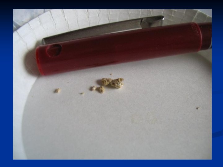
Clinical Evaluation n Kidney Stones: n Collection of incomplete kidney filtration n Causes: n n Crystals of uric acid, calcium 1 mm – 2. 5 cm Family history, stress, diet Signs: n n Pain with urination Pain (stone passed from bladder through urethra)

Clinical Evaluation n Urinary Tract Infections: n n n Bacterial infections of bladder or urethra Similar signs/symptoms of kidney stones Dysuria → frequent need to urinate Hematuria (abnormal urine color) Urethritis: n n n Inflammation of urethra Causes: chlamydia, gonorrhea, syphilis More common in males

Clinical Evaluation n Appendicitis and Appendix Rupture: Anatomy n n n Location: Lower Right Quadrant of Abdomen Elongated tube connected to the cecum (pouch-like structure of the colon) Function of the human appendix is unknown n Considered to be a remnant of a portion of the digestive tract which was once more functional and is now in the process of evolutionary regression

Clinical Evaluation n Appendicitis: n Cause: n n Inflammation caused by fecal obstruction, lymph swelling, tumor High incidence in males (ages 15 – 25) If bursts can bleed into peritoneal cavity and cause bacterial infection Signs and Symptoms: n n n Mild to severe pain in lower abdomen Nausea, vomiting, fever, cramping, abdominal rigidity, point tenderness Mc. Burney’s Point – between ASIS and umbilicus
: able to absorb")
Clinical Evaluation n Hollow Organ Rupture: n n Blunt trauma (non-rupture): able to absorb forces (deform/return to original shape without permanent injury) Rupture: n n Can be fatal (secondary to hemorrhage, peritoneal contamination) MOI and Signs/Symptoms: n n n Blow to abdomen Abdominal pain, possible nausea Palpation reveals guarding, rigidity, tenderness (point, rebound) Bowel sounds are absent (auscultation) Blood in stool

Clinical Evaluation n Gastritis: n Inflammation of stomach lining n Causes: n n Esophageal Reflux: n Backflow of gastric juices into esophagus n n n Aspirin or anti-inflammatory medications Alcohol Infection, bile entering stomach Heartburn, regurgitation of stomach acid Ulcer-like pain Intestinal Ulcers: n Irritation of duodenum (peptic ulcer) n n Abdominal pain, nausea, vomiting, dark stools, fatigue Causes: n n Bacteria Long-term use of aspirin or anti-inflammatory medications

Clinical Evaluation n Dyspepsia: n n Pain in upper abdomen Common causes: Gastroesophageal reflux disease (GERD), stomach ulcers n GERD – stomach acid splashes out of upper valve onto walls of esophagus n n Burning pain in mid-upper abdomen / heartburn Stomach Ulcers – wounds in lining of stomach n n Common causes: Stress, virus, diet Potential for bleeding if ulcers go untreated (open wounds)

Clinical Evaluation n Colitis: n Inflammation of the large intestine n Symptoms: n n n Causes: n n Disease, irritation of bowel, ulcers, ischemia, bacteria, stress Regional Enteritis (Crohn’s Disease): n n n Frequent diarrhea Abdominal pain, increased bowel sounds, fever, painful defecation, nausea, vomiting Affects the ileum Produces LRQ pain, cramping Irritable Bowel Syndrome: n n Alters motility of the muscles of large intestine Alternating bouts of diarrhea and constipation Abdominal pain Gas build-up, nausea, vomiting

Clinical Evaluation n Testicular Contusion: n n MOI: Direct blow Inspection: n Patient instructed to inspect for normal size/consistency n n Ruptured testicle – soft, inconsistent texture Testicular Torsion: n n Spermatic cord and testicle twisted within scrotum Symptoms: Acute testicular pain, swelling, tenderness Note: Immediate referral needed n
 n Female Athlete Triad: n")
Clinical Evaluation n Menstrual Irregularities: (associated with physical activity) n Female Athlete Triad: n Combination: n n n Disordered eating Amenorrhea Osteoporosis n Disorder n n n that often goes unrecognized Lost bone mineral density Premature osteoporotic fractures Lost bone mineral density may never be regained

Clinical Evaluation n Female Athlete Triad: n Disordered Eating: n n Anorexia, Bulimia, ENDOS Amenorrhea: n Related to athlete training/weight fluctuation is caused by changes in the hypothalamus n Result: n Decreased levels of Estrogen Primary Amenorrhea: n No spontaneous uterine bleeding: n n By the age of 14 without development of 20 sexual characteristics By the age of 16 with otherwise normal development

Clinical Evaluation n Female Athlete Triad: n Amenorrhea: n n Secondary Amenorrhea: n 6 -month absence of menstrual bleeding in a woman with primary regular menses n 12 -month absence with previous oligomenorrhea Osteoporosis: n n Loss of bone mineral density and inadequate formation of bone Premature osteoporosis: n Risk for stress fractures n Fx of hip, vertebral column

Hypertrophic Cardiomyopathy Heart muscle thickens reducing the size of the left ventricle n Increased risk of ventricular arrhythmia n

Marfan’s Syndrome Congenital connective tissue disorder that results in weakening of aorta and heart valves. n Abnormally tall and slender n

Commotio Cordis n Blunt trauma to the chest resulting in cardiac arrest.

Anomalous Origin of Coronary Artery n Results in abnormal or obstructed blood flow to the heart

Normal Changes in an Aerobically Trained Heart Increased L ventricle n Decreased resting HR n Systolic Murmer n Extra Heart Sounds n Decreased BP n
- Slides: 75