Thoraco Lumbar Fascia Nerve plexuses DR BINDHU S

Thoraco Lumbar Fascia &Nerve plexuses DR. BINDHU. S

�Fascia enclosing the deep muscles of the back. Extension- posterior layer covers the loin and is continued upwards on the back of the thorax and neck. Anterior &middle layers are confined to the lumbar region.

Anterior layer Middle layer Posterior layer

THORACO LUMBAR FASCIA posterior layer

Attachments � Posterior layer Medially – tips of lumbar and sacral spines Laterally- blends with middle layer Superiorly- continues on the back of the thorax where it is attached to the vertebral spines and angles of ribs. Inferiorly – outer lip of iliac crest.

Middle layer �Medially- tips of lumbar transverse process Laterally – blends with anterior layer Superiorly- lower border of 12 th rib and lumbocostal ligament Inferiorly- intermediate area of iliac crest.

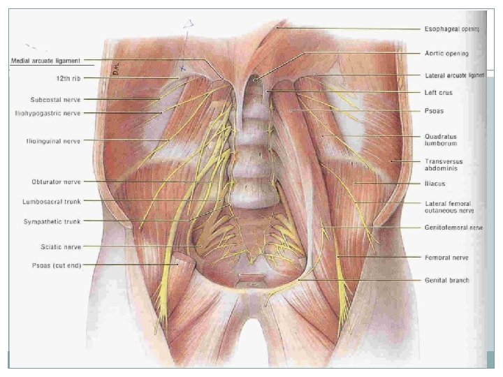
Anterior layer Medially – lumbar transverse processes. �Laterally- blends with middle layer �Superiorly – lateral arcuate ligament �Inferiorly- innerlip of iliac crest.

Anterior Layer

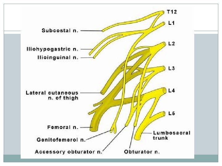
NERVE PLEXUSES �SOMATIC PLEXUSES – lumbar plexus, sacral plexus and coccygeal plexus. �AUTONOMIC PLEXUSES.
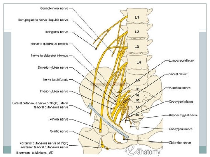

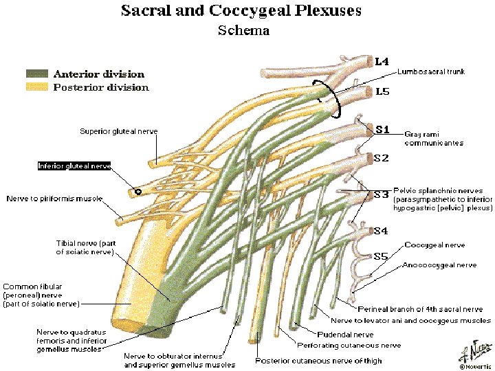
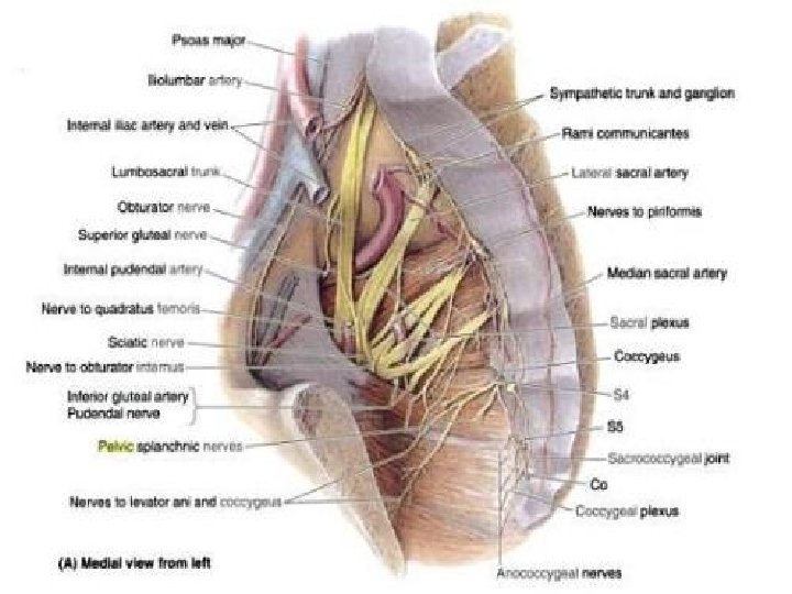
Sacral plexus formation branches �By lumboscral trunk �Sciatic nerve �Ventral rami of S 1 - S 4. �Posterior cut. nerve of thigh �Sup. gluteal, inf. gluteal nerve, nerve ti piriformis, �Quadratusfemoris, obt. int ernus, pudental nerve, br. to levator ani, pelvic splanchnic nerves.

AUTONOMIC NERVE PLEXUSES �SYMPATHETIC: Lumbar sympathetic trunk, Coeliac plexus from Splanchnic nerves. �PARASYMPATHETIC: Vagus to Coeliac plexus, Pelvic Splanchnic nerves from Inferior hypogastric plexus.

Lumbar sympathetic trunk 4 IN NUMBER. 1 st and 2 nd fused. Accessory ganglia may be present. Branches along lumbar nerves. Lies at the medial border of psoas major On the rt side it is overlaped by IVC and left side by lateral aortic nodes. � Ends by becoming continous with sacral part of symp. trunk. � Branches – grey rami carry fibers which are distributed to lower abdominal wall and lower limb. � 4 medial lumbar splanchnic nerves which form coeliac, aortic, and superior hypogastric plexus. � � �

Pelvic splanchnic nerves /nervi erigentes �Represents the sacral outflow of the parasympathetic nervous system �The nerves arise from ventral rami of S 2 -S 4. �They join the inferior hypogasric plexus and are distributed to the pelvic organs.

Coeliac ganglia and plexus �Largest ganglion in the body �Situated on each side of coeliac trunk �Larger upper part – receives greater splanchnic nerves �Lower smaller part – lesser splanchnic nerves

Coelic plexus formation �Coelia plexus is closely �Preganglionic fibers related to coeliac ganglion �Situated on the aorta around coeliac trunk and sup. mesenteric artery. �Overlaped by IVC. from greater and lesser splanchnic nerves �Post ganglionic fibers from coelic ganglion �Vagal fibers �sensory fibers from diaphragm.

Coeliac plexus branches �Phrenic plexus �Hepatic plexus �Left gastric �Suprarenal plexus �Renal plexus �Superior mesenteric plexus �Abdominal aortic plexus �Inferior mesenteric plexus

DISTRIBUTION

SUPERIOR HYPOGASTRIC PLEXUS formation �Lies in front of �Fibers from aortic bifurcation of aorta, left common iliac vein, L 5 �Also called presacral nerve. plexus � 3 rd and 4 th lumbar splanchnic nerve. �Pelvic splanchnic nerves

SUPERIOR HYPOGASTRIC PLEXUS �Branches- right and left hypogastric nerves �Ureteric, testicular and ovarian , common iliac plexus.
- Slides: 24