Thoracic Wall Dr Dawar Husain Skeleton of the


.")

 Origin : From the inner surface of the")


 artery Arises from the 1 st part of subclavian artery.")


- Slides: 35
Thoracic Wall Dr. Dawar Husain
Skeleton of the thoracic wall True ribs 1 -7 False ribs 8 -10 Floating ribs 11&12
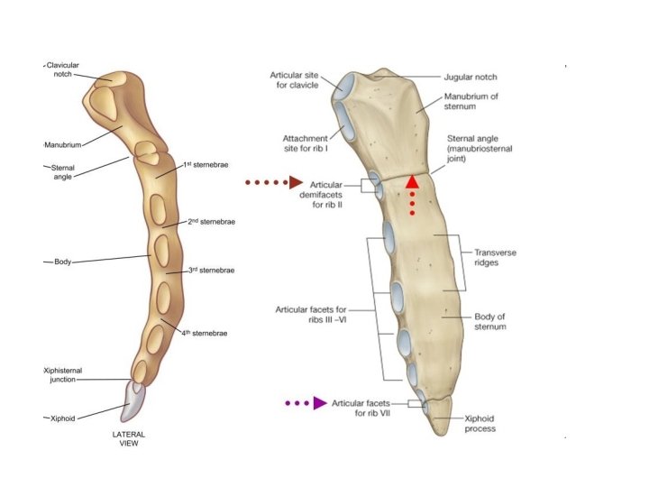
Joints related to the sternum
Superior thoracic aperture Thoracic inlet • The superior thoracic aperture consists of the body of vertebra TI posteriorly, the medial margin of rib I on each side, and the manubrium anteriorly. • The superior margin of the manubrium is in approximately the same horizontal plane as the intervertebral disc between vertebrae TII and TIII. • The first ribs slope inferiorly from their posterior articulation with vertebra TI to their anterior attachment to the manubrium. .
Inferior thoracic aperture Thoracic outlet • The inferior thoracic aperture is large and expandable. Bone, cartilage, and ligaments form its margin • The inferior thoracic aperture is closed by the diaphragm, and structures passing between the abdomen and thorax pierce or pass posteriorly to the diaphragm. • Skeletal elements of the inferior thoracic aperture are: • the body of vertebra T XII posteriorly; • rib XII and the distal end of rib XI posterolaterally; • the distal cartilaginous ends of ribs VII to X, which unite to form the costal margin anterolaterally; and the xiphoid process anteriorly.
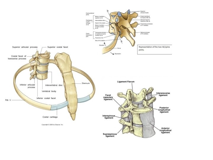
Joints of Skeleton of the thoracic wall
The intervertebral disc in between T 2 and T 3 vertebrae T 3 T 4 The intervertebral disc in between T 4 and T 5 vertebrae T 5 T 6 T 7 T 8 The Upper border of 9 th Thoracic vertebra T 9
Intercostal spaces
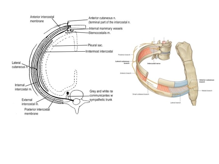
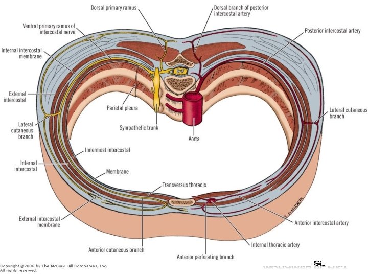
Thoracic wall Layers of thoracic w all : 1 skin 2 superficial fascia Intercostal 3 -intercostal muscles 4 - pleura 5 - lung . Vein Artery Nerve Collateral branches
Intercostal muscles 1 -They are arranged in three layers. 2 - Are supplied by intercostal and subcostal nerves. 3 - Act in respiratory movements External intercostal Internal intercostal Transversus thoracis
Intercostal muscles
External intercostal muscle Origin: From the sharp lower border of the rib above. Insertion: In the blunt upper border of the rib below. Direction of fibers : Downwards and forwards. Extension : From the tubercle of the rib behind to the costo-chondral junction where it is replaced by the external (anterior) intercostal membrane. Action: Inspiratory.
Internal intercostal muscle Origin: From the above rib the inner (lateral surface to groove). Insertion: In the blunt upper border of the rib below. Direction of fibres: Downwards and backwards. Extension: From the lateral margin of the sternum infront to the angle of the rib, where it is replaced by the internal (posterior) intercostal membrane. Action : Expiratory. the of costal
Transversus thoracis muscle Represented by 3 sheets of muscles : • sterno-costalis • innermost intercostal • Subcostales , A- Sterno-costalis Origin: From the lower part of the internal surface of the body of the sternum and xiphoid process. Direction of fibres: Form 5 digitations, which diverge from the origin upwards and laterally. Insertion: Inner surface of the 2 nd to 6 th costal cartilages.
B- Innermost intercostals ( Intercostalis intimis) Origin : From the inner surface of the rib above (medial to the costal groove). Direction of fibers : Downwards and backwards crossing more than one intercostal space. Insertion: Into the upper border of the 2 nd or 3 rd rib below.
Subcostalis Origin: From the inner surface of the rib Direction of fibres: Downwards & backwards crossing more than one space Insertion : Into the upper border of the 2 nd or 3 rd rib below. Action of transversus thoracis : Support the sterno-costal & costo- transverse joints during movements of ribs.
Intercostal Nerves There are 11 pairs of intercostal nerves and one pair below 12 th rib (subcostal nerve). Typical intercostal nerves From 3 rd to 6 th Atypical intercostal nerves 1 1 st 2 2 nd 3 lower five intercostal and subcostal
Typical Intercostal Nerves C o u r s e & relations: u Posteriorly : There are 11 pairs of intercostal nerves and one pair below 12 th rib (subcostal nerve). The nerve lies between the parietal pleura and posterior intercostal membrane It crosses behind the intercostal vessels. Laterally : (in the costal groove). o It runs in the costal groove. o It lies between the internal intercostal and the innermost intercostal muscles. o It runs below the intercostal vessels. From above downwards the arrangement is vein, artery & nerve (VAN). u. Anteriorly : (medial to costo-chondral junction). It crosses in front of the internal thoracic artery. • u Terminally: It is directed forwards piercing the internal intercostal muscle, then the external intercostal membrane to end in the skin as the anterior cutaneous nerve.
1. Branches: 1 - Rami communicantes: A white and grey rami to the corresponding thoracic ganglion. 2. Collateral branch: Runs parallel to the main nerve along the upper border of the next rib It supplies parietal pleura, periosteum of the rib and muscles of the space. It has no cutaneous branch. 3 Lateral cutaneous branch: Pierces the internal and external intercostals and come out in the mid-axillary line. Each divides into anterior and posterior divisions. 4 Anterior cutaneous branch: It is the termination of the intercostal nerve. Pierces the internal intercostal muscle and external intercostal membrane Each divides into medial and lateral divisions. 5 Muscular branches: Supply the intercostal muscles.
5 - Anterior branch Medial branch Lateral branch Sternum Sternocostalis Anterior branch 1 - Rami communicants 3 - Collateral Branch 4 - Lateral cutaneous branch Posterior branch 2 - Muscular branches
Atypical Intercostal Nerves 1 1 st intercostal nerve: Its main part ascends in front of the neck of the 1 st rib to join lower trunk of the brachial plexus. 2 2 nd intercostal nerve: Its course and branches are similar to the typical intercostal nerves , but it differs in that its lateral cutaneous branch does not divide into anterior and posterior branches, but directed posteriorly and crosses the axilla to supply the skin of the upper part of the medial side of the arm, so called intercosto- brachial nerv. e 3 Lower 5 intercostal nerves (7 th to 11 th ): • They supply the anterior abdominal wall. • The 10 th nerve supplies the skin at the level of the umbilicus. 4 - Subcostal nerve (12 th) • Runs below the 12 th rib and ends above the symphysis pubis.
The intercostobrachial nerve of the Second Intercostal Nerve
3 - The Lower 5 Intercostal Nerves The Lower 5 intercosal nerves
The Intercostal Arteries The Anterior and The Posterior Intercostal Arteries Costocervical Trunk Superior Intercostal Artery Left Subclavian Artery Aorta Internal Thoracic (mammary) Artery 1 1 collateral 2 collateral 3 4 collateral 4 5 collateral 5 6 collateral Superior Epigastric Artery 3 2 7 7 collateral 8 8 Musculophrenic Artery 9 collateral 9
Anterior intercostal arteries Subclavian artery Internal mammary artery Two small arteries in each space except the last 2 spaces which have no anterior arteries. Origin : Upper 6 spaces from the internal mammary artery. 7 th, 8 th & 9 th spaces from the musculophrenic artery. 10 th & 11 th spaces there are no anterior arteries. Superior epigasteric artery Musclophrenic artery
1. Internal thoracic (mammary) artery Arises from the 1 st part of subclavian artery. Descends vertically one finger breadth lateral to the sternum. Accompanied by venae commitantes from 3 rd to 6 th space. It ends in the 6 th space by giving two terminal branches; musculophrenic & superior epigastric arteries. Branches: 1 - Pericardiaco-phrenic artery 2 - Anterior intercostal arteries: 2 in each of the upper 6 spaces. 3 - Perforating branches: They accompany the anterior cutaneous branches of the intercostal nerves 4 Musculo-phrenic artery: Passes along the costal margin. 5 Superior epigastric artery: Descends vertically and ends by anastomosing with the inferior epigastric artery
1. Posterior intercostal arteries 1. Each space contains 2 anterior and 1 posterior arteries. 2. Each posterior artery anastomoses with the 2 anterior arteries. 1 st and 2 nd arteries: Arise from the superior intercostal artery, a branch of the costo- cervical trunk of the 2 nd part of subclavian artery. It descends in front of the neck of the 1 st rib. Lower 9 intercostal and subcostal arteries: Arise from descending thoracic aorta. Branches: · Dorsal: - Arises at the neck of the rib to supply the back. - It gives off a spinal branch for the spinal cord. · Collateral: Arises near the angle of the rib. · Lateral cutaneous: Arises near the angle of the rib.
commitantes of the musculo-phrenic artery. From the 6 th to 3 rd spaces; end in the venae commitantes of the internal thoracic artery. From the 1 st & 2 nd spaces; end in the internal thoracic vein which runs medial to the artery. venae commitantes of the musculo-phrenic artery. venae commitantes of the superior epigasteric Internal mammry vein From the 9 th, 8 th and 7 th spaces, end in the venae superior epigasteric 9 in number in each space. venae commitantes of the Anterior intercostal veins
Posterior intercostal veins - There are 11 pairs arranged as follows: - 1 st space drains into corresponding brachio-cephalic vein. -2 nd & 3 rd spaces drain into superior intercostal vein which: On the right side ends in the arch of azygos vein. On the left side ends in the left brachiocephalic vein. • - 4 th to 11 th spaces. • - On the right side: end in the azygos vein. • - On the left side: o From 4 th to 8 th join the accessory hemi-azygos vein. o From 9 th to 11 th join the hemi-azygos. -The 12 th vein ( subcostal vein) unites with the ascending lumbar vein.
AZYGOS VEIN It arises in the abdomen from the back of inferior vena cava. It enters the thorax with the aorta, where it ascends to its right border. At the level of the sternal angle, it forms an arch which ends in the superior vena cava. Runs on vertebral bodies on the right side of the descending thoracic aorta and then behind the right border of the oesophagus. Ascends behind the root of right lung and then curves forwards above the root, forming the arch of vena azygos which crosses on the right side of oesophagus, trachea and right vagus nerve to end in the middle of the superior vena cava. Tributaries : o Right ascending lumbar vein in the abdomen. o Right subcostal vein. o. Right posterior intercostal veins from the 4 th to 11 th. o Right superior intercostal vein. o Superior and inferior hemi-azygos veins. o Bronchial veins from right lung. o Some oesophageal and pericardial veins. N. B. All venous blood from thoracic wall drains into superior vena cava.
Hemi-azygos veins These 2 veins lie on the left side of the vertebral column and drain the left side of the thoracic wall. They are the superior and inferior hemi-azygos veins : 1. Superior hemi-azygos vein It receives the left posterior intercostal veins from the 4 th to 8 th spaces. It crosses the median plane to the right side at the 7 th thoracic vertebra 3. behind the aorta, oesophagus and thoracic duct to join the azygos vein. 1. Inferior hemi-azygos vein It arise either from the back of the left renal vein or from union of the left ascending lumber and left subcostal veins It enters the thorax by piercing left crus of diaphragm It receives the lower left posterior intercostals veins from 9 th to 11 th It receives also oesophageal and mediastinal veins It crosses the median plane to the right side at 8 th thoracic vertebra to join the azgos vein.