THORACIC WALL ABDOMINAL REGION VERTEBRAL COLUMN MUSCLES Salvador

THORACIC WALL ABDOMINAL REGION VERTEBRAL COLUMN MUSCLES Salvador Dali - Anthropomorphic Chest of Drawers, 1936 Kaan Yücel M. D. , Ph. D. 9. March. 2015 MONDAY

1. REGIONS/T ERMS Thoracic cavity between neck and abdomen protected by the thoracic wall. Thoracic wall bounds the thoracic cavity. formed by the skin, bones, fasciae, and muscles. Thoracic cage bony portion of the thoracic wall thoracic skeleton

1. 2. SURFACES OF THE THORAX anteriorly 12 THORACIC VERTEBRAE & POST. RIBS posteriorly RIBS & INTERCOSTAL SPACES laterally STERNUM & COSTAL CARTILAGES Posterior surface 12 thoracic vertebræ & posterior parts of the ribs Anterior surface sternum & costal cartilages Lateral surfaces ribs, separated by the intercostal spaces

1. 3. BOUNDARIES OF THE THORAX Superior • • • Jugular notch Sternoclavicular joint Superior border of clavicle Acromion Spinous processes of C 7 Inferior • • Xiphoid process Costal arch 12 th and 11 th ribs Vertebra T 12

1. 4. CONTENTS OF THE THORAX Organs of the cardiovascular, respiratory, digestive, reproductive, immune, and nervous systems
 muscles between the ribs skin")
2. THORACIC WALL • • • thoracic cage (skeleton) muscles between the ribs skin subcutaneous tissue muscles, and fascia covering its anterolateral aspect. The mammary glands of the breasts lie within the subcutaneous tissue of the thoracic wall.
 Protects vital thoracic and abdominal organs")
2. 1. FUNCTIONS OF THE THORACIC WALL 1) Protects vital thoracic and abdominal organs 2) Resists the negative (sub-atmospheric) internal pressures generated by the elastic recoil of the lungs and inspiratory movements. 3) Provides attachment for and support the weight of the upper limbs. 4) Provides the origins of many of the muscles that move and maintain the position of the upper limbs relative to the trunk. 5) Provides attachments for muscles of the abdomen, neck, back, and respiration.
 12 pairs of ribs and associated costal")
3. SKELETON OF THE THORACIC WALL 1) 12 pairs of ribs and associated costal cartilages 2) 12 thoracic vertebrae and the intervertebral (IV) discs interposed between them 3) Sternum

4. THORACIC APERTURES ‘Thoracic inlet’ ‘Thoracic outlet’

4. 1. Superior thoracic aperture “doorway” between the thoracic cavity and the neck and upper limb bounded: Posteriorly vertebra T 1 Laterally 1 st pair of ribs and their costal cartilages Anteriorly superior border of the manubrium q Trachea q Esophagus q nerves, and vessels that supply and drain the head, neck, and upper limbs.

4. 2. Inferior thoracic aperture By closing the inferior thoracic aperture, the diaphragm separates the thoracic and abdominal cavities almost completely. bounded: Posteriorly 12 th thoracic vertebra Posterolaterally 11 th and 12 th pairs of ribs Anterolaterally joined costal cartilages of ribs 7 -10 costal margins Anteriorly xiphisternal joint

6. MUSCLES OF THE THORACIC WALL Serratus posterior Levator costarum Intercostal muscles(External, internal and innermost) Subcostal Transverse thoracic

6. MUSCLES OF THE THORACIC WALL intercostal muscles three flat muscles found in each intercostal space external intercostal muscles most superficial internal intercostal muscles between external & innermost muscles

6. MUSCLES OF THE THORACIC WALL external intercostal muscles extend from the inferior margins of the ribs above to the superior margins of the ribs below pass obliquely anteroinferiorly

6. MUSCLES OF THE THORACIC WALL internal intercostal muscles most active during expiration between most inferior lateral edge of costal grooves of the ribs above, to the superior margins of ribs below in the opposite direction to those of the external intercostal muscles obliquely posteroinferiorly Attachment to interosseus parts move down during expiration!

6. MUSCLES OF THE THORACIC WALL innermost intercostal muscles least distinct of the intercostal muscles same orientation as the internal intercostals neurovascular bundles (V. A. N. ) in the costal grooves in a plane between innermost & internal intercostal muscles.

6. MUSCLES OF THE THORACIC WALL transversus thoracis muscles on the deep surface of the anterior thoracic wall & @ same plane as innermost intercostals lie deep to the internal thoracic vessels secure these vessels to the wall.

6. MUSCLES OF THE THORACIC WALL subcostales @ same plane as innermost intercostals Fibers parallel the course of the internal intercostal muscles. Extend from the angle of the ribs to more medial positions on the ribs below.

6. 1. Accessory muscles of respiration v upper accessory muscles assist with inspiration. v upper chest, and abdominal muscles assist with expiration.

9. VASCULATURE OF THE THORACIC WALL Mainly posterior and anterior intercostal arteries pass between adjacent ribs in intercostal spaces.

9. 1. ARTERIES OF THE THORACIC WALL Arterial supply to the thoracic wall derives from Thoracic aorta [posterior intercostal & subcostal arteries] Subclavian artery [internal thoracic & supreme intercostal arteries] Axillary artery [superior & lateral thoracic arteries]

intercostal arteries course through the thoracic wall between the ribs. Each intercostal space is supplied by • a large posterior intercostal artery • small pair of anterior intercostal arteries Exception last two intercostal spaces

• Near the angles of the ribs, the nerves pass between internal intercostal & innermost intercostal muscles. V. A. N. • Neurovascular bundles sheltered by the inferior margins of the overlying rib.

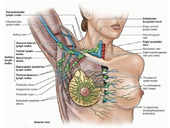
11. BREASTS Reproduction, back pain Aesthetics, and breast cancer Mammary glands & associated skin -connective tissues. modified sweat glands in the superficial fascia anterior to the pectoral muscles and the anterior thoracic wall.
 Axillary lymph nodes Most of the remaining (medial breast quadrants)")
75% (lateral breast quadrants) Axillary lymph nodes Most of the remaining (medial breast quadrants) parasternal lymph nodes or to the opposite breast Lymph from inferior quadrants may pass deeply to abdominal lymph nodes.

The 20 -30 axillary nodes are divided into 5 groups - on the basis of location. The groups are arranged in a manner that reflects the pyramidal shape of the axilla. Humeral (lateral) nodes Pectoral (anterior) nodes Subscapular (posterior) nodes Central nodes Apical nodes

1. Where is the pectoral region? The pectoral region is external to the anterior thoracic wall and anchors the upper limb to the trunk. 28

2. What does the pectoral region consist of? Superficial compartment Contains skin, superficial fascia, breasts Deep compartment Contains muscles & associated structures 29

3. What are the muscles of the pectoral region? Four pectoral muscles move the pectoral girdle Pectoralis major Pectoralis minor Serratus anterior Subclavius Clavicular head: Medial half of clavicle Medial process border of Coracoid of scapula Sternocostal head: Anterior surface of sternum, superior six intertubercular sulcus of costal cartilages, their costal medially humerus Protracts cartilages & rotates aponeurosis of external rotates Lateral parts of oblique muscle scapula humerus 1 st-8 th ribs Stabilizes Lateral lip of scapula 3 rd-5 thand ribs near Adducts 30

Serratus anterior Pectoralis major Pectoralis minor 31

4. What movements does the pectoralis major muscle ? Pectoralis major powerful adduction medial rotation of the arm clavicular head flexing the humerus sternocostal head extending it back 32

33

34

35

MUSCLES OF THE BACK Extrinsic back muscles Superficial group Movements of the upper limb Intermediate group Attached to the ribs May serve as a respiratory function. Intrinsic (deep) back muscles Act on the vertebral column Its movements Maintain posture

SUPERFICIAL GROUP OF BACK MUSCLES Produce and control limb movements. Trapezius Latissimus dorsi Rhomboid major Rhomboid minor Levator scapulae
 Apley scratch test. can")
LATTISIUMUS DORSI Extend Adduct Medially rotate humerus (arm, upper limb) Apley scratch test. can also depress the shoulder, preventing its upward movement

LATTISIUMUS DORSI In combination with pectoralis major powerful adductor of the humerus plays a major role in downward rotation of the scapula in association with this movement

LEVATOR SCAPULAE acts with the descending part of the trapezius to elevate the scapula, or fix it! resists forces that would depress it, as when carrying a load.

RHOMBOID MAJOR RHOMBOID MINOR Deep to trapezius, inferior to levator scapulae Broad parallel bands Pass inferolaterally from vertebrae to medial border of scapulae


INTERMEDIATE GROUP OF BACK MUSCLES

SERRATUS POSTERIOR SUPERIOR SERRATUS POSTERIOR INFERIOR Deep to the muscles in the superficial group. Related to the movements of the thoracic cage Superficial respiratory muscles More likely proprioceptive rather than motor in function

SUBOCCIPITAL REGION muscle “compartment” deep to the superior part of the posterior cervical region under trapezius, sternocleidomastoid, splenius, and semispinalis muscles

SUBOCCIPITAL REGION pyramidal space inferior to the external occipital prominence includes the posterior aspects of vertebrae C 1 and C 2

MUSCLES 4 small muscles deep to the semispinalis capitis muscles two rectus capitis posterior (major and minor) two obliquus capitis (superior and inferior) muscles inferior nuchael line

Do not get confused! Superior surface of transverse process of atlas Inferior surface of jugular process of occipital bone Flexes head laterally to same side PREVERTEBRAL MUSCLES Anterior surface of lateral part of atlas and its transverse process Inferior surface of basilar part of occipital bone Flexes head at atlanto-occipital joint Branches from anterior rami of C 1, C 2

Suboccipital Triangle A region of the neck bounded by the following 3 suboccipital muscles: Superomedial boundary: Rectus capitis posterior major muscle Superolateral boundary: Obliquus capitis superior muscle Inferolateral boundary: Obliquus capitis inferior muscle

Suboccipital Triangle Floor: Posterior atlanto-occipital membrane and posterior arch of vertebra C 1 Roof: Semispinalis capitis muscle
 Third part of vertebral artery 2) Dorsal ramus")
Contents of the suboccipital triangle 1) Third part of vertebral artery 2) Dorsal ramus of nerve C 1 -suboccipital nerve 3) Suboccipital venous plexus

DEEP MUSCLES OF THE BACK

MUSCLES OF THE BACK Extrinsic back muscles Superficial back muscles – produce & control limb movements Intermediate back muscles – produce & control respiratory movements Intrinsic (deep) back muscles specifically act on the vertebral column producing its movements and maintaining posture.

INTRINSIC BACK MUSCLES muscles of back proper, deep back muscles innervated by the posterior rami of spinal nerves act to maintain posture and control movements of the vertebral column

INTRINSIC BACK MUSCLES according to their relationship to 1. Superficialthe surface Splenius muscles Splenius capitis et. cervicis 2. Intermediate Erector spinae iliocostalis (lateral column) longissimus (intermediate column) spinalis (medial column) 3. Deep layers semispinalis multifidus rotatores

ERECTOR SPINAE lie in a “groove” on each side of the vertebral column between spinous processes centrally & angles of the ribs laterally.

transversospinales muscle group DEEP LAYER 1. Semispinalis- superficial group arises from the half of the vertebral column according to sup. attachments Semispinalis capitis/thoracis/cervicis responsible for the longitudinal bulge in the back of the neck 2. Multifidus middle layer short, triangular muscular bundles thickest in the lumbar region 3. Rotatores deepest layer best developed in the thoracic region.

The interspinales, intertransversarii and levatores costarum are minor deep back muscles that are relatively sparse in the thoracic region. The interspinal and intertransversarii muscles connect spinous and transverse processes, respectively. The elevators of the ribs represent the posterior intertransversarii muscles of the neck.
- Slides: 58