Thoracic Surgery OnLine Part 4 Pneumothorax Pneumothorax Tension


- Slides: 15

Thoracic Surgery On-Line Part 4 Pneumothorax

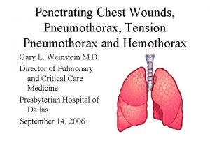
Pneumothorax • Tension Pneumothorax – the air builds up and pushes the heart across, the ribs are expanded because of the large volume of air that can’t escape, and the diaphragm gets pushed down. • An urgent diagnosis and chest drain are required to release these effects.

Pneumothorax • All patients who have had a tension pneumothorax should be considered for a pleurodesis. • Patients fall into 3 groups: – Young patients with otherwise normal lungs – Elderley COPD patients with many risk factors. – Patients with complicated lung diseases.

Pneumothorax • Clinical patterns vary with COPD patients almost asphyxiating from a small pneumothorax because of poor lung function • Young patients can have a total pneumothorax with minimal pain or shortness of breath.

Pneumothorax • After confirming the diagnosis there are options for draining the air – – Needle aspiration – Pigtail catheter to underwater seal drain – Chest drain to underwater seal drain. – Decision is based on ease of the procedure and the likelihood that it will be satisfactory for the pnumothorax being treated.

Pneumothorax • The unknown is the degree of air leak. • Sometimes a pigtail is inserted but the lung doesn’t expand because its bore is too small for effective evacuation of the air building up • Also even though suction is applied, the bore may be too small for effective clearance of the air and a pneumothorax persists despite the procedure.

Pneumothorax • A Heimlich valve or similar, can be attached to the drain for mobility as long as a Chest X ray shows the lung completely expanded with the system connected. • Drains are removed 24 hrs after any air leak stops as long as the lung is expanded.

Pneumothorax • Suction is applied to the drain by connecting directly to the wall suction control and dialling up a negative pressure which remains negative for the whole respiratory cycle, despite the air leak

Pneumothorax • Pleurodesis – Patients may need a pleurodesis and this can be achieved by various methods: • Talc slurry up drain – usually for COPD patients • Transaxillary thoracotomy and apical pleurectomy with stapling of apical blebs/cysts. • Video Assisted Thoracotomy, pleural abrasion, stapling of apical blebs/cysts.

Pneumothorax • COPD • Mix 1 -2 bottles of sterile talc with 60 cc sterile saline, draw into Toomey Syringe • Premedicate patient • Facemask of oxygen • May preferably be done in theatre to have anaesthetic control of sedation/analgesia • Clamp tube, break junction of drain and tubing. Insert syringe into drain, release clamp and inject talc up drain. Reclamp, withdraw syringe and reattach tubing to drain and release clamp.

Pneumothorax • Position patient on side if possible, and hold tubing up, to stop talc coming back, but leaving tubing unclamped to allow air to bubble out. • Lie patient from right side to left side for ¼ hr each side, for 1 hour ie r side, L side, R side, L side, - then lower tubing and sit patient up. • This hopefully coats the chest wall with talc for a pleurodesis. • If Lung remains expanded for 5 days, usually any continuing air leak won’t matter. Clamp the tube and monitor clinical and Chest Xray progress. • Remove tube if lung stays up, or 24 hrs after air leak stops.

Pneumothorax • Surgery • In DBRCT’s there was no difference between VATS and open transaxillary pleurodesis. • Recurrence rates are in the order of 3 -4% • Transaxillary approach – Patient positioned on side with arm on a bar.

Pneumothorax • Transaxillary thoracotomy – procedure starts with a stripping of the apical pleura-from internal mammary artery anteriorly to sympathetic chain posteriorly and over the apex of the chest, then down to the oblique fissure. • After this the lung is examined any apical scar/bleb/cyst is stapled off using Titanium staples – sample sent to Path to exclude rare lung disease. • (Titanium staples allows patients to have MRI scans any time if necessary)

Pneumothorax • 28 F or 32 F drain is inserted and chest is closed. • No sutures are put around the ribs – to minimise pain in the breast or nipple. • Drains are removed when no air leak for 24 hrs, and drainage is <100 cc/24 hrs. • Pt goes home when pain controlled with oral medication, no fever or complication and is mobile for bathroom and shower.

Pneumothorax • Surgery is usually never recommended for the first pneumothorax, but may be considered if the patient has a complicating factor eg Cystic fibrosis, lives in a remote area, or has infected pleural effusion or haemothorax as well • Surgery is usually recommended for recurrent pnumothoraces. • Post op- pts are advised they can’t scuba.
 Difference between simple and tension pneumothorax
 Hemothorax
 Flail chest symptoms
 Massive hemothorax
 Tension pneumothorax triad
 Tension pneumothorax triad
 Tension pneumothorax
 Paradoxical rib movement
 Glasgow coma scale 15 punkte
 5 point auscultation
 Thoracic surgery
 High surface tension vs low surface tension
 Countertransference
 Pengertian tegangan antarmuka
 Redroofs surgery
 Thorax
