THIS IS A STUDY GUIDE NOT AN ALL


")


 Dura mater")

- Slides: 24
� THIS IS A STUDY GUIDE, NOT AN ALL INCLUSIVE REVIEW. � THERE MIGHT BE THINGS NOT COVERED BY THIS STUDY GUIDE THAT MIGHT BE ASKED IN YOUR QUIZZES and PRACTICAL TESTS. � STUDENTS ARE RESPONSIBLE FOR STUDYING THEIR MANUAL AND FOR ALL THE MATERIAL COVERED DURING THE LABORATORY PERIOD, AS PER THE COURSE SYLLABUS
Lab # 11 Nervous System I Neuron and Neuroglia Brain and Spinal Cord
The Anatomical Divisions of the Nervous System Brain Spinal cord Central Nervous System (CNS) It consists of the brain and spinal cord enclosed by cranium and vertebral column. It is responsible for integrating, processing and coordinating sensory data and motor commands. Tract: It is a bundle of nerve fibers (axons) in the CNS (white matter). Nucleus: It is a concentration of neuron cell bodies in the CNS (gray mater) Ganglion Nerve Peripheral Nervous System (CNS) It is all the nervous system except the brain and spinal cord. It consists of nerves and ganglia. It deliveries sensory information to the CNS and carries motor commands to peripheral tissues and system. Nerve: It is a bundle of nerve fibers (axons) wrapped in fibrous connective tissue in the PNS Ganglion: It is a knot-like swelling in a nerve where neuron cell bodies are concentrated in the PNS
NEURON STRUCTURE Histology of the Nervous Tissue 1 - Neurons 2 - Neuroglial cells -In the CNS -In the PNS Glial cell (astrocyte) Neurons are large cells that transmit and generate messages in the form of nerve impulses or neuronal action potentials. The glial cells are supporting cells, which are associated to the neurons and provide a supportive scaffolding for neurons.
Neuroglial Cells 1 - Schwann cells: They wind repeatedly around a nerve fiber and produce a myelin sheath similar to the ones produced by oligodendrocytes in CNS. 2 - Satellite cells: They surround the bodies of the neurons in the PNS. 3 - Astrocytes: They are the most abundant glial cell in CNS covering the entire brain surface. They anchor the neurons and blood vessels in place and regulate the extracellular environment of the brain. . They facilitate the formation of the blood-brain barrier. 4 - Oligodendrocytes: They wrap around the axons of neurons of the CNS to form myelin sheaths. Each arm-like process wraps around a nerve fiber forming an insulating layer that speeds up signal conduction. 5 - Microglial cells: They are small phagocytic cells that clean up debris surrounding the neurons. 6 - Ependymal cells: They form the ciliated cuboidal epithelium that lines the hollow spaces of the brain and spinal cord. They assist in forming the cerebrospinal fluid (CSF).
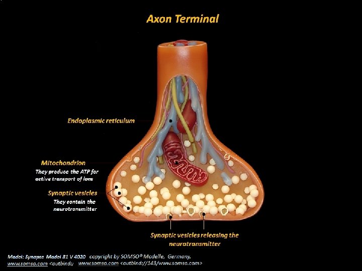
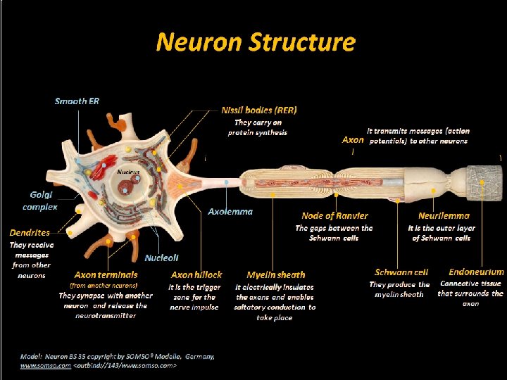
NEURON STRUCTURE The Structure of Neurons Cell body It is the biosynthetic center of or Soma the neuron Telodendria Nucleus Cytoplasm with organelles Dendrite Axon terminal , synaptic terminal, or synaptic knob Dendrites They receive the messages from other neurons. R. E. R. Free ribosomes Nissl bodies Protein synthesis It transmits messages Axon (action potentials) to other neurons Neurofilaments Presynaptic neuron It contains synaptic vesicles with the neurotransmitters. Synaptic cleft Axon hillock It is the trigger zone for the nerve impulse. Axon terminal Telodendria It is the space between the pre and postsynaptic neurons Postsynaptic neuron
Axoplasm It is the cytosol of the axon. It is the outer layer of the Axolemma Neurilemma Schwann cells. It is the plasma membrane of the axon. Node of The gaps between the Ranvier Schwann cells. Schwann cell They surround the axon and produce the myelin sheet. Internode The myelin covered segments between the nodes. Myelin sheath It electrically insulates the axon and enables saltatory conduction.
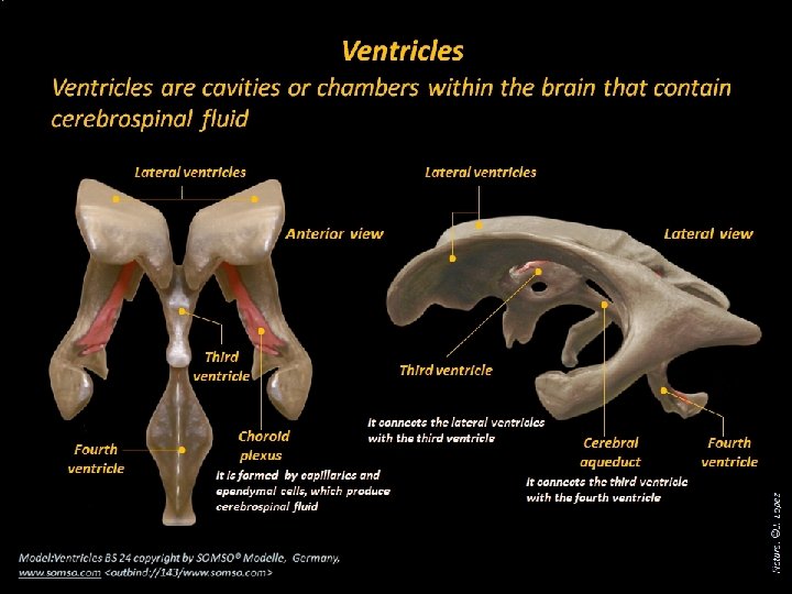
Ventricles Lateral ventricles Interventricular foramen Third ventricle Cerebral aqueduct Fourth ventricle Central canal
Cranial Meninges Skull Arachnoid villus Dura mater: Periosteal layer Meningeal layer Arachnoid mater Pia mater Superior sagittal sinus (contains blood) Subarachnoid Space (contains CSF) Falx cerebri (in longitudinal fissure only)
The Spinal Cord Cervical enlargement C 7 Foramen magnum of occipital Cervical region Medullary cone Thoracic region Cauda equina Lumbar enlargement T 12 Lumbar region Terminal filum It attaches the spinal cord to the coccyx and prevents vertical movements L 5 Sacral region
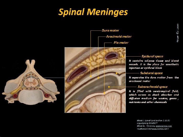
Meninges of the Spinal Cord Epidural space (Space for the epidural anesthesia) Dura mater Subdural space Arachnoid mater Subarachnoid space (It contains cerebrospinal fluid)) Pia mater Arachnoid mater Dura mater
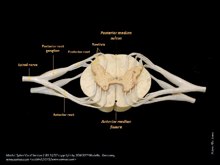
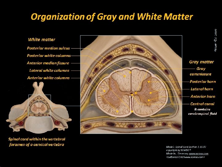
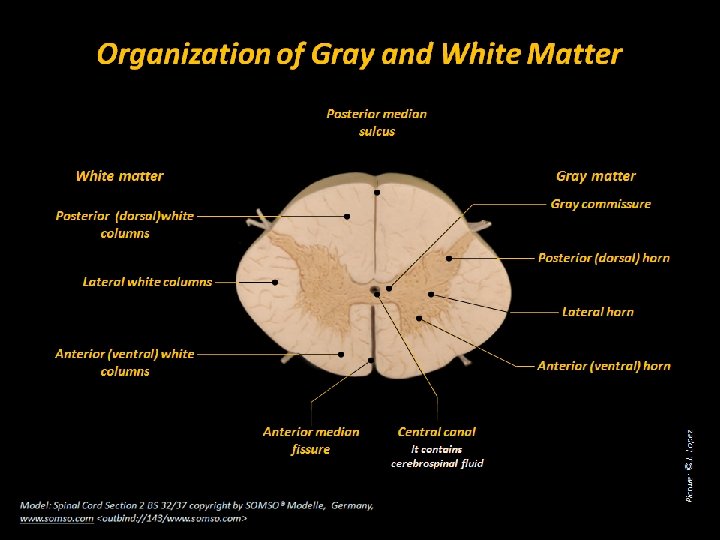
Cross Sectional Anatomy of the Spinal Cord Gray matter It contains the bodies and dendrites of neurons. Therefore, it contains little myelin. It is composed of bundles of axons called tracts. Therefore, it White matter contains abundance of myelin. They carry sensory Posterior (dorsal) information root of spinal nerve Posterior root ganglion They contain the bodies of sensory neurons Posterior They contain bodies of gray horns sensory somatic neurons and sensory visceral neurons Lateral gray horns They contain bodies of visceral motor neurons Spinal nerve They carry both sensory and motor information Anterior (ventral) root of spinal nerve They carry motor information Anterior They contain bodies of gray horns somatic motor neurons