This is a normal glomerulus by light microscopy


This is a normal glomerulus by light microscopy. The glomerular capillary loops are thin and delicate. Endothelial and mesangial cells are normal in number. The surrounding tubules are normal. Life is good.


This is Berger's disease, or Ig. A nephropathy. The Ig. A is deposited mainly in mesangium, which then increases mesangial cellularity as shown at the arrow. Patients with Ig. A nephropathy usually present with hematuria.

This immunofluorescence micrograph demonstrates positivity with antibody to Ig. A. Note that the pattern is that of mesangial staining. This is Ig. A nephropathy.

. Those that are idiopathic are divided into types I")
This is membranoproliferative glomerulonephritis (MPGN). Those that are idiopathic are divided into types I and II by pathologic findings. As seen here, the glomerulus has increased overall cellularity, mainly mesangial.

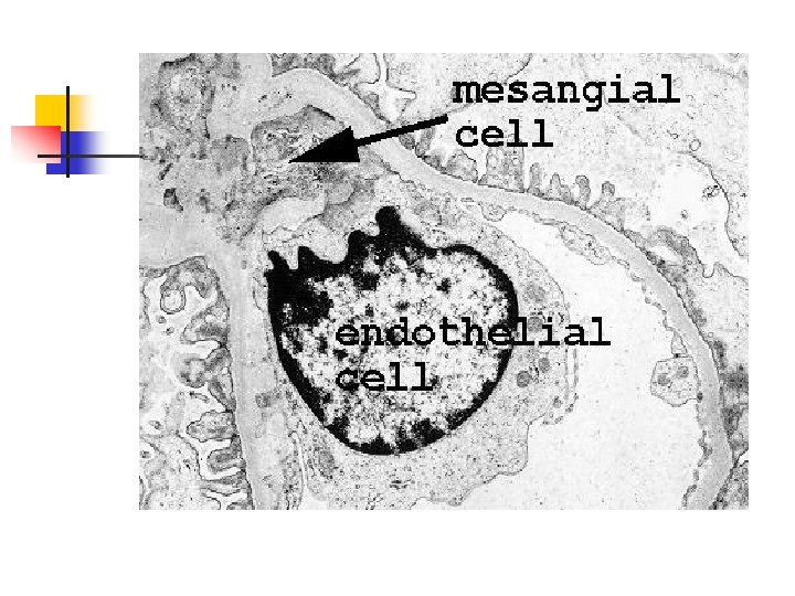
This electron micrograph demonstrates a mesangial cell at the lower left that is interposing its cytoplasm at the arrow into the basement membrane, leading to splitting and reduplication of basement membrane that is piled up above the mesangial cytoplasm in this micrograph. This is MPGN type I. These characteristic EM changes occur when the mesangial cell (which has a macrophage-like function) goes after subendothelial immune deposits, but makes a mess of the basement membrane in the process.

This electron micrograph demonstrates the dense deposits in the basement membrane of MPGN type II. There are dark electron dense deposits within the basement membrane that often coalesce to form a ribbon-like mass of deposits.

- Slides: 21