THIRD EDITION HUMAN PHYSIOLOGY AN INTEGRATED APPROACH Dee


 • Thinner walls")


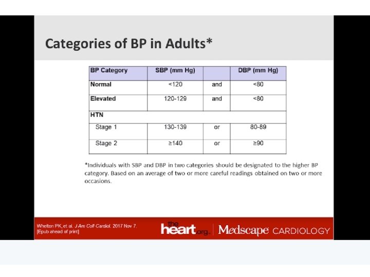
: Measurements • \"Blood pressure\" • Systolic over diastolic • About 120/80")
: Measurements Figure 15 -7: Measurement of arterial blood pressure Copyright ©")


 • Angiotensin II antagonists •")


 • Lymphatic structures • Capillaries with valves •")
 Copyright © 2004 Pearson Education, Inc. , publishing")


 •")


- Slides: 55
THIRD EDITION HUMAN PHYSIOLOGY AN INTEGRATED APPROACH Dee Unglaub Silverthorn, Ph. D. Chapter 15 Blood Flow and the Control of Blood Pressure Power. Point® Lecture Slide Presentation by Dr. Howard D. Booth, Professor of Biology, Eastern Michigan University Copyright © 2004 Pearson Education, Inc. , publishing as Benjamin Cummings
About this Chapter • How various blood vessels are constructed and role in circulation • Components of "blood pressure", role and measurement • Product exchange at the capillary beds • Lymph vessels, distribution and role in circulation • How blood pressure and circulation are regulated • Key components of cardiovascular disease Copyright © 2004 Pearson Education, Inc. , publishing as Benjamin Cummings
The Blood Vessels and the Cardiovascular System • Arteries: blood from heart • Strong & Elastic • Conduct blood to capillaries • Sphincters • Capillaries: exchange with cells • Veins • Return blood to heart • Valves Copyright © 2004 Pearson Education, Inc. , publishing as Benjamin Cummings
The Blood Vessels and the Cardiovascular System Figure 15 -1: Functional model of the cardiovascular system Copyright © 2004 Pearson Education, Inc. , publishing as Benjamin Cummings
Make Up of Blood Vessels: Arteries and Arterioles • Endothelium • Elastic tissues • Rebounds • Evens flow • Smooth muscles • Fibrous tissue • Tough • Resists stretch Copyright © 2004 Pearson Education, Inc. , publishing as Benjamin Cummings Figure 15 -2: Blood vessels
Make Up of Blood Vessels: Capillaries • Endothelium: one cell thick • Continuous • Fenestrated • Basement membrane Copyright © 2004 Pearson Education, Inc. , publishing as Benjamin Cummings
Make Up of Blood Vessels: Capillaries Copyright © 2004 Pearson Education, Inc. , publishing as Benjamin Cummings Figure 15 -16: Types of capillaries
Make Up of Blood Vessels: Veins and Venules (Contrasted to Arteries) • Thinner walls • Larger diameter • Closer to skin • Less muscle • Less elastic Copyright © 2004 Pearson Education, Inc. , publishing as Benjamin Cummings Figure 15 -3: Metarterioles
Angiogenesis: Growth of New Blood Vessels • Normal body maturation and growth • Endometrium • Endurance training • Abnormal growth to service cancerous tissue • Wound repair and consequences • Failure to regrow in heart tissues after heart attack • Failure to regrow in brain after stroke Copyright © 2004 Pearson Education, Inc. , publishing as Benjamin Cummings
Blood Pressure: Generated by Ventricular Contraction • Pulsatile: surges in arteries • Elastic rebound evens & maintains pressure Copyright © 2004 Pearson Education, Inc. , publishing as Benjamin Cummings
Blood Pressure: Generated by Ventricular Contraction Figure 15 -4: Elastic recoil in the arteries Copyright © 2004 Pearson Education, Inc. , publishing as Benjamin Cummings
Blood Pressure (BP): Measurements • "Blood pressure" • Systolic over diastolic • About 120/80 mm. Hg • Sphygmomanometer • "Estimate of pressure" • Korotkoff sounds Copyright © 2004 Pearson Education, Inc. , publishing as Benjamin Cummings
Blood Pressure (BP): Measurements Figure 15 -7: Measurement of arterial blood pressure Copyright © 2004 Pearson Education, Inc. , publishing as Benjamin Cummings
More Blood Pressures: Pulse and Mean Arterial Pressures • Pulse pressure = Systolic–Diastolic • Mean arterial pressure (MAP) = Diastolic + 1/3 pulse pressure Copyright © 2004 Pearson Education, Inc. , publishing as Benjamin Cummings
More Blood Pressures: Pulse and Mean Arterial Pressures Figure 15 -5: Pressure throughout the systemic circulation Copyright © 2004 Pearson Education, Inc. , publishing as Benjamin Cummings
Factors Controlling MAP : The Driving Pressure for Blood Flow • Blood volume • Cardiac output • Resistance • Distribution Copyright © 2004 Pearson Education, Inc. , publishing as Benjamin Cummings
Factors Controlling MAP : The Driving Pressure for Blood Flow Figure 15 -10: Factors that influence mean arterial pressure Copyright © 2004 Pearson Education, Inc. , publishing as Benjamin Cummings
Antihypertensive Drug Classes: Action Sites Blood Pressure = Cardiac Output A n t i h y p ert en s i v e Dru g C l a s s es -Blockers Total Peripheral Resistance -Blockers ACE Inhibitors AT 1 Blockers Direct renin inhibitors 1 -Blockers Non-DHP CCBs 2 -Agonists Diuretics All CCBs Sympatholytics Vasodilators ACE = angiotensin-converting enzyme; AT 1 = angiotensin type 1; CCBs = calcium channel blockers; DHP = dihydropyridine Slide Source Hypertension Online www. hypertensiononline. org
Classes of Antihypertensive Drugs • Aldosterone receptor antagonists (blockers) • Angiotensin II antagonists • Angiotensin-converting enzyme inhibitors • -Blockers – 1 -Selective – Nonselective • -Blockers – -1/ -2 – -1 predominant – / – Intrinsic sympathomimetic activity • Calcium channel antagonists – Nondihydropyridine – Dihydropyridine • Central 2 agonists • Direct renin inhibitors • Direct vasodilators • Diuretics – Thiazide-type – Loop-type – Potassium-sparing • Ganglionic blockers Slide Source Hypertension Online www. hypertensiononline. org
Arteriole Resistance: Control of Local Blood Flow • Myogenic auto regulation • Paracrines: • Hyperemia • Sympathetic nerves – CNS Copyright © 2004 Pearson Education, Inc. , publishing as Benjamin Cummings
Distribution of Blood in the Body Organs Copyright © 2004 Pearson Education, Inc. , publishing as Benjamin Cummings Figure 15 -13: Distribution of blood in the body at rest
Capillary Blood Flow: Greatest Total Cross Sectional Area • Lowest Velocity • Hydrostatic pressure drops Copyright © 2004 Pearson Education, Inc. , publishing as Benjamin Cummings Figure 15 -17: The velocity of flow depends on the total crosssectional area
Capillary Exchange: Colloidal Osmotic Pressure is Constant • Proteins stay in capillary • Water, oxygen, glucose – move out • CO 2, N wastes, water – move in • Bulk flow out on arterial side, in on venous side Copyright © 2004 Pearson Education, Inc. , publishing as Benjamin Cummings
Capillary Exchange: Hydrostatic Pressure Declines • High on arterial side – bulk flow out • Low on venous side – bulk flow in • Fenestrations &/or leaky joints speed exchange Copyright © 2004 Pearson Education, Inc. , publishing as Benjamin Cummings Figure 15 -18 a: Fluid exchange at the capillary
Net Out Flow Into ECF • Net filtration – net absorption = net out flow • About 2 L/day collected by lymph vessels Copyright © 2004 Pearson Education, Inc. , publishing as Benjamin Cummings Figure 15 -18 b: Fluid exchange at the capillary
Lymphatic System: Structure and Roles (overview) • Lymphatic structures • Capillaries with valves • Lymph vessels • Lymph nodes & organs • Immune defense: lymphocytes • Transport of fats • Collects excess ECF • Returns to plasma • Edema Copyright © 2004 Pearson Education, Inc. , publishing as Benjamin Cummings
Lymphatic System: Structure and Roles (overview) Copyright © 2004 Pearson Education, Inc. , publishing as Benjamin Cummings Figure 15 -19: The lymphatic system
Lymphatic System: Overview • Consists of two semi-independent parts • A meandering network of lymphatic vessels • Lymphoid tissues and organs scattered throughout the body • Returns interstitial fluid and leaked plasma proteins back to the blood • Lymph – interstitial fluid once it has entered lymphatic vessels Copyright © 2004 Pearson Education, Inc. , publishing as Benjamin Cummings
Figure 20. 2 a Copyright © 2004 Pearson Education, Inc. , publishing as Benjamin Cummings
Lymphatic System: Overview Figure 20. 1 a Copyright © 2004 Pearson Education, Inc. , publishing as Benjamin Cummings
Lymphatic Capillaries • Similar to blood capillaries, with modifications • Remarkably permeable • Loosely joined endothelial minivalves • Withstand interstitial pressure and remain open • The minivalves function as one-way gates that: • Allow interstitial fluid to enter lymph capillaries • Do not allow lymph to escape from the capillaries Copyright © 2004 Pearson Education, Inc. , publishing as Benjamin Cummings
Lymphatic Capillaries • During inflammation, lymph capillaries can absorb: • Cell debris • Pathogens • Cancer cells • Cells in the lymph nodes: • Cleanse and “examine” this debris • Lacteals – specialized lymph capillaries present in intestinal mucosa • Absorb digested fat and deliver chyle to the blood Copyright © 2004 Pearson Education, Inc. , publishing as Benjamin Cummings
Lymphatic Trunks • Lymph is delivered into one of two large trunks • Right lymphatic duct – drains the right upper arm and the right side of the head and thorax • Thoracic duct – arises from the cisterna chyli and drains the rest of the body Copyright © 2004 Pearson Education, Inc. , publishing as Benjamin Cummings
Lymph Transport • The lymphatic system lacks an organ that acts as a pump • Vessels are low-pressure conduits • Uses the same methods as veins to propel lymph • Pulsations of nearby arteries • Contractions of smooth muscle in the walls of the lymphatics Copyright © 2004 Pearson Education, Inc. , publishing as Benjamin Cummings
Regulation of Blood Pressure and Heart Rate • Medullary cardiac control center (Brainstem) • Cardioacceleratory Center • Cardioinhibitory Center • Baroreceptor reflex • Carotid • Aortic • Kidney: blood volume • Hypothalamus & Cortex: stress, blushing, etc. Copyright © 2004 Pearson Education, Inc. , publishing as Benjamin Cummings
Copyright © 2004 Pearson Education, Inc. , publishing as Benjamin Cummings
Copyright © 2004 Pearson Education, Inc. , publishing as Benjamin Cummings
Copyright © 2004 Pearson Education, Inc. , publishing as Benjamin Cummings
Copyright © 2004 Pearson Education, Inc. , publishing as Benjamin Cummings
Copyright © 2004 Pearson Education, Inc. , publishing as Benjamin Cummings
Copyright © 2004 Pearson Education, Inc. , publishing as Benjamin Cummings
Copyright © 2004 Pearson Education, Inc. , publishing as Benjamin Cummings
Copyright © 2004 Pearson Education, Inc. , publishing as Benjamin Cummings
Regulation of Blood Pressure Figure 15 -22: The baroreceptor reflex: the response to increased blood pressure Copyright © 2004 Pearson Education, Inc. , publishing as Benjamin Cummings
Copyright © 2004 Pearson Education, Inc. , publishing as Benjamin Cummings
Copyright © 2004 Pearson Education, Inc. , publishing as Benjamin Cummings
Copyright © 2004 Pearson Education, Inc. , publishing as Benjamin Cummings
Copyright © 2004 Pearson Education, Inc. , publishing as Benjamin Cummings
Cardiovascular Diseases: #1 killer • Risk Factors: • Smoking • Obesity • Diabetes • Genes • Diseases: • Hypertension • Stroke • "Heart Attack" Copyright © 2004 Pearson Education, Inc. , publishing as Benjamin Cummings
Mechanism of Atherosclerosis • LDL build up • Plaque • Flow • Rupture • Clot • Blocked flow • Tissue death Copyright © 2004 Pearson Education, Inc. , publishing as Benjamin Cummings
How Atherosclerosis Develops We now understand that atherosclerosis is a chronic inflammation of arteries, which develops over decades in response to the biologic effects of risk factors. Atherogenesis begins as a qualitative change to intact endothelial cells; when subjected to oxidative, hemodynamic, or biochemical stimuli (from smoking, hypertension, or dyslipidemia) and inflammatory factors, they change their permeability to promote the entry and retention of blood-borne monocytes and cholesterol-containing LDL particles. Inflammation and biochemical modifications ensue, causing endothelial and smooth-muscle cells to proliferate, produce extracellular matrix molecules, and form a fibrous cap over the developing atheromatous plaque. Plaques lead to clinical symptoms by producing flow-limiting stenoses (causing stable angina) or by provoking thrombi that interrupt blood flow on either a temporary basis (causing unstable angina) or a permanent one (causing myocardial infarction). Physical disruption (rupture) of the plaque exposes procoagulant material within the core of the plaque to coagulation proteins and platelets, triggering clotting. Copyright © 2004 Pearson Education, Inc. , publishing as Benjamin Cummings
Summary • Blood vessels, anatomy & role in circulation • Measuring blood pressures, MAP & pulse pressure • Role of resistance in BP and distribution of blood • Autoregulation, baroreceptros, medullary cardiac control center and CNS regulation of blood pressure & distribution Copyright © 2004 Pearson Education, Inc. , publishing as Benjamin Cummings
Summary • Hydrostatic & colloidal osmotic pressures direct bulk flow in capillary exchange by diffusion, fenestrations & leaky joints • Role of lymphatic system to return excess ECF to plasma • Atherosclerosis common to several cardiovascular diseases Copyright © 2004 Pearson Education, Inc. , publishing as Benjamin Cummings