There are three basic types of muscle Found


There are three basic types of muscle: Found only in heart tissue Found in voluntary muscle tissue e. g. biceps Found in involuntary muscle tissue e. g. stomach, artery walls

• Also called voluntary muscle as it is • under conscious control • also called striated muscle because of its striped appearance.
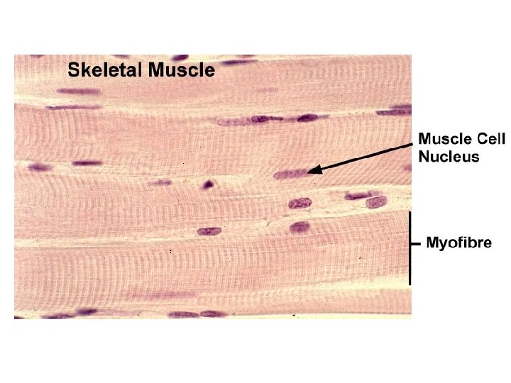

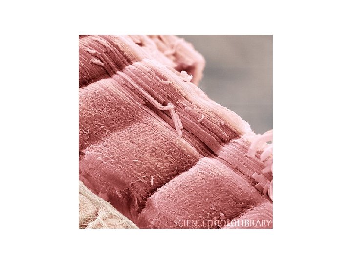
• made up of hundreds of specialised cells called muscle fibres, which are several cms long • the surface membrane of each muscle fibre is called the sarcolemma • the cytoplasm is called the sarcoplasm and contains many nuclei and mitochondria

sarcolemma sarcoplasm myofibril nuclei

myofilaments muscle fibre made of actin myosin myofibril made of myofilament myofibril

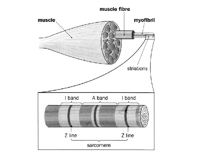
• Within each muscle fibre are many parallel myofibrils • Each myofibril consists of overlapping thick and thin myofilaments • Thick myofilaments are made of the large protein myosin, a fibrous protein from which globular heads project • Thin myofilaments are composed of the smaller protein actin

• The filaments are arranged in a very specific order, forming distinct striations/stripes along the muscle fibre • The stripes are caused by the thick and thin filaments and areas of overlap

M
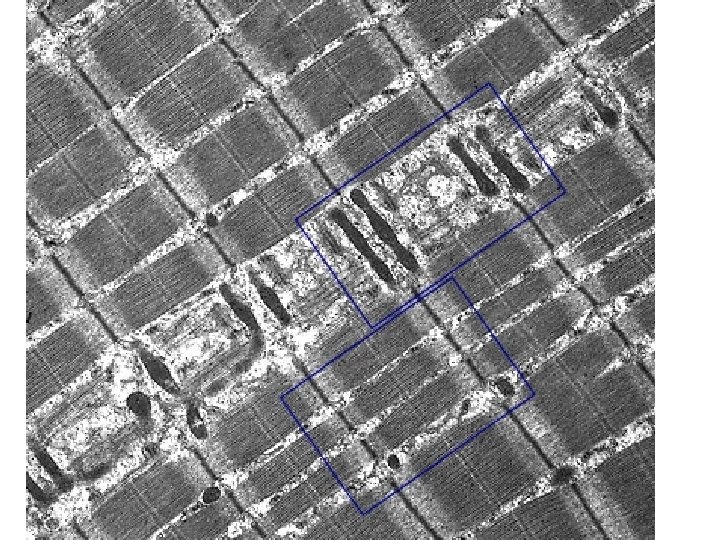

• The thicker myosin filaments create the darker striations called the A-band • Within the A-band is a slightly lighter band, called the H-zone • At the centre of the H-zone is the M-line • The thinner actin filaments create a light band called the I-band • At the centre of the I-band is the Z-line • The distance between adjacent Z-lines is called a sarcomere

A band Width of myosin filament, contains H band dark region either side where myosin and actin filaments overlap H zone region within the A band containing only myosin filaments I band lighter region with only actin Z Line occurs at the centre of the I band marks the end of the one sarcomere and the beginning of the next
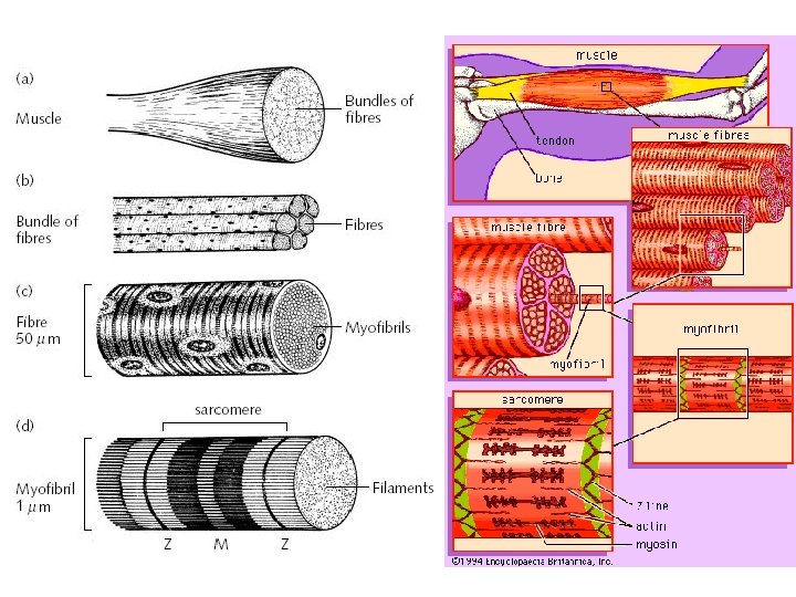

Muscle contraction When a muscle contracts the actin filaments are pulled over the mysoin filaments (towards the M line) by myosin heads. This causes the following changes: • The Z lines become closer together, so the sarcomere shortens • The I bands become shorter • The H zone becomes narrower However the A band (length of myosin) does not change in length

H zone Myosin heads

H zone Myosin heads

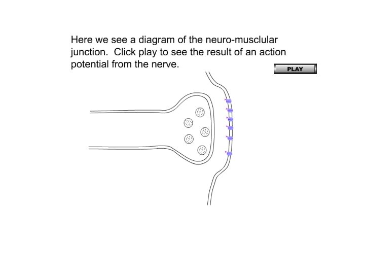
• The synapse between the axon of a motor neurone and a muscle is called the neuromuscular junction

Muscle fibres are innervated by a motor neurone Motor end plate

Motor end plate

myelin sheath synaptic vesicle cleft axon terminal presynaptic membrane postsynaptic membrane Myofibril contains repeating dark and light bands, responsible for the striped appearance mitochondria sarcolemma sarcoplasmic reticulum A band Dark band I band Light band

• An action potential arriving at the neuromuscular junction causes the release of neurotransmitter that crosses the synaptic cleft • This causes the depolarisation of the sarcolemma (post-synaptic membrane of the muscle fibre) • resulting in an action potential which propagates along the entire length of the sarcolemma as an impulse

sarcolemma AP AP AP Ca 2+ myofibril T tubule sarcoplasmic reticulum

• The sarcolemma is indented forming T tubules deep into the cell and surrounding the myofibrils. • These tubules lie close to the sarcoplasmic reticulum.

• The T-tubules and sarcoplasmic reticulum carry the impulse through the muscle fibre • and causes the sarcoplasmic reticulum to release calcium ions into the sarcoplasm • Ca 2+ cause ancillary proteins, covering binding sites on the actin filaments to be displaced, so the binding sites are uncovered. • The globular myosin heads on the myosin filaments attach to the actin forming actomyosin cross-bridges

• The myosin heads rotate back pulling the thin actin filaments over the thick myosin filaments • Energy from ATP is needed to detach the heads • The detached heads regain their original position, and attach to another actin binding site further along the filament • The cycle of attachment, rotation, detachment is repeated in a ratchet type mechanism • The process continues as long as APs are propagated through the muscle fibre • As the thin filaments are pulled over the thick filaments the sarcomeres and myofibrils shorten

QUESTION • Froggy p 348 Q 7

REVIEW

- Slides: 33