The Wrist Hand Forearm Elbow Chapter 19 Anatomy

The Wrist, Hand, Forearm & Elbow Chapter 19

Anatomy of the Wrist and Hand Bones of the Wrist Bones of the Hand ◦ 8 Carpal Bones ◦ 5 Metacarpals ◦ 14 Phalanx Bones Ligaments: ◦ Complex series of ligaments that bind the carpal bones to one another, to the ulna & radius, and to the proximal metacarpals ◦ Wrist: UCL & RCL ◦ Volar aspect of wrist is Transverse Carpal Ligament ◦ Each Joint in the fingers has MCL & LCL ligaments

Muscles of the Hand Extrinsic muscles: Intrinsic Muscles: Motions of the Wrist originate outside of the hand Motions of the Fingers originate inside of the hand Flexion Extension Radial Deviation Ulnar Deviation Flexion Extension Abduction Adduction Motions of the Thumb Opposition Flexion Extension Abduction Adduction

Metacarpal Fracture Cause: ◦ Direct Axial force ◦ Trauma ◦ Contact Signs & Symptoms: ◦ Pain ◦ Point Tenderness ◦ Defect ◦ Rapid Swelling Care: ◦ Physician referral for x-ray & evaluation ◦ If the fracture is not displaced, they injury is casted ◦ If the fracture is displaced, surgical reduction may be necessary The most common metacarpal fracture is a 5 th metacarpal fracture, aka Boxer’s Fracture

Metacarpal Fracture

Boxer’s Fracture of the 5 th Metacarpal

Mallet Finger Rupture of the Extensor Tendon at the Distal Interphalangeal Joint Bony Avulsion of that tendon Must be splinted in extension splint for 6 -8 weeks, 24 hours a day

Mallet Finger

Boutonniere Deformity Rupture of the extensor tendon over the middle phalanx at the Proximal Interphalangeal Joint Caused by forcing the DIP into extension & the PIP into flexion Splint in extension for 6 -8 weeks If not treated properly a boutonniere deformity will be permanent

Boutonniere Deformity

Jersey Finger Cause: ◦ Rupture of the Flexor Digitorum Profundus from distal phalanx (DIP Joint) ◦ May occur with an avulsion Signs & Symptoms: ◦ Finger cannot be flexed ◦ Finger stuck in extension ◦ Pain & point tenderness over the distal phalanx Care: ◦ Surgery is required for flexion to be restored ◦ Surgery must be done within 7 -10 days. Recovery is approx 12 weeks

Jammed Finger Cause: forced axial load to the tip of the finger. Incredibly common in sports. Signs & Symptoms: severe point tenderness, lateral instability, swelling Care: x-ray to rule out fracture, ice, & splint, tape to return to play

Collateral Ligament Sprain “Jammed Finger”

Gamekeeper’s Thumb Cause: ◦ Ulnar Collateral Ligament Strain at MCP Joint Signs & Symptoms: ◦ Pain over UCL ◦ Weakness & pain with pinch ◦ Swelling over medial aspect of thumb ◦ Joint instability Care: ◦ X-ray to rule out fracture ◦ Splint or Cast 4 -6 weeks ◦ Surgery can be required if not protected properly

Gamekeeper’s Thumb

Dislocated Finger Cause: Blow to the tip of the finger Signs & Symptoms: Deformity, tearing of capsule resulting in hemorrhage, swelling Care: x-ray to rule out fracture, reduction of dislocation, splint for 3 -4 weeks, tape to return to play

Phalanx Fracture Cause: Trauma such as being stepped on, hit, mashed between two objects, twisting, forced dislocation Signs & Symptoms: pain, swelling, point tenderness, deformity Care: Splint, refer for x-ray & physician evaluation

Phalanx Fracture

Subungual Hematoma Cause: ◦ Contusion or crushing injury to the first phalanx that causes blood to accumulate under the nail Signs & Symptoms: ◦ Extreme pain, blood under nail, more than 25% fracture needs to be ruled out Care: ◦ Drain within 12 to 24 hours, ice, elevate, clean once drilled, may have to be drilled twice, keep clean & covered. DO NOT DRILL THE NAIL IF A FRACTURE IS SUSPECTED!!!

Wrist Sprain Cause: a wrist sprain is caused by Hyperextension most often, but it can be caused by violent flexion or torsion Make sure the athlete gets an x-ray to rule out a fracture. Care: ice, splinting & analgesics, strengthening exercises, & taping to return to play
 Cause: repetitive wrist Signs & Symptoms: Care: acceleration & deceleration Pain")
Wrist Tenosynovitis (Tendinitis) Cause: repetitive wrist Signs & Symptoms: Care: acceleration & deceleration Pain with use or pain with passive stretching. Point tender with swelling over the tendon Ice massage, NSAIDs, rest, & splint injured tendon

Carpal Tunnel Syndrome Cause: ◦ Carpal Tunnel is caused by inflammation of the flexor tendons that compress the median nerve Signs & Symptoms: ◦ Sensory & motor deficits ◦ Tingling & numbness ◦ Paresthesia of thumb, index & middle fingers. ◦ Weakness in thumb, index & middle fingers ◦ Aching in hand Care: ◦ Rest ◦ Wrist splint ◦ NSAIDS ◦ Injection of corticosteroid ◦ Surgery

Carpal Tunnel Syndrome

Dislocated Lunate Caused by forceful hyperextension Not very common in sports Pain, swelling, deformity, difficulty with flexion May have numbness or paralysis due to pressure on the median nerve See physician immediately for x-ray and reduction. Cast for 4 -8 weeks

Dislocated Lunate

Scaphoid Fracture Scaphoid is found in the anatomical snuff box Typically caused by falling on outstretched hand Signs & symptoms include swelling & severe point tenderness, loss of grip strength, weakness with thumb movement Athlete should be x-rayed immediately, immobilized in a cast ◦ Rexray athlete every 2 weeks, & remain in splint if fx. Shows up. ◦ If a fracture is present, immobilization period is typically around 6 weeks ◦ The scaphoid doesn’t have good blood supply, which can result in a non-union ◦ Non-union fracture healing must be repaired with surgery ◦ When athlete returns to play once out of cast, they must be taped for 8 -12 weeks

Scaphoid Fracture

Hamate Fracture Cause: ◦ A fall ◦ Contact while holding a sports implement such as the handle of a tennis racket, bat, golf club. Signs & Symptoms: ◦ Wrist pain & weakness ◦ Point tenderness ◦ Numbness & tingling (ulnar nerve) Care: ◦ Cast the wrist ◦ Doughnut pad over hook of hamate

Hamate Fracture

Wrist Ganglion Cause: ◦ Damage to joint capsule, synovial sheath or a cyst ◦ Typically occurs after a trauma ◦ Usually found on dorsal side of wrist Signs & Symptoms: ◦ Pain ◦ Lump at the site of pain that is palpable ◦ Pain with wrist extension Care: ◦ Old: Pressure pad ◦ New: Combined treatment. Aspiration, Cauterization, pressure pad ◦ Only sure way to remove ganglion cyst is surgery

Wrist Ganglion

Anatomy of the Forearm Bones: Radius & Ulna Joints: Superior, Middle & Distal Radioulnar Joint Soft Tissue: Interosseous Membrane
: Muscles (posterior): ◦ Biceps Brachii ◦ Triceps Brachii")
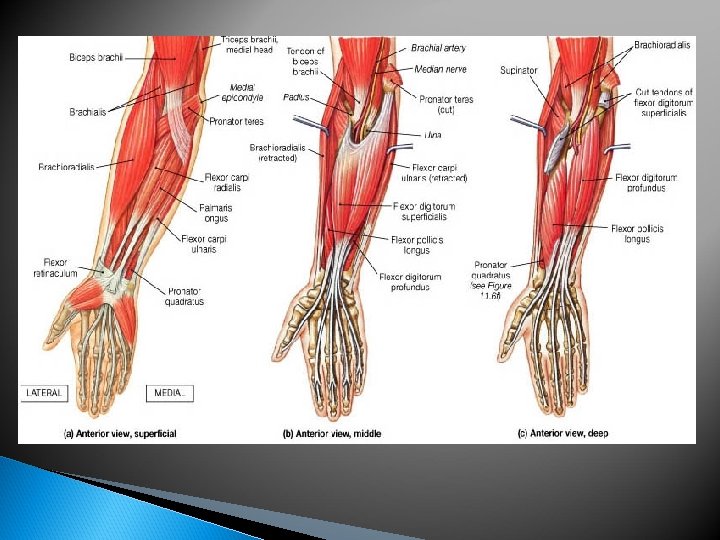
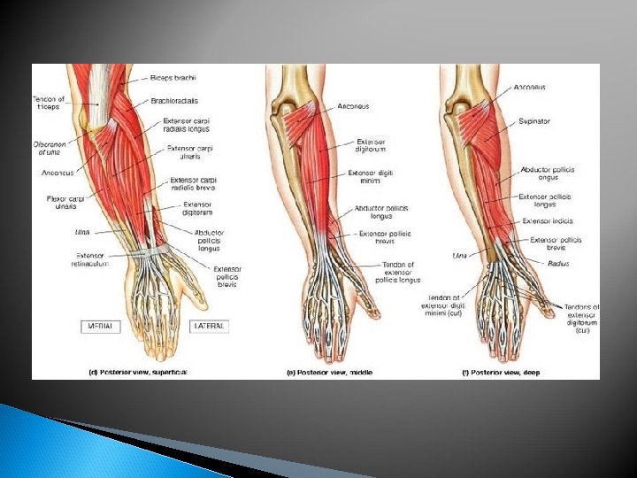
Muscles of the Forearm Muscles (anterior): Muscles (posterior): ◦ Biceps Brachii ◦ Triceps Brachii ◦ Brachialis ◦ Anconeous ◦ Pronator Teres ◦ Flexor Carpi Ulnaris ◦ Brachioradialis ◦ Extensor Carpi Ulnaris ◦ Flexor Carpi Radialis ◦ Extensor Digiti Minimi ◦ Palmaris Longus ◦ Extensor Carpi Radialis Longus ◦ Flexor Carpi Ulnaris ◦ Extensor Carpi Radialis Brevis ◦ Flexor Pollicis Longus ◦ Extensor Digitiorum ◦ Pronator Quadratus ◦ Abductor Pollicis Longus ◦ Extensor Pollicis Brevis ◦ Extensor Pollicis Longus

Muscles Flexors Extensors

Forearm Injury Evaluation History: how did it happen, where does it hurt, Observation: swelling, deformity, discoloration, Palpation: tenderness, edema, deformity, skin prior injury, loss of function, symptoms skin defects, motion temperature, bone fragments, continuity of bone

Contusion Cause: repeated blows to Signs & Symptoms: Care: P. R. I. C. E. , stretching, the forearm or a blow with significant force. Typically occur on the ulnar side bruising, pain, swelling, hematoma, myositis ossificans may develop if the injury is severe Range of motion exercises, Pad/Protect to return to play

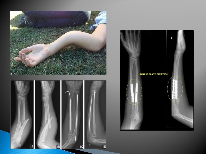
Forearm Fracture Cause: typically caused by a Signs & Symptoms: Pain, Care: Splint & immediately blow or falling on an outstretched hand swelling, deformity, ‘false joint’. There is a danger of soft tissue damage if the fractured bones move significantly refer for x-ray & physician consult. Ice. Typically casted for 6 -8 weeks, surgery may be required if the deformity is significant. Take Tylenol for pain, NOT Ibuprofen can delay fracture healing by hindering the inflammatory response phase

Forearm Fracture

Colles’ Fracture Cause: fall on outstretched hand forces the wrist into hyperextension. The most common forearm fracture. Distal Radius Fracture. Signs & Symptoms: visible deformity, pain, swelling, quick accumulation of blood. Median nerve damage is possible if the radius shifts forward enough Care: Splint & immediately refer for x-ray & physician consult. Ice. Typically casted for 4 -8 weeks. Fracture deformity is usually reduced under anesthesia.

Anatomy of the Elbow Bones: Humerus Radius Ulna Ligaments: ◦ Ulnar Collateral Ligament Connects the ulnar & humerus. Protects the elbow against valgus stress ◦ Annular Ligament Surrounds the radial head. Allows rotation of the radius. ◦ Radial Collateral Ligament Connects the humerus and radius. Protects the elbow against varus stress

Elbow Flexors Elbow Flexors: Biceps Brachii Brachialis Brachioradialis

Elbow Extensors Elbow Extensors: Triceps Brachii
 : Pronator Teres Pronator Quadratus")
Elbow Pronators Elbow Pronation (forearm) : Pronator Teres Pronator Quadratus
: Biceps brachii Supinator muscles")
Elbow Supinators (forearm): Biceps brachii Supinator muscles

Injury Evaluation History ◦ Ask questions: How, where, when, who Observation ◦ What do you see? Swelling, bruising, deformity Palpation ◦ What do you feel? Instability, deformity, defects Special Tests ◦ What structures are damaged? ◦ How stable is the joint? ◦ Are they point tender?

Injury Prevention Acute injuries usually occur from trauma or falling Wear protective padding to reduce the force of a blow Learning how to fall correctly Chronic injuries are very common in the elbow, forearm and wrist Limit # of repetitions throwing or hitting a tennis ball Make sure of proper mechanics Maintain appropriate strength and endurance levels Stretch to warm up & stretch to cool down Don’t overlook chronic injuries. REST if needed!

Olecranon Bursitis Cause: Can be caused by a Signs & Symptoms: Care: Ice, rest, compression direct blow, falling on the tip of the elbow or excessive flexion/extension. This injury can be acute or chronic Excessive swelling that appears to be superficial, redness, warmth sleeve. On rare occasions, the elbow is drained. Pad when return to play. Athlete may take NSAIDs to reduce swelling. Ultrasound may be used if this is a chronic condition

Olecranon Bursitis

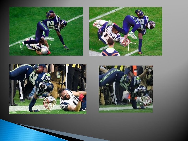
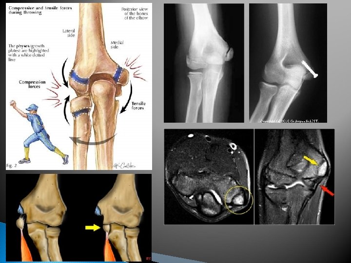
Ulnar Collateral Ligament Sprain Cause: Hyperextension or excessive Valgus Force. This injury usually occurs during the cocking phase of the throwing motion Signs & Symptoms: Pain, Care: Ice, compression Instability, numbness & tingling, Point tenderness over the UCL sleeve, sling, rest. Follow up with a physician for evaluation, x-ray & MRI if necessary. Surgery is required to repair the injury. Recovery after surgery is approximately 9 months During rehabilitation throwing is restricted for the first 12 weeks. After 12 weeks, throwing is controlled by limiting & gradually progressing the number of throws per day

UCL Sprain Ulnar Collateral Ligament

Stress on the UCL

Ulnar Collateral Ligament Repair

Ulnar Collateral Ligament Repair Using Palmaris Longus Tendon

Ulnar Nerve Injury Cause: Typically happens as a result of a weak or torn UCL. It can become strained, chronically dislocated, or impinged Signs & Symptoms: Paresthesia or burning in fourth & fifth fingers, weakness in forearm Care: treat conservatively by rest & restricting aggravation motions. Surgery may be required. Do not compress the nerve with outside pressure

Ulnar Nerve Injury

Lateral Epicondylitis Cause: repetitive wrist Signs & Symptoms: extension. Wrist extensor tendon become inflammed at the attachment. AKA Tennis Elbow Swelling, Pain with wrist extension, aching, weakness, point tenderness over lateral epicondyle Care: P. R. I. C. E, NSAIDs, Ice, Range of Motion exs, stretching, strengthening, Counterforce brace

Lateral Epicondylitis

Medial Epicondylitis Cause: repetitive wrist flexion & elbow flexion. AKA as Little Leaguer’s elbow, Pitcher’s Elbow & Golfer’s Elbow Signs & Symptoms: Point tenderness, Swelling over medial epicondyle, Pain with wrist Flexion, Pain radiating down the arm, aching, weakness, point tenderness over medial epicondyle Care: Rest, NSAIDs, Ice, Range of Motion exs, stretching, strengthening, Counterforce brace. Return to play through throwing program if athlete is an overhead thrower, Severe cases may require splinting

Medial Epicondylitis

Elbow Dislocation Cause: Fall on outstretched hand with elbow in hyperextension or a severe twist with elbow flexion Signs & Symptoms: bones can be displace anterior, posterior, or laterally, most noticeable deformity is displaced olecranon, widening of joint, loss or ROM, nerve injury, severe pain, swelling Care: Check pulses, immobilize injury, refer for emergency reduction, X-ray & splinting

Elbow Dislocation

Elbow Fracture Cause: Fall on outstretched hand, fall on flexed elbow, direct blow or trauma to elbow Signs & Symptoms: May not see a visible deformity. Hemorrhage, swelling, muscle spasms Care: Ice, sling or splint, immediate referral to ER for xray & cast. Casting (immobilization) is typically limited to 3 weeks. After 3 wks. , patient is moved to splint/brace that stabilizes but allows motion

Volkmann’s Contracture A fractured elbow is associated with rapid swelling that may cause a Volkmann’s contracture. Volkmann's contracture results from acute ischemia/necrosis of the muscle fibers of the flexor muscles in the forearm caused by obstruction of the brachial artery near the elbow. The obstruction may be caused by improper tourniquet use, improper casting, compartment syndrome, or profuse bleeding of fractured bones. The two most affected muscles are the flexor digitorum profundus & flexor pollicis longus.

Elbow Fracture

Elbow Fracture

Bracing
- Slides: 72