The visual system ey thalamu e s The


 Copyright © 2002 Wadsworth Group. Wadsworth is an imprint of")
 Copyright © 2002 Wadsworth Group. Wadsworth is an imprint of")


 Specialized for Visual Motion Lesion Experiments motion-blind patient -")
 Specialized for Visual Motion Newsome and others 100% correlation")
 - Complex form Processing Tanaka, et al. , 1991: receptive fields")
 have cells that respond to stimuli that")
 of the Inferotemporal (IT) Cortex: cells responsive to faces")


 -Greebles Copyright © 2002 Wadsworth")

 – measurement of FFA to non-face, but experienced stimuli? Cells")
 – measurement of FFA in individuals diagnosed with")


: Ring a bell Unconditioned response")

- Slides: 26
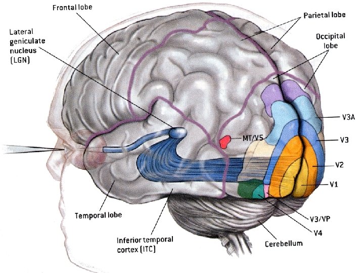
The visual system ey thalamu e s The visual system s visual cortex lateral geniculate nucleus (LGN)
Extrastriate cortex Occipital Striate Cortex Extrastriate Cortex: secondary cortical areas communicated to & from striatal cortex (V 1 - primary receptive areas)
Ungerleider and Mishkin (1981) Copyright © 2002 Wadsworth Group. Wadsworth is an imprint of the Wadsworth Group, a division of Thomson Learning Top = Dorsal Bottom = Ventral From “Object Vision and Spatial Vision: Two Central Pathways, ” by M. Mishkin, L. G. Ungerleider & K. A. Makco, 1983, Trends in Neuroscience, 6, 414 -417, figure 1. Copyright © 1983 Elsevier Science Publishers B. V. Reprinted by permission.
Ungerleider and Mishkin (1981) Copyright © 2002 Wadsworth Group. Wadsworth is an imprint of the Wadsworth Group, a division of Thomson Learning Remove Parietal lobe = couldn’t identify location Remove Temporal lobe = couldn’t identify object
Figure 4. 26 The two types of discrimination tasks used by Ungerleider and Mishkin. (a) Object discrimination: Pick the correct shape. Lesioning the temporal lobe (shaded area) makes this task difficult. (b) Landmark discrimination: Pick the food well closer to the cylinder. Lesioning the parietal lobe makes this task difficult. (From Mishkin, Ungerleider, & Macko, 1983. )
See pages 93 -96 in chapter 4 of text object discrimination monkeys' performance impaired after lesion to temporal lobe (“What pathway”), but not after lesion to parietal lobe (“Where pathway”) landmark task monkeys' performance impaired after lesion to parietal lobe (“Where pathway”), but not after temporal lesion (“What pathway”)
Processing in Extrastriate Visual Cortex Milner and Goodale - patient "D. F. " - damage to ventral stream can not recognize objects, = visual agnosia but can interact with them - patient "V. K. " - damage to the dorsal stream - can identify objects, but did not interact with them appropriately temporal stream parietal stream - "What" pathway Both “Where” & "How" pathway
Figure 4. 8, page 114 Visual pathway Movement Copyright © 2002 Wadsworth Group. Wadsworth is an imprint of the Wadsworth Group, a division of Thomson Learning Medial Temporal Cortex (Both Temporal Lobe) Form, depth, color Inferotemporal Cortex V 1 = Striate Cortex
Medial temporal Cortex (Area MT) Specialized for Visual Motion Lesion Experiments motion-blind patient - motion agnosia or Akinetopsia http: //www. sockshare. com/file/8971 FC 52 E 214244 A# • Thank you Leigh. Ann
Medial temporal Cortex (Area MT) Specialized for Visual Motion Newsome and others 100% correlation 50% correlation normal threshold = 1 -2% threshold after lesions of MT = 10 -20%
Inferotemporal cortex (IT) - Complex form Processing Tanaka, et al. , 1991: receptive fields “respond best” to different specific forms Two types of cells found in the IT: 1. “Primary Cells”: respond to slits, spots, ellipses & squares 2. “Elaborate Cells”: respond to specific complex shapes (and influenced by color & texture)
Neighboring columns in the Inferotemporal Cortex (IT) have cells that respond to stimuli that share similar features
“Fusiform Face Area” (FFA) of the Inferotemporal (IT) Cortex: cells responsive to faces
Some cells respond best to profiles of faces = view-specific cells (other cells showed view-invariance)
Identity!!!
Figure 4. 24, page 129 Gauthier et al. (1999) -Greebles Copyright © 2002 Wadsworth Group. Wadsworth is an imprint of the Wadsworth Group, a division of Thomson Learning Human adults come to recognized specific Greeble faces with training. f. MRI activity correlates Fusiform Face Area of Inferotemporal lobe with Greeble recognition
IT neuron properties; built in or experience? - probably both - five week old infant monkeys have faceselective neurons (“built in” through Natural Selection) - Logothetis and Pauls - trained monkeys to complex shapes of a particular orientation -subsequently found cells in IT that were tuned to that object and that orientation
Tarr & Gauthier (2000) – measurement of FFA to non-face, but experienced stimuli? Cells fired to car pictures for car experts Cells fired to bird pictures for bird experts
Pierce, Haist, Sedaghat, & Courchesne (2004) – measurement of FFA in individuals diagnosed with Autism? • Initial finding: firing patterns in the FFA (with f. MRI) are different from “typical” control subjects – Question about do they not process the face because of damage to FFA, or… – Do they not have typical firing in FFA because they don’t look at faces (chicken and egg problem)? • Tested FFA activity to highly familiar faces (their mom) – FFA activation looked like controls to mom and other highly familiar faces (i. e. , co-workers) – When novel faces were mixed in with familiar faces, the FFA activated more like “typical controls” • Conclusion: cortical areas of the brain (i. e. , IT-FFA) are “experience dependent” – they re-organizes with experience (referred to as “brain plasticity”)
Summary of the Inferotemporal lobe and “object recognition” • Organized in columns • Cells in each column have similar “preferences” for activation • Primary cells (slits, spots, squares of light) • Elaborate cells (complex shape, color, texture) • Some columns respond to faces (call FFA) • FFA may have an evolutionary “edge, ” but experience is essential – FFA cells can be “taught” to fire to previously novel stimuli with experience – FFA cells are “expert” cells
Putting the parts together: The “Binding problem” • Seeing red car in motion? – Ganglion cells from cones (fovea) and/or rods (periphery) offer color, movement, location information – LGN cells from differing eyes, movement, form, color & where on the retina – V 1 striate cortex magnify & locate (retinotopic) the car, and offer form (i. e. , spatial frequency) information – Inferotemporal lobe processing FORM – Medial temporal lobe processing MOTION – Extrastriatal area V 4 processing COLOR – Multiple sites are simultaneously activated at each level (Ganglion, LGN, V 1, IT, MT, V 4, etc. ) Ø How do we put this all together to perceive a red CAR driving down the road? ? ?
Figure 4. 8, page 114 Visual pathway Moving Car: all areas are “humming at once” Copyright © 2002 Wadsworth Group. Wadsworth is an imprint of the Wadsworth Group, a division of Thomson Learning Medial Temporal Cortex (Both Temporal Lobe) Inferotemporal Cortex
Associative Learning Classical conditioning: Pavlov’s dog Unconditioned stimulus (US): Ring a bell Unconditioned response (UR): salivation when dog eats Conditioned stimulus (CS): Pair together: Ring the bell just before giving the food Ring a bell Conditioned response (CR): salivation when dog hears bell
We've Pulled the Stimulus Apart, How do we put the Perception of the “event” together? synchronous oscillations Two neurons firing in an “out of synch” manner A neuron bursting at regular intervals = oscillation Two neurons “oscillating” (bursting) synchronously