
The Urinary System Lecture for medical students Department

- Slides: 54
The Urinary System Lecture for medical students Department of histology, cytology and embryology Kharkiv National Medical University Ukraine 2012
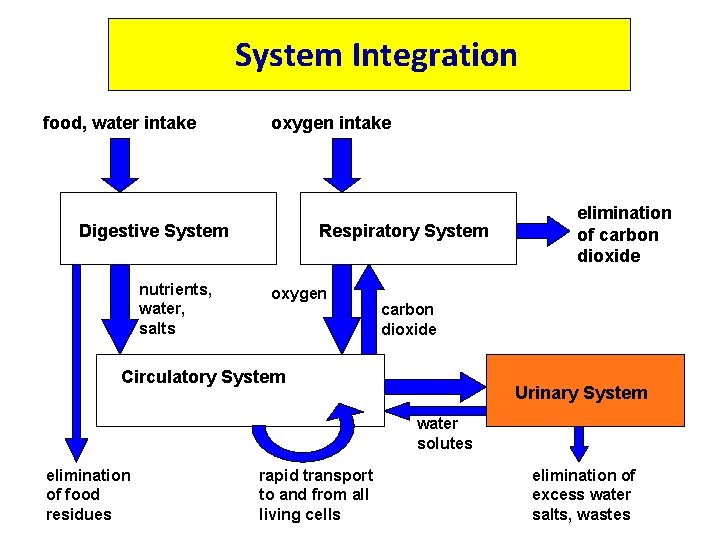
System Integration food, water intake oxygen intake Digestive System nutrients, water, salts Respiratory System oxygen elimination of carbon dioxide Circulatory System Urinary System water solutes elimination of food residues rapid transport to and from all living cells elimination of excess water salts, wastes
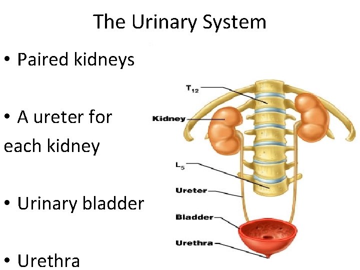
The Urinary System • Paired kidneys • A ureter for each kidney • Urinary bladder • Urethra
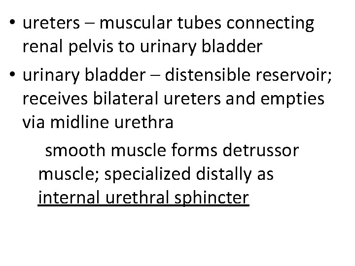
• ureters – muscular tubes connecting renal pelvis to urinary bladder • urinary bladder – distensible reservoir; receives bilateral ureters and empties via midline urethra smooth muscle forms detrussor muscle; specialized distally as internal urethral sphincter
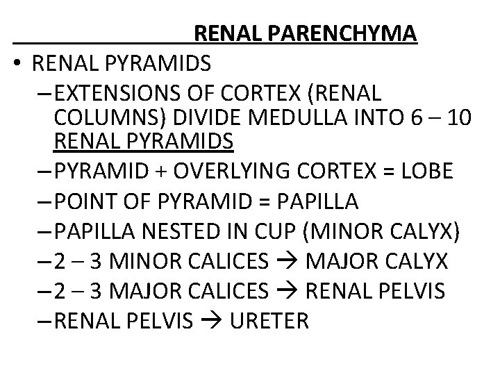
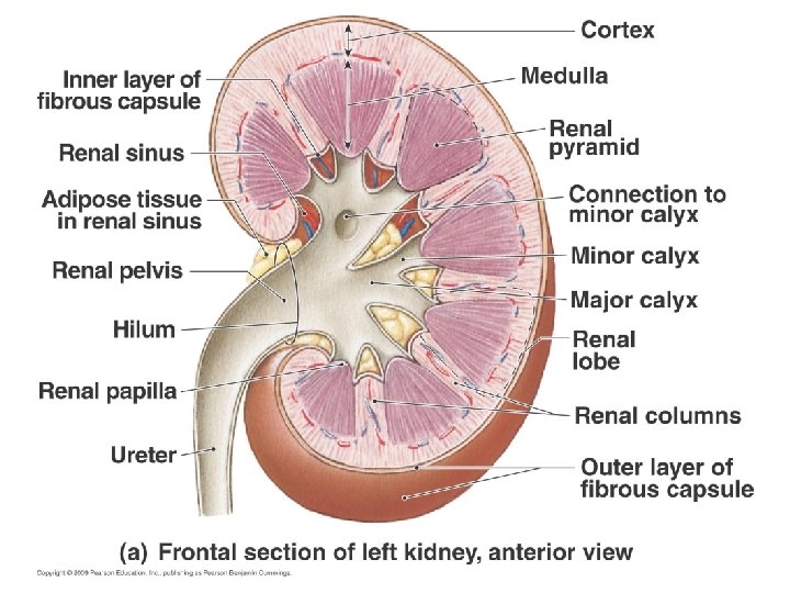
RENAL PARENCHYMA • RENAL PYRAMIDS – EXTENSIONS OF CORTEX (RENAL COLUMNS) DIVIDE MEDULLA INTO 6 – 10 RENAL PYRAMIDS – PYRAMID + OVERLYING CORTEX = LOBE – POINT OF PYRAMID = PAPILLA – PAPILLA NESTED IN CUP (MINOR CALYX) – 2 – 3 MINOR CALICES MAJOR CALYX – 2 – 3 MAJOR CALICES RENAL PELVIS – RENAL PELVIS URETER
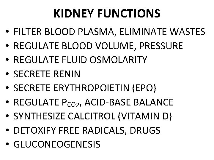
KIDNEY FUNCTIONS • • • FILTER BLOOD PLASMA, ELIMINATE WASTES REGULATE BLOOD VOLUME, PRESSURE REGULATE FLUID OSMOLARITY SECRETE RENIN SECRETE ERYTHROPOIETIN (EPO) REGULATE PCO 2, ACID-BASE BALANCE SYNTHESIZE CALCITROL (VITAMIN D) DETOXIFY FREE RADICALS, DRUGS GLUCONEOGENESIS
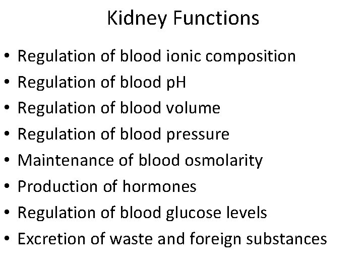
Kidney Functions • • Regulation of blood ionic composition Regulation of blood p. H Regulation of blood volume Regulation of blood pressure Maintenance of blood osmolarity Production of hormones Regulation of blood glucose levels Excretion of waste and foreign substances
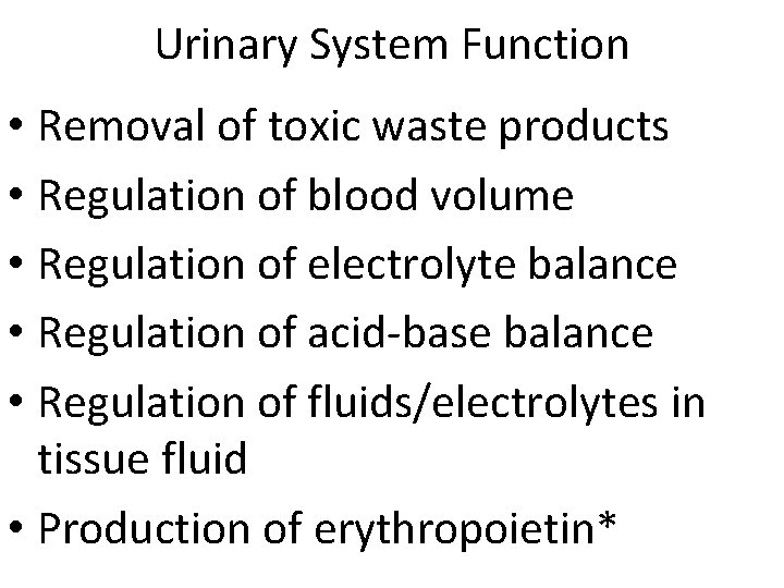
Urinary System Function • Removal of toxic waste products • Regulation of blood volume • Regulation of electrolyte balance • Regulation of acid-base balance • Regulation of fluids/electrolytes in tissue fluid • Production of erythropoietin*
9
Cortex Outer, divisible grossly into alternating : (2) Cortical labyrinths (1) Medullary rays cortical labyrinths – rich in (striated) : : medullary rays - rich (a) renal corpuscles, in (a) straight tubules and (b) convoluted tubules and collecting ducts (c)collecting tubules
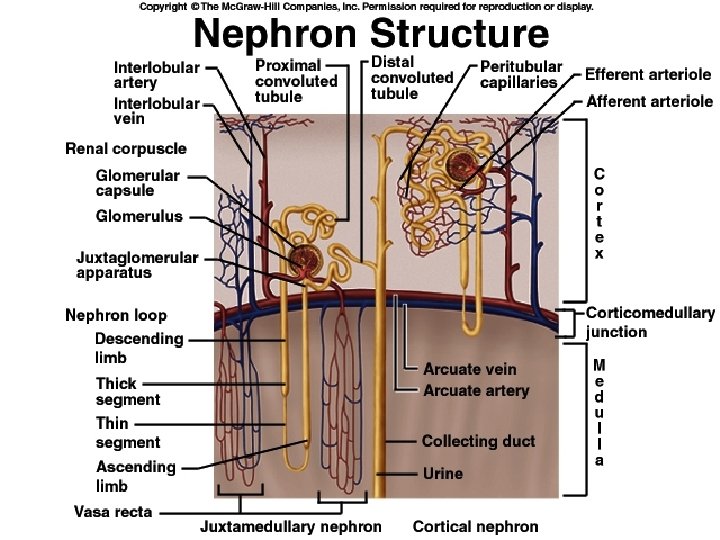
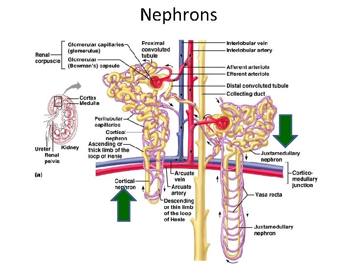
NEPHRONS • FUNCTIONAL UNITS OF KIDNEY • ~1. 2 MILLION PER KIDNEY • THREE MAIN PARTS –BLOOD VESSELS –RENAL CORPUSCLE –RENAL TUBULE
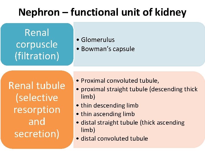
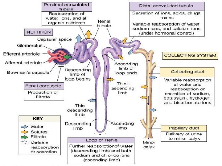
Nephron – functional unit of kidney Renal corpuscle (filtration) Renal tubule (selective resorption and secretion) • Glomerulus • Bowman’s capsule • Proximal convoluted tubule, • proximal straight tubule (descending thick limb) • thin descending limb • thin ascending limb • distal straight tubule (thick ascending limb) • distal convoluted tubule
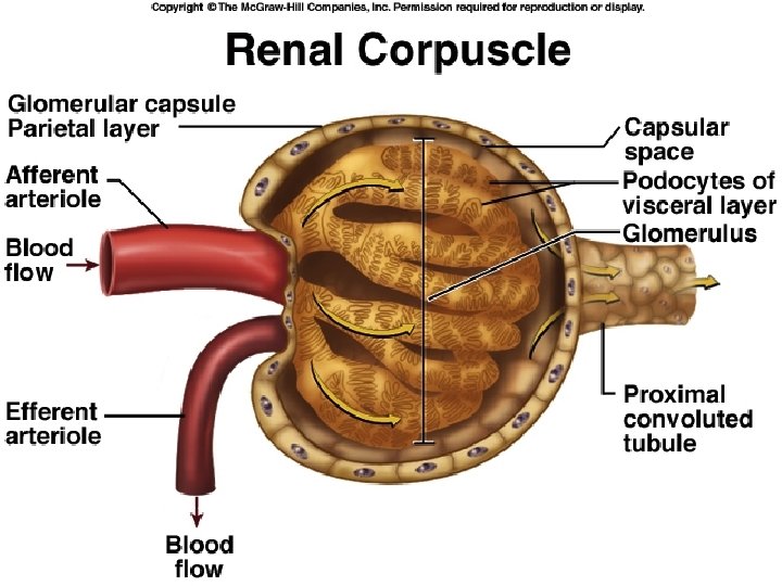
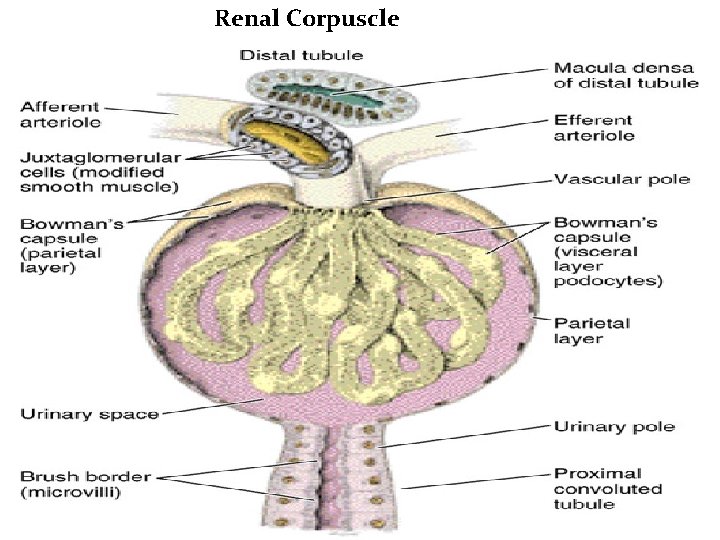
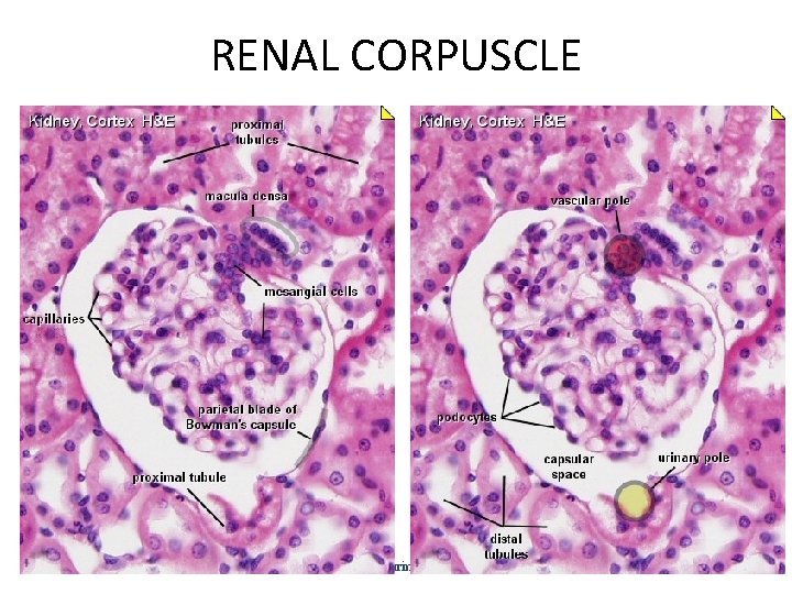
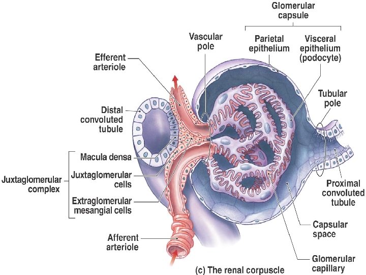
• • • RENAL CORPUSCLE GLOMERULUS PLUS CAPSULE GLOMERULUS ENCLOSED IN TWOLAYERED GLOMERULAR CAPSULE –“BOWMAN’S CAPSULE” FLUID FILTERS FROM GLOMERULAR CAPILLARIES –“GLOMERULAR FILTRATE” FLUID COLLECTS IN CAPSULAR SPACE FLUID FLOWS INTO RENAL TUBULE
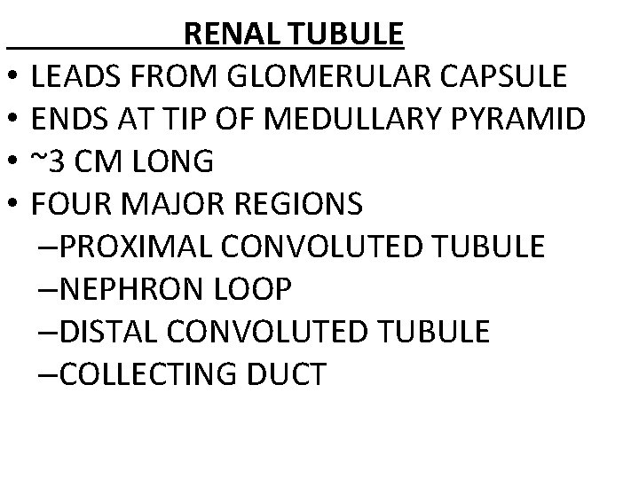
• • RENAL TUBULE LEADS FROM GLOMERULAR CAPSULE ENDS AT TIP OF MEDULLARY PYRAMID ~3 CM LONG FOUR MAJOR REGIONS –PROXIMAL CONVOLUTED TUBULE –NEPHRON LOOP –DISTAL CONVOLUTED TUBULE –COLLECTING DUCT
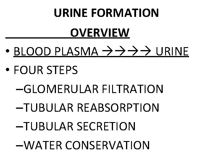
URINE FORMATION OVERVIEW • BLOOD PLASMA URINE • FOUR STEPS –GLOMERULAR FILTRATION –TUBULAR REABSORPTION –TUBULAR SECRETION –WATER CONSERVATION
Renal Corpuscle
RENAL CORPUSCLE histology of urinary system 19
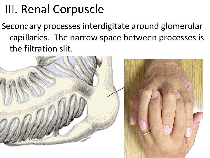
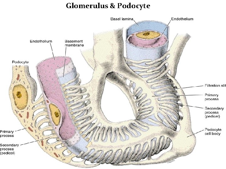
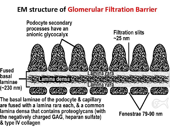
III. Renal Corpuscle Secondary processes interdigitate around glomerular capillaries. The narrow space between processes is the filtration slit.
Glomerulus & Podocyte
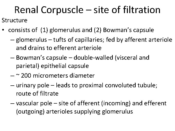
Renal Corpuscle – site of filtration Structure • consists of (1) glomerulus and (2) Bowman’s capsule – glomerulus – tufts of capillaries; fed by afferent arteriole and drains to efferent arteriole – Bowman’s capsule – double-walled (visceral and parietal) epithelial capsule – ~ 200 micrometers diameter – urinary pole – leads to proximal convoluted tubule; route of filtrate – vascular pole – site of afferent (incoming) and efferent (outgoing) arterioles supplying glomerulus
EM structure of Glomerular Filtration Barrier
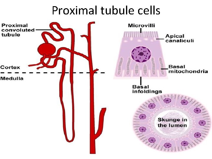
Proximal tubule cells
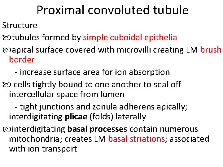
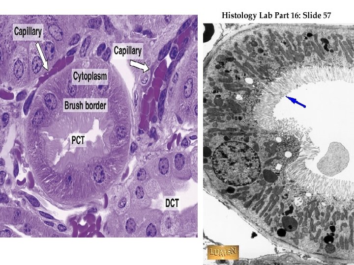
Proximal convoluted tubule Structure tubules formed by simple cuboidal epithelia apical surface covered with microvilli creating LM brush border - increase surface area for ion absorption cells tightly bound to one another to seal off intercellular space from lumen - tight junctions and zonula adherens apically; interdigitating plicae (folds) laterally interdigitating basal processes contain numerous mitochondria; creates LM basal striations; associated with ion transport
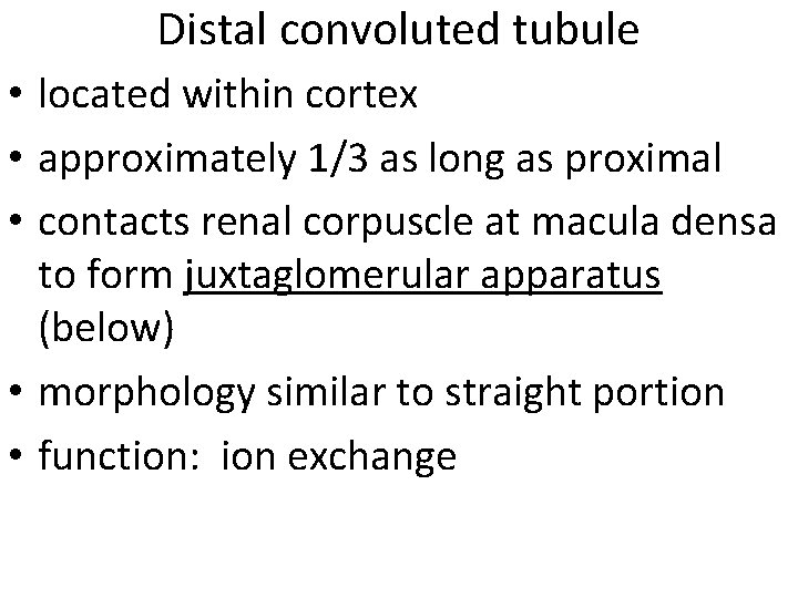
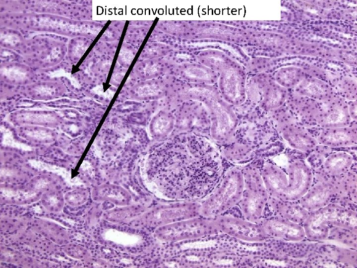
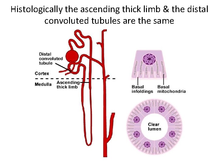
Distal convoluted tubule • located within cortex • approximately 1/3 as long as proximal • contacts renal corpuscle at macula densa to form juxtaglomerular apparatus (below) • morphology similar to straight portion • function: ion exchange
Distal convoluted (shorter)
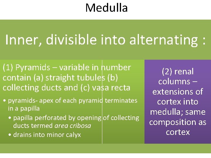
Medulla Inner, divisible into alternating : (1) Pyramids – variable in number contain (a) straight tubules (b) collecting ducts and (c) vasa recta • pyramids- apex of each pyramid terminates in a papilla • papilla perforated by opening of collecting ducts termed area cribosa • drains into minor calyx (2) renal columns – extensions of cortex into medulla; same composition as cortex
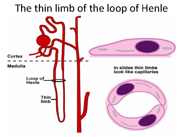
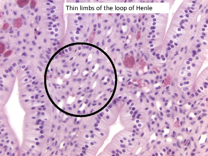
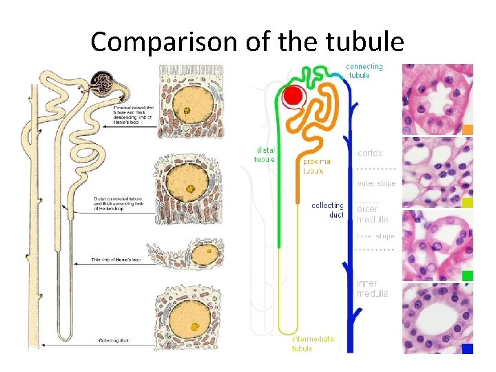
The thin limb of the loop of Henle
Histologically the ascending thick limb & the distal convoluted tubules are the same
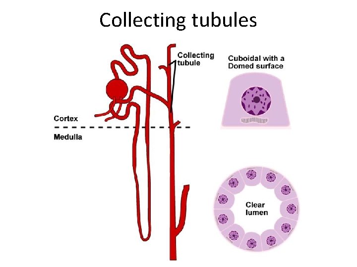
Collecting tubules
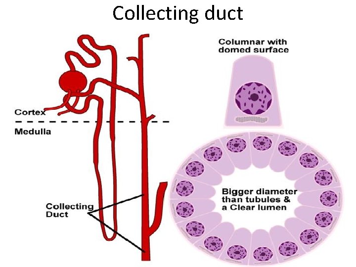
Collecting duct
Thin limbs of the loop of Henle
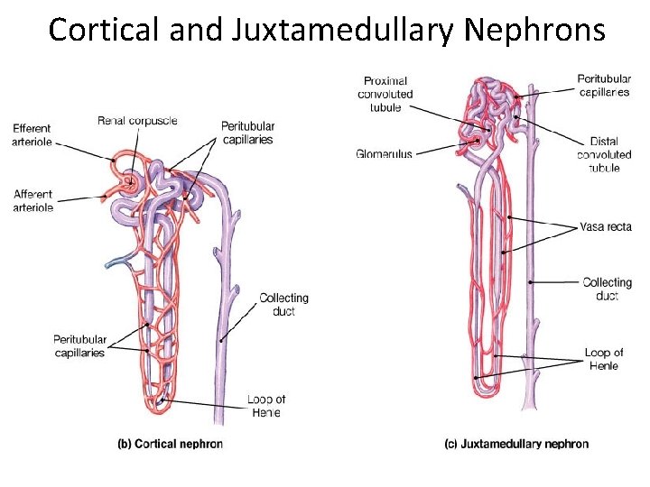
Cortical and Juxtamedullary Nephrons
Nephrons histology of urinary system 38
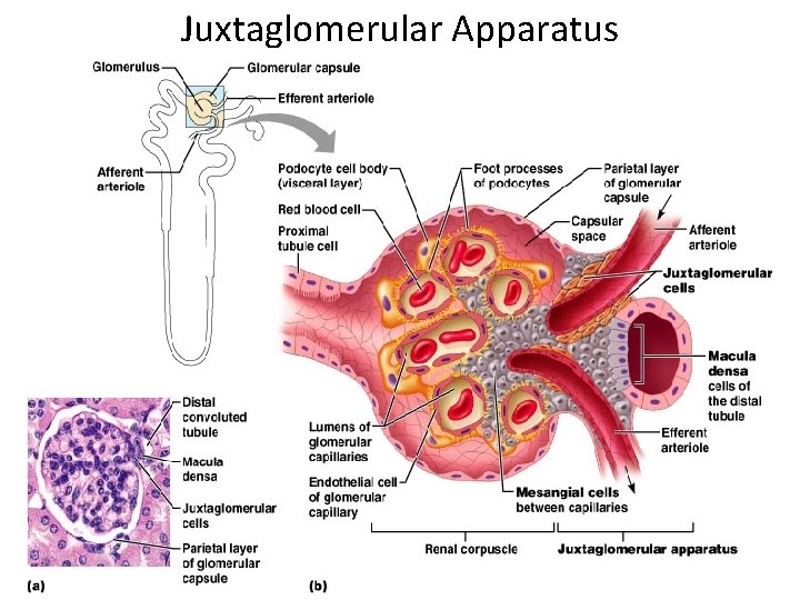
Juxtaglomerular Apparatus
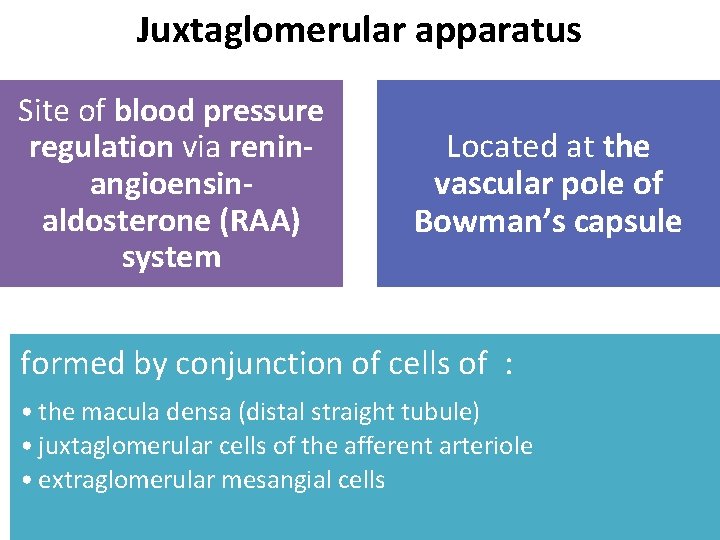
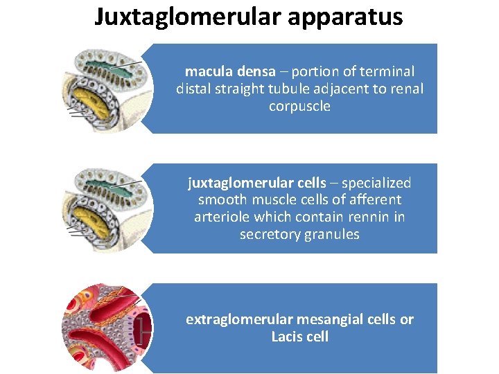
Juxtaglomerular apparatus Site of blood pressure regulation via reninangioensinaldosterone (RAA) system Located at the vascular pole of Bowman’s capsule formed by conjunction of cells of : • the macula densa (distal straight tubule) • juxtaglomerular cells of the afferent arteriole • extraglomerular mesangial cells
Juxtaglomerular apparatus macula densa – portion of terminal distal straight tubule adjacent to renal corpuscle juxtaglomerular cells – specialized smooth muscle cells of afferent arteriole which contain rennin in secretory granules extraglomerular mesangial cells or Lacis cell
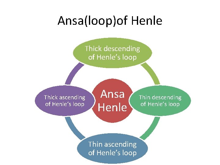
Ansa(loop)of Henle Thick descending of Henle’s loop Thick ascending of Henle’s loop Ansa Henle Thin ascending of Henle’s loop Thin descending of Henle’s loop
Comparison of the tubule
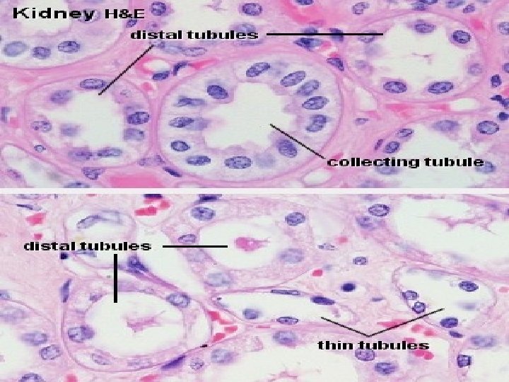
Collecting tubules / ducts • Start in cortex and descend through medulla • as ducts coalesce and increase in size, cells of tubes change from somewhat squamous to cuboidal to columnar • terminate at tip of renal pyramid where urine enters into minor calyx • distinguishable cell margins • central nuclei with poorly staining cytoplasm • At EM level many possess single cilium and sparse microvilli
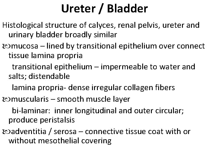
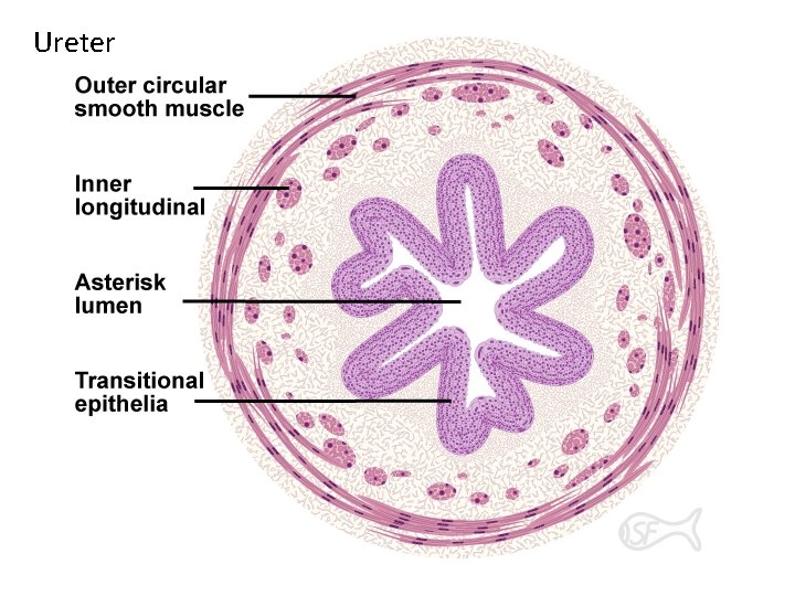
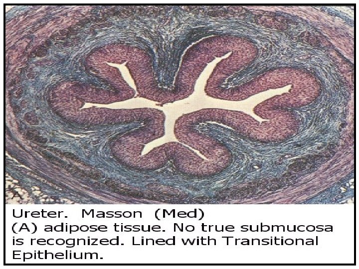
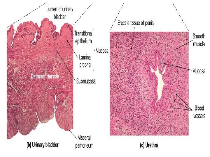
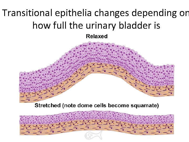
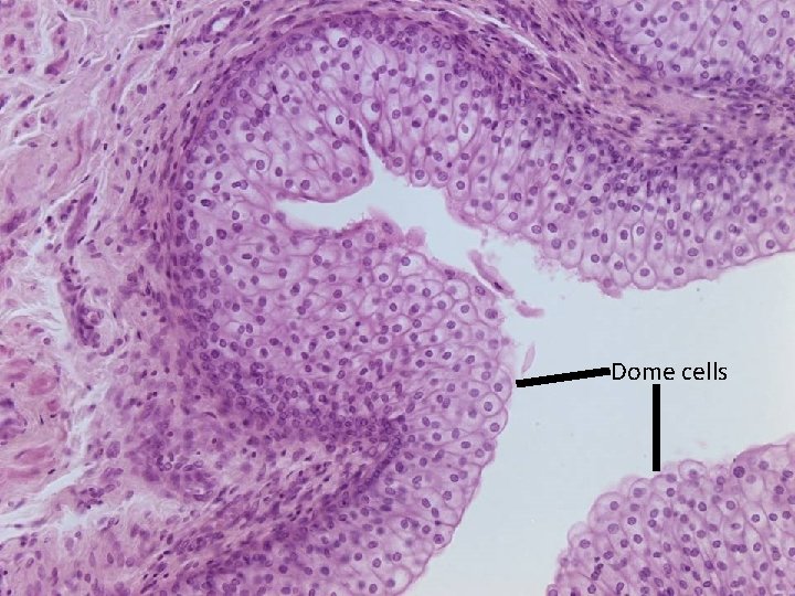
Ureter / Bladder Histological structure of calyces, renal pelvis, ureter and urinary bladder broadly similar mucosa – lined by transitional epithelium over connect tissue lamina propria transitional epithelium – impermeable to water and salts; distendable lamina propria- dense irregular collagen fibers muscularis – smooth muscle layer bi-laminar: inner longitudinal and outer circular; produce peristalsis adventitia / serosa – connective tissue coat with or without mesothelial covering
Ureter
Transitional epithelia changes depending on how full the urinary bladder is
Dome cells
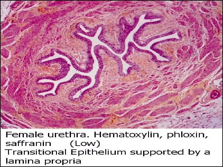
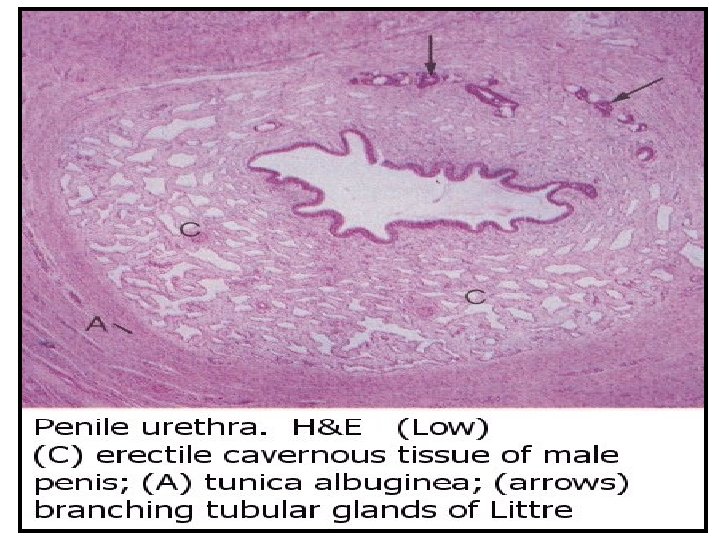
Urethra • fibromuscular tube connecting bladder to external urethral orifice • sexually dimorphic males – terminal duct for both urinary and genital systems females – urinary system only • lining – in both sexes grades from transitional epithelium adjacent to bladder to stratified squamous at orifice males have stratified columnar or pseudostratified columnar in middle portion