
The Urinary System Chapter 26 Video http www



- Slides: 72
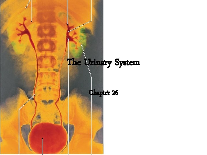
The Urinary System Chapter 26
Video • http: //www. youtube. com/watch? v=a. QZa. NXN ro. VY
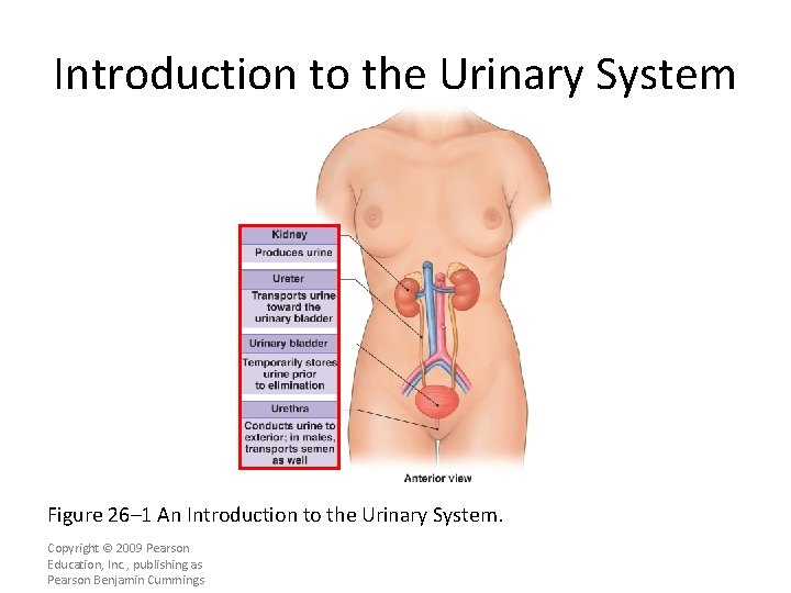
Introduction to the Urinary System Figure 26– 1 An Introduction to the Urinary System. Copyright © 2009 Pearson Education, Inc. , publishing as Pearson Benjamin Cummings
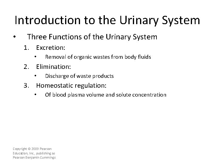
Introduction to the Urinary System • Three Functions of the Urinary System 1. Excretion: • Removal of organic wastes from body fluids 2. Elimination: • Discharge of waste products 3. Homeostatic regulation: • Of blood plasma volume and solute concentration Copyright © 2009 Pearson Education, Inc. , publishing as Pearson Benjamin Cummings
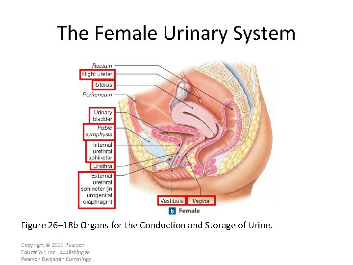
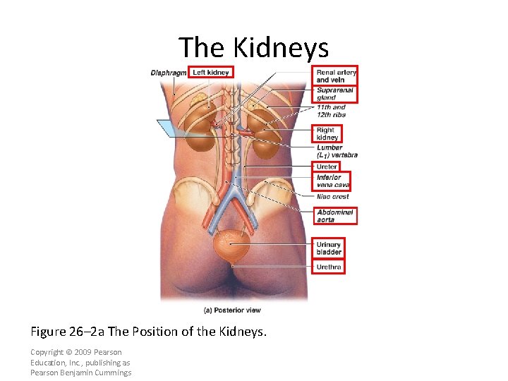
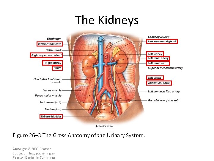
Introduction to the Urinary System • Kidneys — organs that produce urine • Urinary tract — organs that eliminate urine – Ureters (paired tubes) – Urinary bladder (muscular sac) – Urethra (exit tube) • Urination or micturition — process of eliminating urine – Contraction of muscular urinary bladder forces urine through urethra, and out of body Copyright © 2009 Pearson Education, Inc. , publishing as Pearson Benjamin Cummings
Introduction to the Urinary System • Five Homeostatic Functions of Urinary System 1. Regulates blood volume and blood pressure: • • By adjusting volume of water lost in urine Releasing erythropoietin and renin 2. Regulates plasma ion concentrations: • • Sodium, potassium, and chloride ions (by controlling quantities lost in urine) Calcium ion levels (through synthesis of calcitriol) Copyright © 2009 Pearson Education, Inc. , publishing as Pearson Benjamin Cummings
Introduction to the Urinary System • Five Homeostatic Functions of Urinary System 3. Helps stabilize blood p. H: • By controlling loss of hydrogen ions and bicarbonate ions in urine 4. Conserves valuable nutrients: • By preventing excretion while excreting organic waste products 5. Assists liver in detoxifying poisons Copyright © 2009 Pearson Education, Inc. , publishing as Pearson Benjamin Cummings
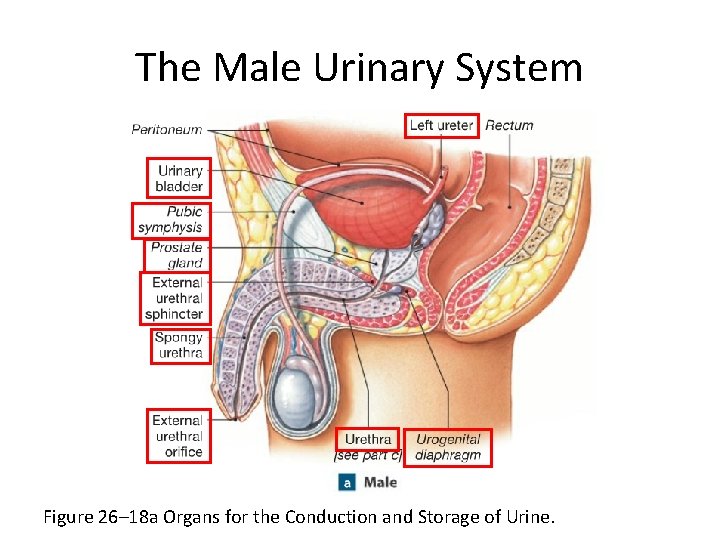
The Male Urinary System Figure 26– 18 a Organs for the Conduction and Storage of Urine.
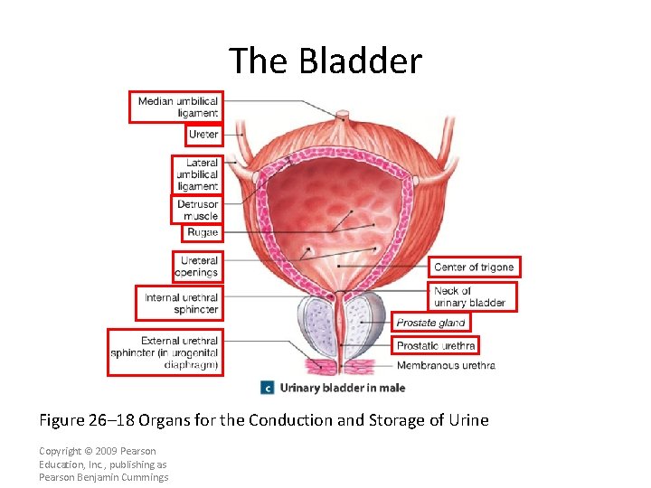
The Bladder Figure 26– 18 Organs for the Conduction and Storage of Urine Copyright © 2009 Pearson Education, Inc. , publishing as Pearson Benjamin Cummings
The Female Urinary System Figure 26– 18 b Organs for the Conduction and Storage of Urine. Copyright © 2009 Pearson Education, Inc. , publishing as Pearson Benjamin Cummings
The Kidneys Figure 26– 2 a The Position of the Kidneys. Copyright © 2009 Pearson Education, Inc. , publishing as Pearson Benjamin Cummings
The Kidneys Figure 26– 3 The Gross Anatomy of the Urinary System. Copyright © 2009 Pearson Education, Inc. , publishing as Pearson Benjamin Cummings
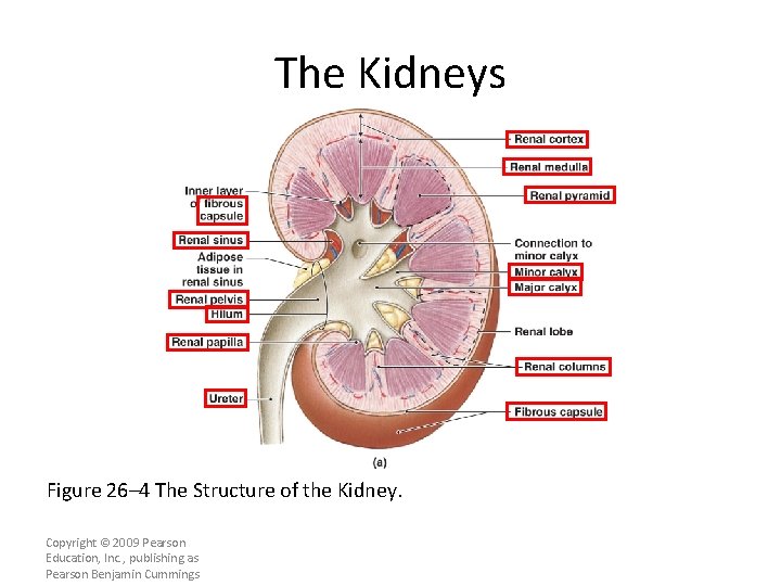
The Kidneys Figure 26– 4 The Structure of the Kidney. Copyright © 2009 Pearson Education, Inc. , publishing as Pearson Benjamin Cummings
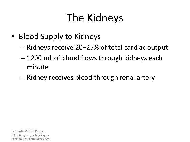
The Kidneys • Blood Supply to Kidneys – Kidneys receive 20– 25% of total cardiac output – 1200 m. L of blood flows through kidneys each minute – Kidney receives blood through renal artery Copyright © 2009 Pearson Education, Inc. , publishing as Pearson Benjamin Cummings
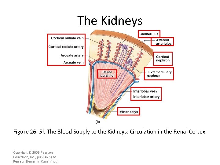
The Kidneys Figure 26– 5 b The Blood Supply to the Kidneys: Circulation in the Renal Cortex. Copyright © 2009 Pearson Education, Inc. , publishing as Pearson Benjamin Cummings
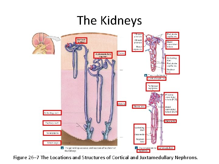
The Kidneys Figure 26– 7 The Locations and Structures of Cortical and Juxtamedullary Nephrons.
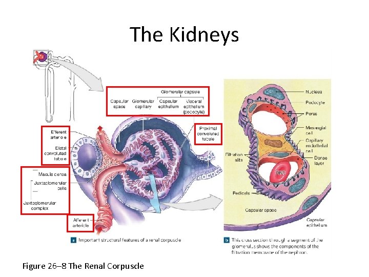
The Kidneys Figure 26– 8 The Renal Corpuscle
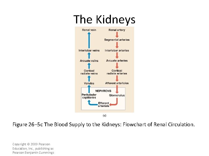
The Kidneys Figure 26– 5 c The Blood Supply to the Kidneys: Flowchart of Renal Circulation. Copyright © 2009 Pearson Education, Inc. , publishing as Pearson Benjamin Cummings
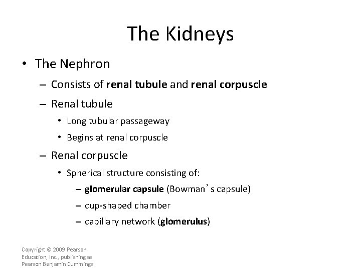
The Kidneys • The Nephron – Consists of renal tubule and renal corpuscle – Renal tubule • Long tubular passageway • Begins at renal corpuscle – Renal corpuscle • Spherical structure consisting of: – glomerular capsule (Bowman’s capsule) – cup-shaped chamber – capillary network (glomerulus) Copyright © 2009 Pearson Education, Inc. , publishing as Pearson Benjamin Cummings
The Kidneys • Glomerulus – Consists of 50 intertwining capillaries – Blood delivered via afferent arteriole – Blood leaves in efferent arteriole • Flows into peritubular capillaries • Which drain into small venules • And return blood to venous system Copyright © 2009 Pearson Education, Inc. , publishing as Pearson Benjamin Cummings
The Kidneys • Filtration – Occurs in renal corpuscle – Blood pressure • Forces water and dissolved solutes out of glomerular capillaries into capsular space • Produces protein-free solution (filtrate) similar to blood plasma Copyright © 2009 Pearson Education, Inc. , publishing as Pearson Benjamin Cummings
The Kidneys • Three regions of Renal Tubule 1. Reabsorb useful organic nutrients that enter filtrate 2. Reabsorb more than 90% of water in filtrate 3. Secrete waste products that failed to enter renal corpuscle through filtration at glomerulus Copyright © 2009 Pearson Education, Inc. , publishing as Pearson Benjamin Cummings
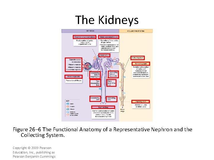
The Kidneys • Segments of Renal Tubule – Located in cortex • Proximal convoluted tubule (PCT) • Distal convoluted tubule (DCT) – Separated by nephron loop (loop of Henle) • U-shaped tube • Extends partially into medulla Copyright © 2009 Pearson Education, Inc. , publishing as Pearson Benjamin Cummings
The Kidneys • Organization of the Nephron – Traveling along tubule, filtrate (tubular fluid) gradually changes composition – Changes vary with activities in each segment of nephron • Each Nephron – Empties into the collecting system: • A series of tubes that carries tubular fluid away from nephron Copyright © 2009 Pearson Education, Inc. , publishing as Pearson Benjamin Cummings
The Kidneys • Collecting Ducts – Receive fluid from many nephrons – Each collecting duct • Begins in cortex • Descends into medulla • Carries fluid to papillary duct that drains into a minor calyx Copyright © 2009 Pearson Education, Inc. , publishing as Pearson Benjamin Cummings
The Kidneys Figure 26– 6 The Functional Anatomy of a Representative Nephron and the Collecting System. Copyright © 2009 Pearson Education, Inc. , publishing as Pearson Benjamin Cummings
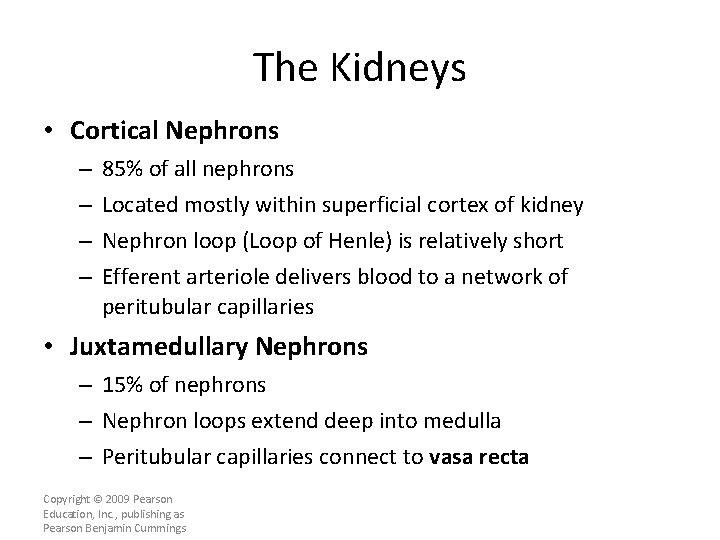
The Kidneys • Cortical Nephrons – – 85% of all nephrons Located mostly within superficial cortex of kidney Nephron loop (Loop of Henle) is relatively short Efferent arteriole delivers blood to a network of peritubular capillaries • Juxtamedullary Nephrons – 15% of nephrons – Nephron loops extend deep into medulla – Peritubular capillaries connect to vasa recta Copyright © 2009 Pearson Education, Inc. , publishing as Pearson Benjamin Cummings
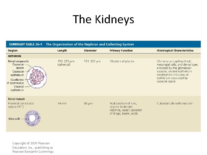
The Kidneys • The Renal Corpuscle – Each renal corpuscle • Is 150– 250 µm in diameter • Glomerular capsule: – is connected to initial segment of renal tubule – forms outer wall of renal corpuscle – encapsulates glomerular capillaries • Glomerulus – knot of capillaries Copyright © 2009 Pearson Education, Inc. , publishing as Pearson Benjamin Cummings
The Kidneys • The Visceral Epithelium – Consists of large cells (podocytes) • With complex processes or “feet” (pedicels) that wrap around specialized lamina densa of glomerular capillaries • Filtration Slits – Are narrow gaps between adjacent pedicels Copyright © 2009 Pearson Education, Inc. , publishing as Pearson Benjamin Cummings
The Kidneys • Filtration – Blood pressure forces water and small solutes across membrane into capsular space – Larger solutes, such as plasma proteins, are excluded • Filtration at Renal Corpuscle – Is passive – Solutes enter capsular space • Metabolic wastes and excess ions • Glucose, free fatty acids, amino acids, and vitamins Copyright © 2009 Pearson Education, Inc. , publishing as Pearson Benjamin Cummings
The Kidneys • Reabsorption – Useful materials are recaptured before filtrate leaves kidneys – Reabsorption occurs in proximal convoluted tubule Copyright © 2009 Pearson Education, Inc. , publishing as Pearson Benjamin Cummings
The Kidneys • The Proximal Convoluted Tubule (PCT) – Is the first segment of renal tubule – Entrance to PCT lies opposite point of connection of afferent and efferent arterioles with glomerulus • Epithelial Lining of PCT – Is simple cuboidal – Has microvilli on apical surfaces – Functions in reabsorption – Secretes substances into lumen Copyright © 2009 Pearson Education, Inc. , publishing as Pearson Benjamin Cummings
The Kidneys • Tubular Cells – Absorb organic nutrients, ions, water, and plasma proteins from tubular fluid – Release them into peritubular fluid (interstitial fluid around renal tubule) Copyright © 2009 Pearson Education, Inc. , publishing as Pearson Benjamin Cummings
The Kidneys • Nephron loop (also called loop of Henle) – Renal tubule turns toward renal medulla • Leads to nephron loop – Descending limb • Fluid flows toward renal pelvis – Ascending limb • Fluid flows toward renal cortex – Each limb contains • Thick segment • Thin segment Copyright © 2009 Pearson Education, Inc. , publishing as Pearson Benjamin Cummings
The Kidneys • The Thick Descending Limb – Has functions similar to PCT • Pumps sodium and chloride ions out of tubular fluid • Ascending Limbs – Of juxtamedullary nephrons in medulla • Create high solute concentrations in peritubular fluid Copyright © 2009 Pearson Education, Inc. , publishing as Pearson Benjamin Cummings
The Kidneys • The Thin Segments – Are freely permeable to water • Not to solutes – Water movement helps concentrate tubular fluid • The Thick Ascending Limb – Ends at a sharp angle near the renal corpuscle • Where DCT begins Copyright © 2009 Pearson Education, Inc. , publishing as Pearson Benjamin Cummings
The Kidneys • The Distal Convoluted Tubule (DCT) – The third segment of the renal tubule – Initial portion passes between afferent and efferent arterioles – Has a smaller diameter than PCT – Epithelial cells lack microvilli Copyright © 2009 Pearson Education, Inc. , publishing as Pearson Benjamin Cummings
The Kidneys • Three Processes at the DCT 1. Active secretion of ions, acids, drugs, and toxins 2. Selective reabsorption of sodium and calcium ions from tubular fluid 3. Selective reabsorption of water: • Concentrates tubular fluid Copyright © 2009 Pearson Education, Inc. , publishing as Pearson Benjamin Cummings
The Kidneys • Juxtaglomerular Complex – An endocrine structure that secretes • Hormone erythropoietin • Enzyme renin – Formed by • Macula densa • Juxtaglomerular cells Copyright © 2009 Pearson Education, Inc. , publishing as Pearson Benjamin Cummings
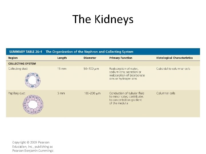
The Kidneys • The Collecting System – The distal convoluted tubule opens into the collecting system – Individual nephrons drain into a nearby collecting duct – Several collecting ducts • Converge into a larger papillary duct • Which empties into a minor calyx – Transports tubular fluid from nephron to renal pelvis – Adjusts fluid composition – Determines final osmotic concentration and volume of urine Copyright © 2009 Pearson Education, Inc. , publishing as Pearson Benjamin Cummings
The Kidneys Copyright © 2009 Pearson Education, Inc. , publishing as Pearson Benjamin Cummings
The Kidneys Copyright © 2009 Pearson Education, Inc. , publishing as Pearson Benjamin Cummings
The Kidneys Copyright © 2009 Pearson Education, Inc. , publishing as Pearson Benjamin Cummings
Renal Physiology • The goal of urine production – Is to maintain homeostasis – By regulating volume and composition of blood – Including excretion of metabolic waste products • The Kidneys – Usually produce concentrated urine • 1200– 1400 m. Osm/L (four times plasma concentration) Copyright © 2009 Pearson Education, Inc. , publishing as Pearson Benjamin Cummings
Renal Physiology • Three Organic Waste Products 1. Urea 2. Creatinine 3. Uric acid – Are dissolved in bloodstream – Are eliminated only while dissolved in urine – Removal is accompanied by water loss Copyright © 2009 Pearson Education, Inc. , publishing as Pearson Benjamin Cummings
Renal Physiology • Kidney Functions – To concentrate filtrate by glomerular filtration • Failure leads to fatal dehydration – Absorbs and retains valuable materials for use by other tissues • Sugars and amino acids Copyright © 2009 Pearson Education, Inc. , publishing as Pearson Benjamin Cummings
Renal Physiology • Renal Threshold – Is the plasma concentration at which • A specific compound or ion begins to appear in urine – Varies with the substance involved Copyright © 2009 Pearson Education, Inc. , publishing as Pearson Benjamin Cummings
Renal Physiology • Renal Threshold for Glucose – Is approximately 180 mg/d. L – If plasma glucose is greater than 180 mg/d. L • Tm of tubular cells is exceeded • Glucose appears in urine: – glycosuria Copyright © 2009 Pearson Education, Inc. , publishing as Pearson Benjamin Cummings
Renal Physiology • Renal Threshold for Amino Acids – Is lower than glucose (65 mg/d. L) – Amino acids commonly appear in urine • After a protein-rich meal • Aminoaciduria Copyright © 2009 Pearson Education, Inc. , publishing as Pearson Benjamin Cummings
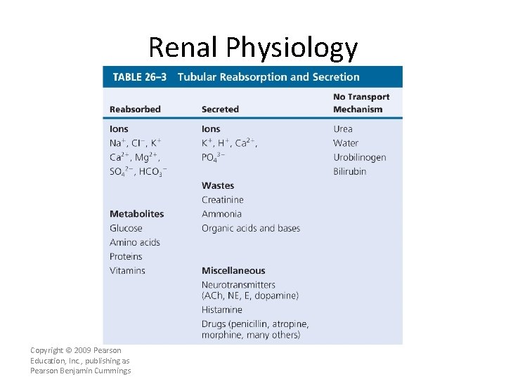
Renal Physiology Copyright © 2009 Pearson Education, Inc. , publishing as Pearson Benjamin Cummings
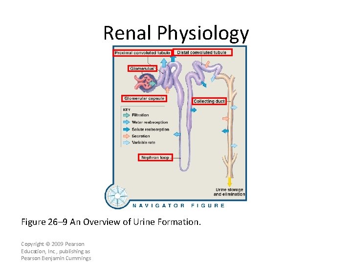
Renal Physiology Figure 26– 9 An Overview of Urine Formation. Copyright © 2009 Pearson Education, Inc. , publishing as Pearson Benjamin Cummings
Glomerular Filtration • Glomerular Filtration Rate (GFR) – Is the amount of filtrate kidneys produce each minute – Averages 125 m. L/min – About 10% of fluid delivered to kidneys • Leaves bloodstream • Enters capsular spaces Copyright © 2009 Pearson Education, Inc. , publishing as Pearson Benjamin Cummings
Glomerular Filtration • Control of the GFR – Autoregulation (local level) – Hormonal regulation (initiated by kidneys) – Autonomic regulation (by sympathetic division of ANS) Copyright © 2009 Pearson Education, Inc. , publishing as Pearson Benjamin Cummings
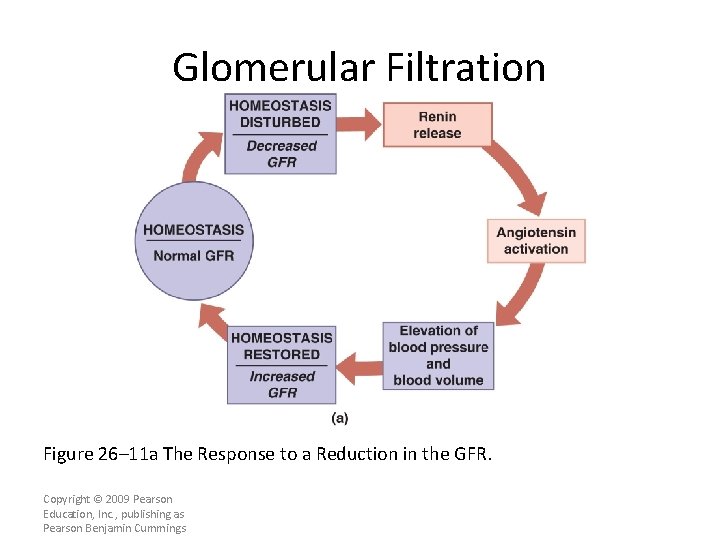
Glomerular Filtration • Hormonal Regulation of the GFR – By hormones of the • Renin–angiotensin system • Natriuretic peptides (ANP and BNP) • The Renin–Angiotensin System – Three stimuli cause the juxtaglomerular complex (JGA) to release renin • Decline in blood pressure at glomerulus • Fall in systemic pressures • Stimulation of juxtaglomerular cells Copyright © 2009 Pearson Education, Inc. , publishing as Pearson Benjamin Cummings
Glomerular Filtration Figure 26– 11 a The Response to a Reduction in the GFR. Copyright © 2009 Pearson Education, Inc. , publishing as Pearson Benjamin Cummings
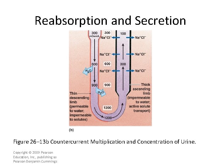
Reabsorption and Secretion • Countercurrent Multiplication – Is exchange that occurs between two parallel segments of loop of Henle • The thin, descending limb • The thick, ascending limb Copyright © 2009 Pearson Education, Inc. , publishing as Pearson Benjamin Cummings
Benefits of Countercurrent Multiplication 1. Efficiently reabsorbs solutes and water before the tubular fluid reaches the DCT and collecting system 2. It establishes a concentration gradient that permits the passive reabsorption of water from the tubular fluid in the collecting system.
Reabsorption and Secretion Figure 26– 13 b Countercurrent Multiplication and Concentration of Urine. Copyright © 2009 Pearson Education, Inc. , publishing as Pearson Benjamin Cummings
Reabsorption and Secretion • Aldosterone – Is a hormone produced by suprarenal cortex – Controls ion pump and channels – Stimulates synthesis and incorporation of Na+ pumps and channels • In plasma membranes along DCT and collecting duct – Reduces Na+ lost in urine Copyright © 2009 Pearson Education, Inc. , publishing as Pearson Benjamin Cummings
Reabsorption and Secretion • Hypokalemia – Produced by prolonged aldosterone stimulation – Dangerously reduces plasma concentration Copyright © 2009 Pearson Education, Inc. , publishing as Pearson Benjamin Cummings
Reabsorption and Secretion • Natriuretic Peptides (ANP and BNP) – Oppose secretion of aldosterone • And its actions on DCT and collecting system • Parathyroid Hormone and Calcitriol – Circulating levels regulate reabsorption at the DCT Copyright © 2009 Pearson Education, Inc. , publishing as Pearson Benjamin Cummings
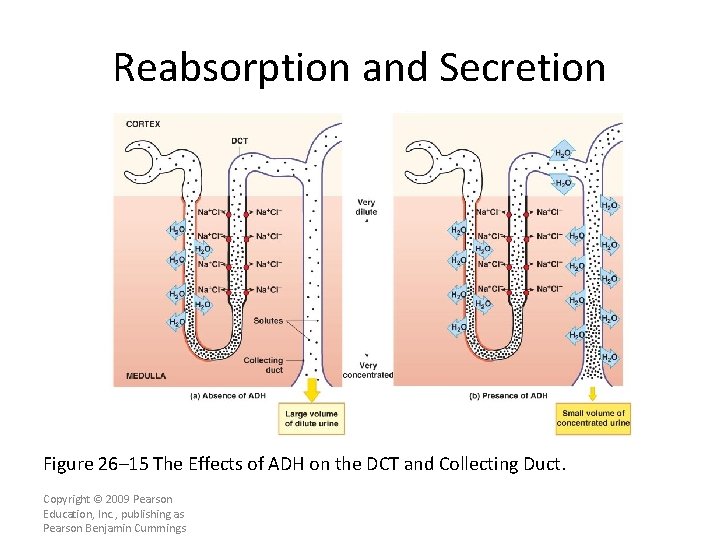
Reabsorption and Secretion • ADH – Hormone that causes special water channels to appear in apical cell membranes – Increases rate of osmotic water movement – Higher levels of ADH increase • Number of water channels • Water permeability of DCT and collecting system Copyright © 2009 Pearson Education, Inc. , publishing as Pearson Benjamin Cummings
Reabsorption and Secretion Figure 26– 15 The Effects of ADH on the DCT and Collecting Duct. Copyright © 2009 Pearson Education, Inc. , publishing as Pearson Benjamin Cummings
Reabsorption and Secretion • Urine Production – A healthy adult produces • 1200 m. L per day (0. 6% of filtrate) • With osmotic concentration of 800– 1000 m. Osm/L Copyright © 2009 Pearson Education, Inc. , publishing as Pearson Benjamin Cummings
Reabsorption and Secretion • The Vasa Recta – Carries water and solutes out of medulla – Balances solute reabsorption and osmosis in medulla Copyright © 2009 Pearson Education, Inc. , publishing as Pearson Benjamin Cummings
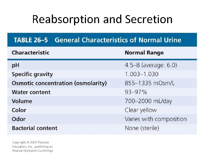
Reabsorption and Secretion • The Composition of Normal Urine – Results from filtration, absorption, and secretion activities of nephrons – Some compounds (such as urea) are neither excreted nor reabsorbed – Organic nutrients are completely reabsorbed • Other compounds missed by filtration process (e. g. , creatinine) are actively secreted into tubular fluid Copyright © 2009 Pearson Education, Inc. , publishing as Pearson Benjamin Cummings
Reabsorption and Secretion • The Composition of Normal Urine – A urine sample depends on osmotic movement of water across walls of tubules and collecting ducts – Is a clear, sterile solution – Yellow color (pigment urobilin) • Generated in kidneys from urobilinogens – Urinalysis, the analysis of a urine sample, is an important diagnostic tool Copyright © 2009 Pearson Education, Inc. , publishing as Pearson Benjamin Cummings
Reabsorption and Secretion Copyright © 2009 Pearson Education, Inc. , publishing as Pearson Benjamin Cummings
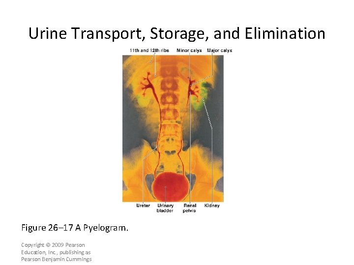
Urine Transport, Storage, and Elimination Figure 26– 17 A Pyelogram. Copyright © 2009 Pearson Education, Inc. , publishing as Pearson Benjamin Cummings
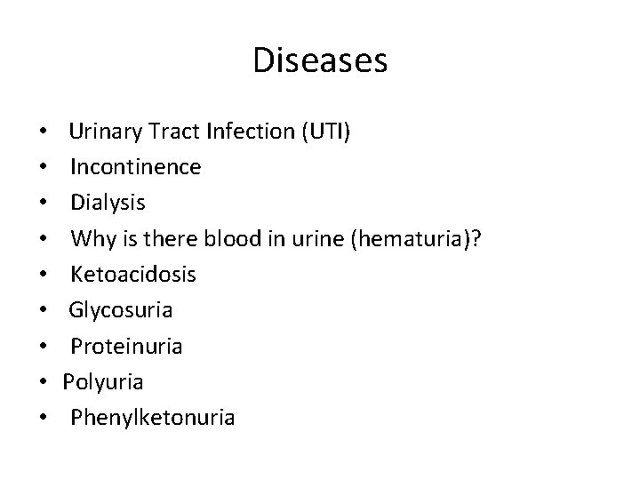
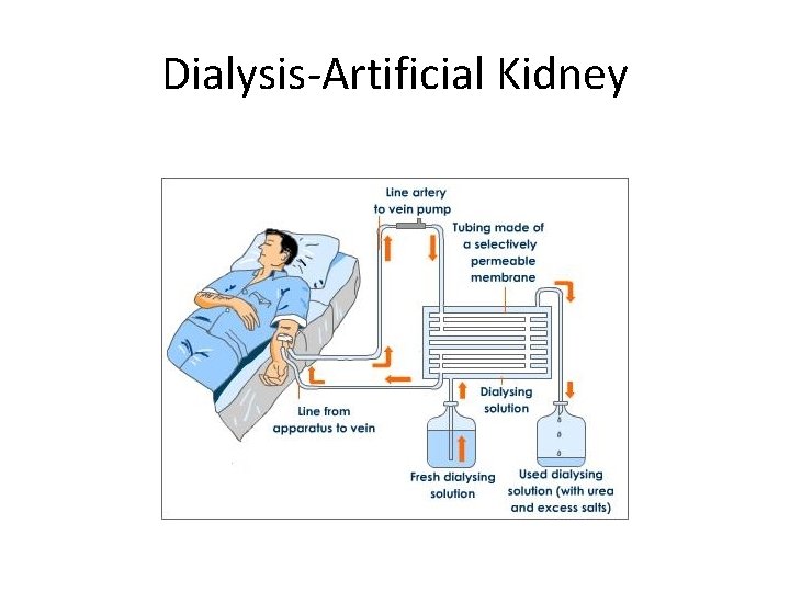
Diseases • • • Urinary Tract Infection (UTI) Incontinence Dialysis Why is there blood in urine (hematuria)? Ketoacidosis Glycosuria Proteinuria Polyuria Phenylketonuria
Dialysis-Artificial Kidney
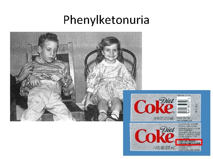
Phenylketonuria