The upper limb Muscles That Move the Pectoral

The upper limb

Muscles That Move the Pectoral Girdle • Originate on the axial skeleton and insert on the clavicle and scapula. • Stabilize the scapula and move it to increase the arm’s angle of movements. • Some of the superficial muscles of the thorax are grouped together according to the scapular movement they direct. – elevation, depression, protraction, or retraction EXTRINSIC MUSCLES

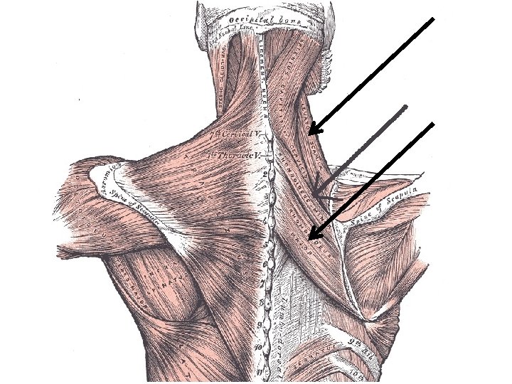
The muscles of back Superficial group • Trapezius - 1 • Latissimus dorsi - 2 • Levator scapulae - 3 • Rhomboidei (minor, major) - 4 Deep group • Erector spinae - 5 • Splenius - 6 • Thoracolumbar fascia - 7 6 3 1 4 7 2 s

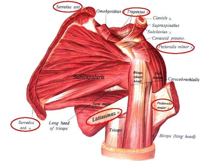
The muscles of thorax Extrinsic muscles • Pectoralis major - 1 • Pectoralis minor - 2 • Serratus anterior - 3 Intrinsic muscles • Intercostales externi • Intercostales intimi 1 2 3

Rhomboid minor Rhomboid major
 • Deltoid • Supraspinatus •")
The Muscles of Upper Limb Muscles of shoulder (intrinsic) • Deltoid • Supraspinatus • Infraspinatus • Teres minor • Teres major • Subscapularis

Deltoid • Origin: lateral third of clavicle, acromion, and spine of scapula • Insertion: deltoid tuberosity of humerus • Action: abducts,flexes and medially rotates, extends, and laterally rotates arm • Nerve supply: axillary n.

Teres major • Origin: dorsal surface of inferior angle of scapula • Insertion: crest of lesser tubercle of humerus • Action: medially rotates and adducts arm • Nerve supply: lower scapular nerve (C 5 -C 8) or thoracodorsal nerve (C 5 -C 7)

Foramen axillare laterale et mediale Mnemonic: A - artery / army is on the bridge N - nerve/ navy is under the bridge

Quadrangular foramen: Axillary nerve Post. humeral circumflex a. , v. QAP Triangular foramen: Circumflex scapular a. , v.
 compartment • Posterior (extensor)")
Arm Muscles That Move the Shoulder/Elbow Joint • Anterior (flexor) compartment • Posterior (extensor) compartment • Anterior compartment – primarily contains shoulder/elbow flexors • Posterior compartment – contains elbow extensors – the principal flexors • biceps brachii, brachialis, and brachioradialis – muscles that extend the elbow joint • triceps brachii and the anconeus
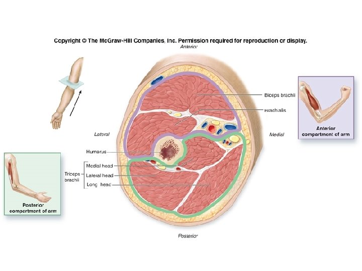

Muscles of arm • Anterior group – 1. Biceps brachii – 2. Coracobrachialis – 3. Brachialis 2 4 • Posterior group � 4. Triceps brachii 1 3

Biceps brachii • Origin: long head, supraglenoid tubercle; short head, coracoid process • Insertion: radial tuberosity • Action: supinator of forearm, flexor of elbow joint, weak flexor of should joint • Nerve supply - musculocutaneus Pronator teres • Origin: medial epicondyle of humerus and deep fascia of forearm • Insertion: middle of lateral surface of radius • Action: pronation of forearm and flexion of elbow

Triceps brachii • Origin: long head, infraglenoid tubercle; lateral head, above groove for radial n. , medial head, below groove for radial n. • Insertion: olecranon of ulna • Action: extends elbow joint), long head can extend adduct shoulder joint • Nerve supply: radial n.

Posterior arm Surface anatomy Triceps brachii long, medial and lateral heads Surface elements Skin thick and movable Subcutaneous tissue well expressed posterior brachial cutaneous nerve inferior lateral brachial cutaneous nerve posterior antebrachial cutaneous nerve

Deep structures А. Brachial fascia - Thick, thins distally B. Neurovascular bundles 1. Upper - in canalis humero-muscularis, between: - sulcus n. radialis - medial and lateral head of m. triceps brachii 1. 1. radial nerve – lies on the humerus - inferior lateral brachial cutaneous nerve - posterior antebrachial cutaneous nerve - muscular branches 1. 2. profunda brachii artery - medial collateral artery - radial collateral artery

Lower neurovascular bundle /behind medial epicondyle/ - ulnar nerve - superior ulnar collateral artery

deep brachial https: //www. earthslab. com/anatomy/inferior-ulnar-collateral-artery/

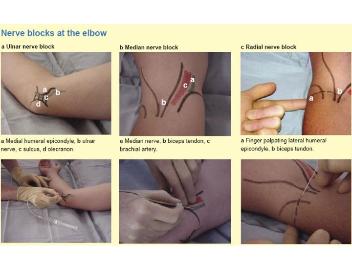
Elbow region

Anterior elbow Surface anatomy Three eminences Two grooves Elements Lateral groove - cephalic vein, lateral antebrachial cutaneous nerve Medial groove – basilic vein, medial antebrachial cutaneous nerve Median cubital vein

Deep structures А. Muscles Flexors of forearm – medial eminence Extensors of forearm – lateral eminence Brachial muscles: mm. biceps, brachialis – middle eminence

Deep structures B. Vessels and nerves Medial bundle - between m. biceps brachii and pronator teres - a. v. brachialis, n. medianus Lateral bundle - between m. brachialis and brachioradialis - n. radialis, a. collateralis radialis, a. recurrens radialis C. Cubital fossa Boundaries - m. brachioradialis (lateral), m. pronator teres (medial) Content - tendo m. brachialis, m. biceps brachii, bursa bicipitoradialis

Layers of the elbow Venous 1 v. cephalica 2 v. basilica 3 v. mediana cubiti Apponeurotic 1 aponeurosis bicipis 2 tendo m. biceps brachii

Neurovascular 1 a. brachialis 2 n. medianus Muscles 1 m. supinator 2 m. brachialis 3 tendo m. biceps brachii Bones 1 humerus 2 radius 3 ulna

Anterior elbow И M

Arteries of upper limb Axillary artery • Continuation of subclavian artery at lateral border of first rib • Becomes brachial artery at lower border of teres major • Divided into three parts by overlying pectoralis minor – First portion, above muscle-gives rise to superior thoracic and thoracoacromial a. – Second portion, behind muscle-gives rise to lateral thoracic a. – Third portion, below muscle-gives rise to subscapular a. • divides into throcodorsal a. • and circumflex scapular a. – anterior and posterior humeral circumflex a. ;

1 4 5 1. Subclavian artery 2. Axillary artery 3. Brachial artery 4. Superior thoracic a. 5. Thoracoacromial a. 6. Lateral thoracic a. 7. Subscapular a. 8. Circumflex scapular a. 9. Thoracodorsal a. 10. Ant. humeral circumflex a. 11. Post. humeral circumflex a. 2 7 3 11 10 8 6 9

2 Brachial artery - 1 • Continuation of axillary artery - 2 • Divides into radial (6)and ulnar (7) arteries at level of neck of radius • Branches 3 – Deep brachial a. accompanies radial nerve - 3 – Superior ulnar collateral a. - 4 accompanies ulnar nerve – Inferior ulnar collateral a. - 5 4 1 5 6 7

Radial artery and branches • Radial recurrent a. 1 • Superficial palmar branch 2 • Principal artery of thumb 3 Ulnar artery and branches • Ulnar recurrent a. 4 • Common interosseous artery 5 1 5 6 7 – Anterior interosseous a. 6 – Posterior interosseous a. 7 • Deep palmar branch 8 4 2 3 8
 and superficial")
5 Superficial palmar arch - 1 • Formed by ulnar artery (2) and superficial palmar branch of radial artery (3) • Curve of arch lies across the palm, level with the distal border of fully extended thumb • Gives rise to three common palmar digital arteries (4) each then divides into two proper palmar digital arteries (5) 4 1 3 2
 and deep palmar")
Deep palmar arch - 1 • Formed by radial artery (2) and deep palmar branch of ulnar artery (3) • Curve of arch lies across upper part of palmar at level with proximal border of extended thumb • Gives rise to three palmar metacarpal arteries (4) 4 3 1 2

Veins of the upper limb Deep veins: accompany the arteries of the same region and bear similar names Superficial veins • Cephalic vein ü Arises from the lateral side of the dorsal venous rete of hand ü Ascends on radial side of the forearm to the elbow and then in the lateral side of biceps brachii furrow, continues up the arm in the deltopectoral groove and then to the infraclavicular fossa, where it pierces clavipectoral fascia to drain into axillary vein

• Basilic vein ü Arises from the medial side of the dorsal venous rete of hand ü Ascends on the ulnar side of forearm to the elbow and then in the medial bicipital furrow to middle of the arm where it pierces the deep fascia and joins the brachial vein or axillary vein • Median cubital vein ü links cephalic vein and basilic vein in the cubital fossa. It is a frequent site for venipuncture to remove a sample of blood or add fluid to the blood

The lymphatic drainage of upper limb Lymphatic vessels • Superficial-follow the superficial veins, drain into supratrochlear and axillary lymph nodes • Deep-accompany main vessels, end in axillary lymph nodes Lymph nodes • Cubital lymph node: lies above medial epicondyle of humerus • Axillary lymph node-arranged in five groups

Axillary lymph nodes Arranged in five groups • Lateral lymph nodes lie around the distal end of axillary vein , receiving drainage from the arm, forearm, and hand • Pectoral lymph nodes lie along lateral thoracic vessels, receive afferents from anterior thoracic wall including central and lateral portion of mamma • Subscapular lymph node along subscapular vessels, receive lymph from nape and scapular region Efferents above three groups pass to central lymph node

• Central lymph nodes – in fat of axillary fossa, receive drainage from all the above nodes, efferents pass to apical lymph node • Apical lymph nodes – In the apex of the axilla, along the proximal end of axillary vessels – Drains central lymph nodes, upper portion of mamma Pectoral; Subscapular; Lateral Central Apical subclavian trunk

Brachial plexus Formation: • Five roots: anterior rami of C 5 -C 8 and T 1 spinal nerves, roots C 5 -C 7 give rise to long thoracic n. • Three trunks – Upper trunk formed by the joining of roots C 5, C 6. – Middle trunk continuation of root C 7. – Lower trunk formed by the joining of roots C 8 and T 1. • Six divisions: above clavicle, trunks form anterior and posterior divisions • Three cords: below clavicle, divisions form three cords that surround the second portion of axillary a.

Position: passes through the scalene fissure to posterosuperior of subclavian artery, then enters the axilla to form lateral, medial and posterior cords Main branches • Lateral cord – Musculocutaneous n. – Lateral root to median n. • Medial cord – Medial root to median n. – Ulnar n. – Medial brachial cutaneous n. – Medial antebrachial cutaneous n.

subscapular • Posterior cord – radial n. - 1 – axillary n. - 2 – thoracodorsal n. - 3 2 3 1

Musculocutaneous nerve Supplies: Muscles -‘BBC nerve’ – Biceps brachii, – Brachalis – Coracobrachialis; skin on the anterior aspect of forearm

Median nerve Supplies: Flexors of forearm except brachioradialis, Flexor carpi ulnaris flexor digitorum profundus - ulnar half Thenar except adductor pollicis, two lateral lumbricals; Skin of thenar, central part of palm, palmar aspect of radial three and one-half fingers, including middle and distal fingers on dorsum • • • Injury: Ape hand • produces sign of benediction, the index and middle fingers cannot be flexed and the thumb cannot be opposed

Ulnar nerve Supplies: • • • flexor carpi ulnaris, ulnar half of flexor digitorum profundus, hypothenar muscles, interossei, 3 rd and 4 th lumbricals and adductor pollicis; skin of hypothenar, palmar surface of ulnar one and one-half fingers, ulnar half of dorsum of hand, posterior aspect of ulnar two and one-half fingers Injury: claw hand

Radial Supplies: – Extensor muscles of arm and forearm, – Brachioradialis; – skin on back of arm, forearm, and radial side of dorsum of hand radial two and onehalf fingers Injury: Wrist drop

Axillary nerve Supplies: – Deltoid and teres minor muscle; – skin over deltoid and upper posterior aspect of arm Injury: – deltoid and teres minor paralysis (loss of shoulder abduction and wheel external rotation) – loss of sensation over the deltoid

Forearm Muscles Supinate and Pronate • Supinator muscle supinates the forearm. • Biceps brachii supinates the forearm. • Pronator teres and pronator quadratus pronate the forearm. Move the Wrist Joint, Hand, and Fingers n n Muscles in the forearm move the hand at the wrist and/or the fingers. Extrinsic muscles of the wrist and hand originate on the forearm, not the wrist or hand. Tendons of forearm muscles typically are surrounded by tendon (synovial) sheaths and held adjacent to the skeletal elements by strong fascial structures. At the wrist, the deep fascia of the forearm forms thickened, fibrous bands termed retinacula.

n The forearm extends from elbow to wrist. n It posses two bones radius laterally & Ulna medially. n The two bones are connected together by the interosseous membrane. This membrane allows movement of Pronation and Supination while the n n two bones are connected together. Also it gives origin for the deep muscles.

Fascial Compartments of the Forearm §Sheath of deep fascia attached to the posterior border of the ulna. §The fascial sheath, interosseous membrane & fibrous intermuscular septa, divide the forearm into 3 compartments, each having its own muscles, nerves, and blood supply.

These muscles: 8 § Act on the elbow & wrist joints and those of the fingers. § Form fleshy masses in the proximal part and become tendinous in the distal part of the forearm. • Arranged in three groups: I-Superficial: 4 Ø Pronator teres Ø Flexor carpi radialis Ø Palmaris longus Ø Flexor carpi ulnaris II-Intermediate: 1 Ø Flexor digitorum superficialis FLEXOR GROUP III- Deep: 3 Ø Flexor digitorum profundus Ø Flexor pollicis longus Ø Pronator quadratus

n n n Superficial Flexors: They arise - more or less- from the common flexor origin (front of medial epicondyle). All are supplied by median nerve except one, flexor carpi ulnaris, FCU (ulnar). All cross the wrist joint except one, pronator teres, (PT).

Muscles of forearm • Superficial layer – – – Brachioradialis - 1 Pronator teres - 2 Flexor carpi radialis - 3 Palmaris longus - 4 Flexor carpi ulnaris - 5 • common flexor origin (front of medial epicondyle) • supplied by median nerve (6) except flexor carpi ulnaris • All cross the wrist joint except pronator teres 1 2 3 6 4 5

Second layer Flexor digitorum superficials Origin: • Common flexor origin, • Coronoid process of ulna; • Anterior surface of radius Insertion: • Base of middle phalanges of 2 nd to 5 th fingers. Action: • Flexes middle and proximal phalanges of 2 nd to 5 th fingers, and the hand
 – Flexor pollicis longus (2)")
• Third layer – Flexor digitorum profundus (1) – Flexor pollicis longus (2) 1 • Fourth layer – Pronator quadratus (3) Action: flex radiocarpal joint and fingers, pronate forearm 2 3
 – Brachioradialis – Extensor carpi radialis longus – Extensor carpi radialis")
Lateral compartment (3) – Brachioradialis – Extensor carpi radialis longus – Extensor carpi radialis brevis • Common extensor origin, (front of lateral epicondyle of the humerus), EXCEPT, 2 (BR & ECRL). • Cross the wrist EXCEPT brachioradialis. • Supplied by deep branch of radial nerve
 Superficial layer (3) Extensor digitorum - 1 Extensor digiti minimi -")
Posterior compartment (8) Superficial layer (3) Extensor digitorum - 1 Extensor digiti minimi - 2 Extensor carpi ulnaris Common extensor origin, (front of lateral epicondyle of the humerus), • Cross the wrist. • Supplied by deep branch of radial nerve • • • 3 1 2
 • Supinator -1 • Abductor pollicis longus - 2 • Extensor")
Deep layer (5) • Supinator -1 • Abductor pollicis longus - 2 • Extensor pollicis brevis - 3 • Extensor pollicis longus - 4 • Extensor indicis - 5 • Action: extend radiocapral joint and fingers, and supinate forearm 1 4 5 2 3 4

INSERTION Extensor carpi radialis brevis: base of 3 rd metacarpal bone. Extensor digitorum: Extensor expansion of the medial 4 fingers. Extensor digiti minimi: Extensor expansion of the little finger. Extensor carpi ulnaris: Base of the 5 th metacarpal bone.

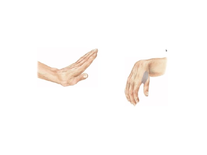
Supination and pronation It occurs in the superior and inferior radioulnar joints; Muscles produce supination § Biceps brachii. § Supinator. Muscles produce pronation § Pronator teres. § Pronator quadratus. NB. Brachioradialis put the forearm in midproneposition.

Human hand – masterpiece of art
 • Lateral group thenar (4) – – Abductor pollicis brevis")
Muscles of hand (palm) • Lateral group thenar (4) – – Abductor pollicis brevis Flexor pollicis brevis Opponens pollicis Adductor pollicis • Action: flex, abduct, adduct and oppose thumb • Medial group hypothenar (3) – Abductor digiti minimi – Flexor digiti minimi brevis – Opponens digiti minimi • Action: flex, abduct , and oppose little finger
 flex fingers at MP joints; extend fingers at IP")
Intermediate group • Lumbricals (4) flex fingers at MP joints; extend fingers at IP joints • Palmar interossei (3) adduct fingers towards middle finger at MP joints • Dorsal interossei (4) abduct fingers away from middle finger at MP joints
- Slides: 66