The transport system What is the transport circulatory

 system? Ø It carries blood and dissolved substances to")


 - secondary center of heart automation - in the")


 one contraction ~ 70 m. L of blood Cardiac output (CO)")


 - ventricles in diastole (relaxed) QRS")


 causes increase in heart rate in fight – or – flight")
")


- Slides: 59
The transport system
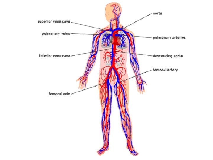
What is the transport (circulatory) system? Ø It carries blood and dissolved substances to and from different places in the body. Ø The Heart and blood vessels together make up the Circulatory System. Ø The Heart pumps blood and substances around the body in tubes called blood vessels.
to deliver oxygen, nutrients and metabolites to cells The anatomic position of the heart: - chest cavity between the lungs - most of the heart is in the middle, 1/3 on the right side of the heart - apex is on the left side
Blood from the heart gets around the body through blood vessels There are 3 types of blood vessels: a. ARTERY b. VEIN c. CAPILLARY
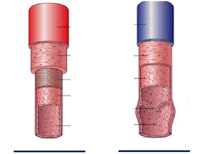
COMPARE AND DESCRIBE DIFFERENCES AND SIMILARITIES IN ARTERY AND VEIN ANATOMY! adventitia
T A S K D I F F E R E N C E S S I M I L A R I T I E S ARTERY VEIN
The ARTERY Arteries carry blood away from the heart. the elastic fibres allow the artery to stretch under pressure -thick muscle and elastic fibres -narrower lumen than veins the thick muscle can contract to push the blood along.
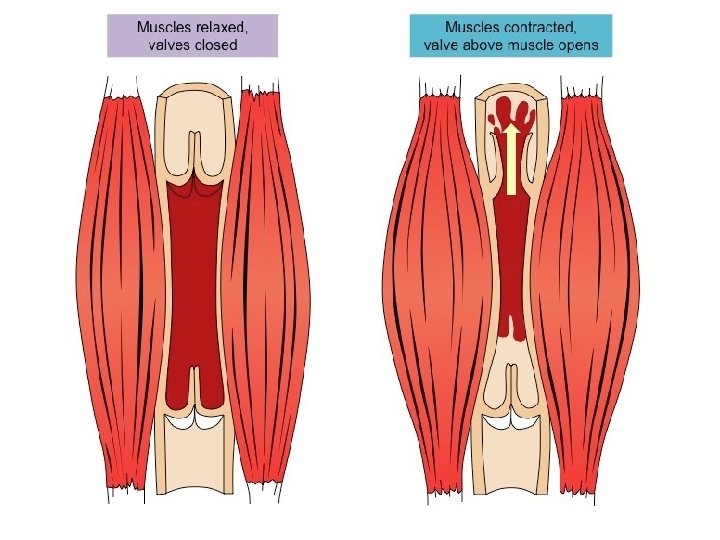
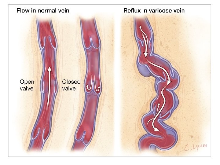
The VEIN Veins carry blood towards from the heart. veins have valves which act to stop the blood from going in the wrong direction. thin muscle and elastic fibres body muscles surround the veins so that when they contract to move the body, they also squeeze the veins and push the blood along the vessel.
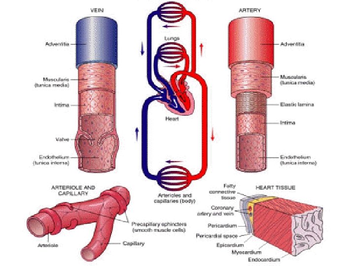
Arteries Pulsating Muscular layer Elastic vasodilation – parasympathetic nerves vasoconstriction - sympathetic nerves Veins Blood flows continuously Thin muscle layer On the surface of the organism VENOUS VALVES (vena cava has none) - pocket valves - three cup shaped flaps
ARTERIAL BLOOD – oxigenated rich with oxygen VENOUS BLOOD poor with oxygen
The CAPILLARY Capillaries link Arteries with Veins Thin and short blood vessels, 8 μm Deliver blood to the cells the wall of a capillary is only one cell thick The exchange of materials between the blood and the body can only occur through capillaries.
The CAPILLARY A collection of capillaries is known as a CAPILLARY BED artery body cell vein capillaries
The human heart
The human heart Pumpes blood ATRIUM VENTRICLE VALVES: ATRIOVENTRICULAR L – BICUSPID R - TRICUSPID VALVE
The human heart VALVES: SEMILUNAR VALVES Where the artery and ventricle are connected Aortic semilunar valve Pulmonary semilunar valve ATRIOVENTRICULAR L – BICUSPID R – TRICUSPID VALVE
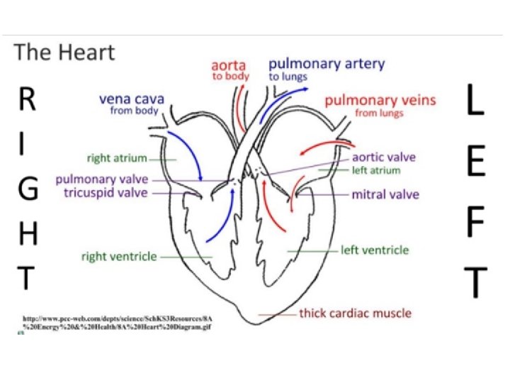
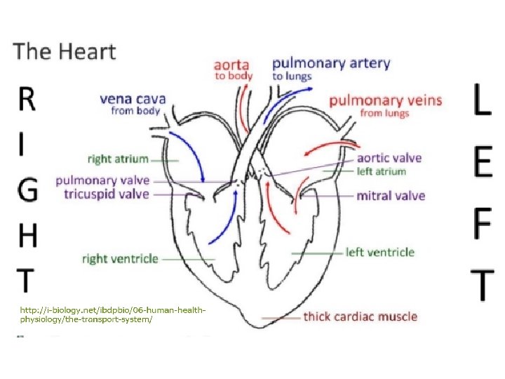
Name the structures! vv VENA CAVA, THICK CARDIAC MUSCLE, PULMONARY VALVE, LEFT VENTRICLE, TRICUSPID VALVE, RIGHT VENTRICLE, RIGHT ATRIUM, MITRAL VALVE, AORTA, PULMONARY VEINS, PULMONARY ARTERY, LEFT ATRIUM
http: //i-biology. net/ibdpbio/06 -human-healthphysiology/the-transport-system/
The human heart – outer anatomy http: //www. learnerstv. com/animation. php? ani=321&cat=biology
How does the Heart work? STEP ONE blood from the body blood from the lungs The heart beat begins when the heart muscles relax and blood flows into the atria.
How does the Heart work? STEP TWO The atria then contract and the valves open to allow blood into the ventricles.
How does the Heart work? STEP THREE The valves close to stop blood flowing backwards. The ventricles contract forcing the blood to leave the heart. At the same time, the atria are relaxing and once again filling with blood. The cycle then repeats itself.
http: //www. phschool. com/science/biology_place/biocoach/cardio 1/cycle. html
LABEL WITH CORRECT TERMS: HIGH PRESSURE, HIGHEST PRESSURE, LOWEST PRESSURE
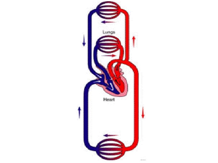
DOUBLE CIRCULATION
http: //www. kscience. co. uk/animations/blood_system. swf
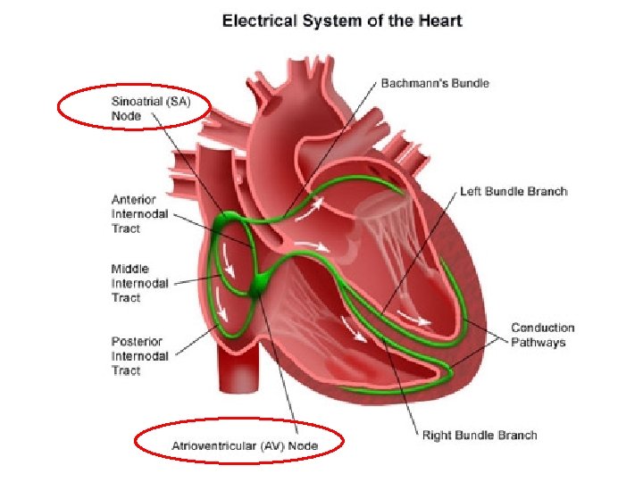
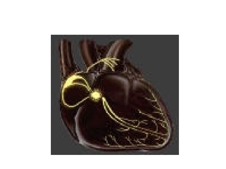
Centers of automation In the heart Manages the work of the heart (centers of automation) 1. SINOATRIAL NODE (S-A node or the PACEMAKER) - The primary center of heart automation - In the right atrium - This is a node in a 70 × min. positively charged, depolarised (Na+ entry through cell membrane) - Action of Na+ / K+ pump (sodium-potassium pump) ejects Na+ so the membrane is negatively charged again, repolarised SA node depolarisation spreads like a wave, and stimulate the muscles Depolarization = contraction
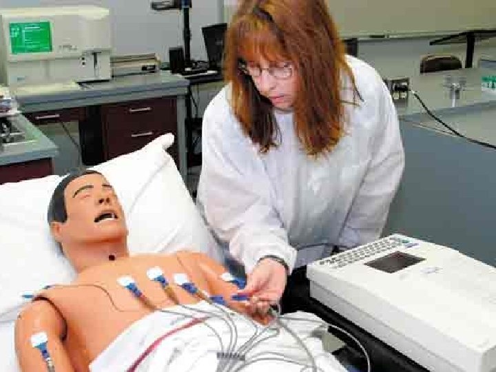
2. ATRIOVENTRICULAR NODE (AV node) - secondary center of heart automation - in the partition between the R atria and R ventricles - continue to transmit stimuli through the conduction pathways (nerves) - electrical stimulus spreads in the surrounding tissue, the electrodes on the chest (properly spaced) receive electrical stimulation Electrocardiograph - device Electrocardiogram (EKG) - recorded electrical potentials
http: //highered. mcgrawhill. com/sites/0072495855/student_view 0/chapter 22/animation__conducting_system_of_the_hear t. html
The heart contracts approximately 70 times× per minute, and then relaxes Systole - contraction Diastole - relaxation Stroke volume (SV) one contraction ~ 70 m. L of blood CHILDREN'S HEART CONTRACTS IN THE FIRST YEAR ~ 130 × PER MINUTE
Stroke volume (SV) one contraction ~ 70 m. L of blood Cardiac output (CO) - the volume of blood being pumped by the heart, in particular by a left or right ventricle, per unit time 70 m. L x 70 = 4900 m. L/min
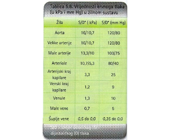
BLOOD PRESSURE – pressure of blood on vessel walls normal 16/10, 7 k. Pa 120/80 mm. Hg = torr = 133, 322 Pa -sistolic – maximal – upper value - when stroke volume gets to aorta -diastolic – lower -remains after stroke volume passes
BLOOD PRESSURE – pressure of blood on vessel walls normal 16/10, 7 k. Pa 120/80 mm. Hg = torr = 133, 322 Pa -sistolic – maximal – upper value - when stroke volume gets to aorta -diastolic – lower -remains after stroke volume passes
wave height - shows the voltage of the wavelength - time during the process
P - wave - systole (atrium contraction, depolarization) - ventricles in diastole (relaxed) QRS - complex - systole (ventricle contraction, depolarization) - ventricles squeeze blood from the heart T - wave - ventricular relaxation (repolarization)
http: //library. med. utah. edu/kw/pharm/hyper_heart 1. html
Homework http: //i-biology. net/ibdpbio/06 -human-health-physiology/the-transport-system/
http: //www. sympatheticnervoussystem. net/ Controlled by the AUTONOMIC NERVOUS SYSTEM – responds automatically to changes in body conditions
http: //www. rci. rutgers. edu/~uzwiak/Anat. Phys/Blood_Vessels. html http: //www. slideshare. net/fullscreen/gurustip/the-transport-system-core-presentation/16
ADRENALINE (epinephrine) causes increase in heart rate in fight – or – flight responses
PACEMAKER NERVES – stimulation THE MEDULA OF THE BRAIN ADRENALINE (EPINEPHRINE)
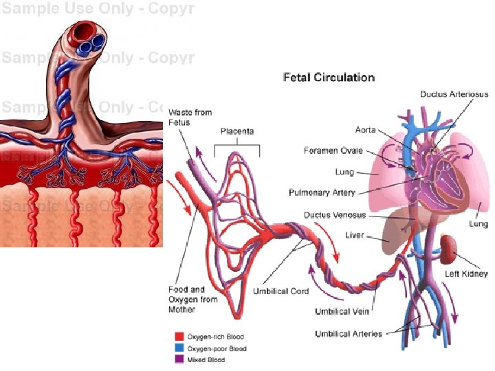
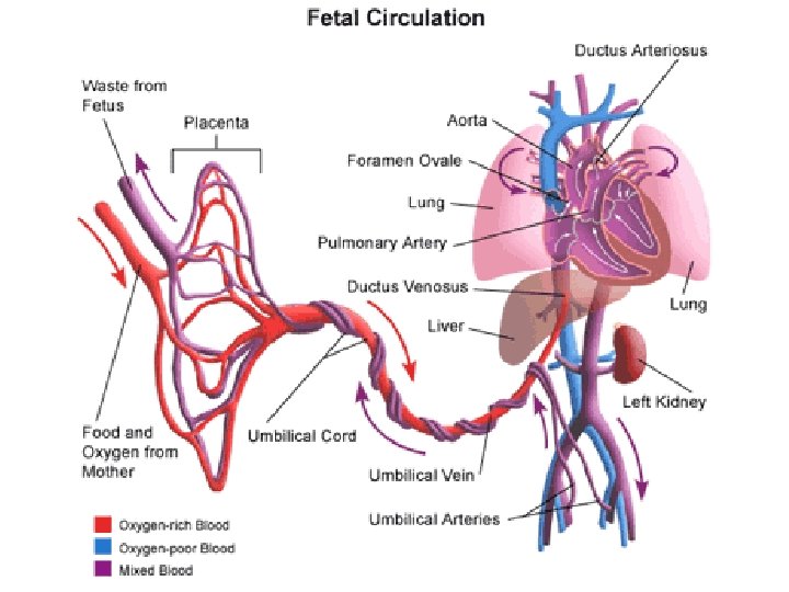
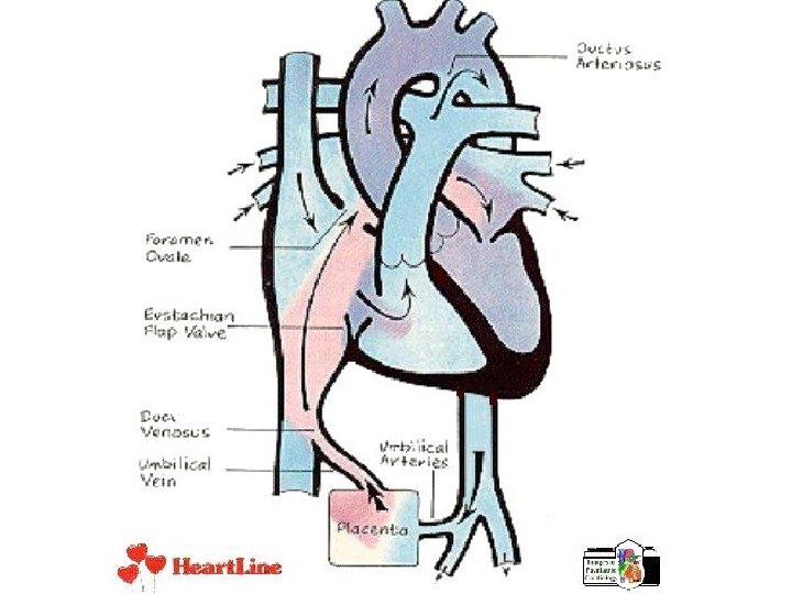
FETALNI KRVOTOK Pupčana vrpca – vena, 2 arterije - u DA veći tlak nego u LA - pupčanom venom oksigenirana krv ulazi u DA - nepotpuno pregrađeni atriji – miješa se arterijska i venska krv - foramen ovale – ovalni procijep (primarni i sekundarni septum) - vodi u LA, te u LV te u aortu - veći dio krvi - ductus arteriosus (d. a. Botalli)– povezuje DA i aortu – manji dio krvi - dio u DV - pluća POROĐAJ – presjecanje pupčane vrpce - porast CO 2 u krvi stimulira inspirijsko središte ; prvi INSPIRIJ - tada se poveća tlak u LA i preklope se primarni i sekundarni septum (70% djece) - zatvara se ductus arteriosus
TRANSLATE NEW TERMS! kapilarni tlak – 2, 7 k. Pa venski tlak - ~ 1, 3 k. Pa HIPERTENZIJA (hipertonija) HIPOTENZIJA (hipotonija) INFARKT MIOKARDA – zbog oštećenja koronarnih žila ATEROSKLEROZA – ovapnjenje krvnih žila (kolesterol) TROMBOZA - tromb – ugrušak (nastane zbog prolaza kroz suženje art. ) PLUĆNA EMBOLIJA – embolus (mali putujući ugrušak) se zaustavi u plućima (nema dovoda hranjivih tvari i kisika)
http: //www. nucleusinc. com/animation 2. php http: //www. kscience. co. uk/animations/blood_system. sw f http: //highered. mcgrawhill. com/sites/0072495855/student_view 0/chapter 22/ani mation__conducting_system_of_the_heart. html http: //library. med. utah. edu/kw/pharm/hyper_heart 1. html http: //i-biology. net/ibdpbio/06 -human-healthphysiology/the-transport-system/ http: //www. phschool. com/science/biology_place/biocoa ch/cardio 1/cycle. html