
The thoracic cavity It may be divided into
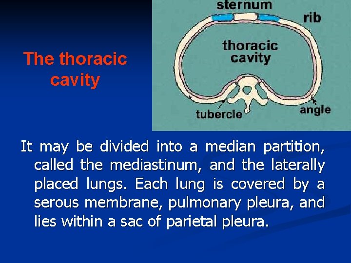
The thoracic cavity It may be divided into a median partition, called the mediastinum, and the laterally placed lungs. Each lung is covered by a serous membrane, pulmonary pleura, and lies within a sac of parietal pleura.
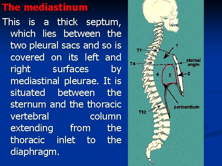
The mediastinum This is a thick septum, which lies between the two pleural sacs and so is covered on its left and right surfaces by mediastinal pleurae. It is situated between the sternum and the thoracic vertebral column extending from the thoracic inlet to the diaphragm.
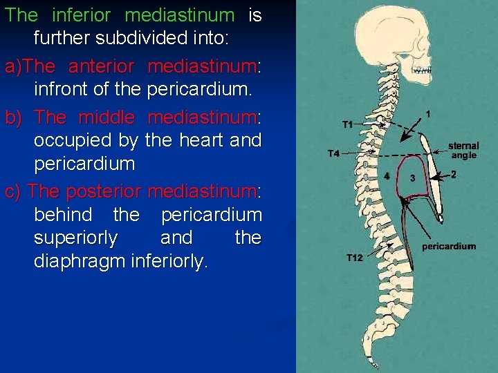
The inferior mediastinum is further subdivided into: a)The anterior mediastinum: infront of the pericardium. b) The middle mediastinum: occupied by the heart and pericardium c) The posterior mediastinum: behind the pericardium superiorly and the diaphragm inferiorly.
Pleura • Definition: It is a serous membrane arranged as a closed invaginated sac surrounding the lung on each side Parts: 1) Parietal pleura, lines the thoracic wall, covers the thoracic surface of the diaphragm and the lateral aspect of the mediastinum, and extends into the root of the neck to line the undersurface of the suprapleural membrane at the thoracic inlet. 2) Visceral pleura, completely covers the outer surfaces of the lungs and extends into the depths of the interlobar fissures
n The two parts of the pleura become continuous with one another by means of a cuff of pleura that surrounds the structures entering and leaving the lung at the lung root. To allow for movement of the lung root during respiration, the pleural cuff hangs down as a loose fold called the pulmonary ligament.
n The parietal and visceral layers of pleura are separated from one another by a slit like space, the pleural cavity. This normally contains fluid, tissue a smallofamount pleural fluid, which covers the surfaces of the pleura as a thin film and permits the two layers to move on each other with the minimum friction.


Pleural recesses These are parts of the pleural cavity which are not occupied by the lung except in full inspiration. They are: ►

a) The costomediastinal recess: a slit like space (on each side) between the costal and mediastinal parietal pleurae along the anterior border of the pleura. They are separated by a capillary layer of pleural fluid. During inspiration and expiration the anterior borders of the lungs slide in and out of the

b)The costodiaphragmatic recess is a slit like space, (on each side) between the costal and diaphragmatic parietal pleurae , along the inferior border of the pleura, that are separated only by a capillary layer of pleural fluid.
n During inspiration the lower margins of the lungs descend into the recesses. During expiration the lower margins of the lungs ascend so that the costal and diaphragmatic pleurae come together again. The recess is 2 inches (5 cm) deep in the scapular line posteriorly, 3 to 31/2 inches (8 -9 cm) in the midaxillary line; and 1 to 11/2 inches (2. 5 -4 cm) in the midclavicular line.
The surface marking of the pleurae The boundaries of the pleural sac can be marked out as lines on the surface of the body. These lines of the parietal pleura where it lies close to the body surface, are referred to as the lines of pleural reflection.
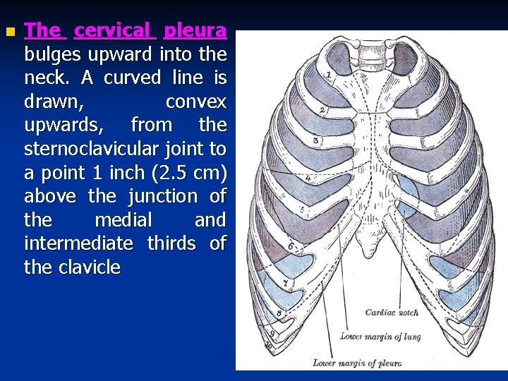
n The cervical pleura bulges upward into the neck. A curved line is drawn, convex upwards, from the sternoclavicular joint to a point 1 inch (2. 5 cm) above the junction of the medial and intermediate thirds of the clavicle
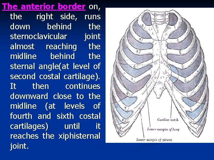
The anterior border on, the right side, runs down behind the sternoclavicular joint almost reaching the midline behind the sternal angle(at level of second costal cartilage). It then continues downward close to the midline (at levels of fourth and sixth costal cartilages) until it reaches the xiphisternal joint.
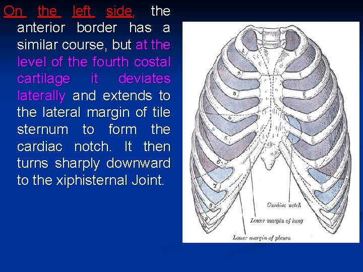
On the left side, the anterior border has a similar course, but at the level of the fourth costal cartilage it deviates laterally and extends to the lateral margin of tile sternum to form the cardiac notch. It then turns sharply downward to the xiphisternal Joint.
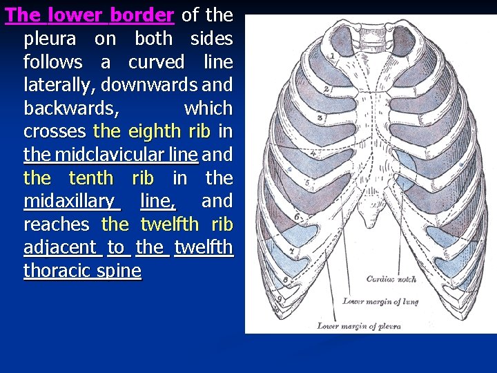
The lower border of the pleura on both sides follows a curved line laterally, downwards and backwards, which crosses the eighth rib in the midclavicular line and the tenth rib in the midaxillary line, and reaches the twelfth rib adjacent to the twelfth thoracic spine

The posterior border is a vertical line extends downwards from the summit of the pleura to the medial end of the twelfth rib opposite the twelfth thoracic spine.
n Nerve Supply of the Pleura: The parietal pleura is supplied as follows: The costal pleura is segmentally supplied by the intercostal nerves; the mediastinal pleura is supplied by the phrenic nerve; and the diaphragmatic pleura is supplied over the domes by the phrenic nerve and around the periphery by the lower five intercostal nerves. The visceral pleura covering the lungs, receives an autonomic vasomotor supply, but is insensitive to common sensations such as pain and touch.

Origin Somatopleure of Splanchnopleure of lateral mesodermal plate. Site Lines the thoracic wall. Surrounds the lung Arterial supply Intercostal arteries Bronchial arteries. internal thoracic artery, musculophrenic artery Nerve supply Intercostal nerves, and Anterior and posterior phrenic nerve. pulmonary plexuses.
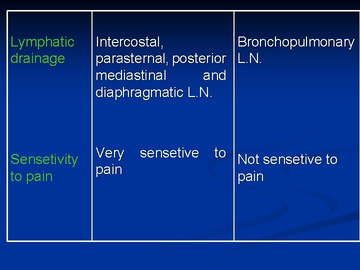
Lymphatic drainage Intercostal, Bronchopulmonary parasternal, posterior L. N. mediastinal and diaphragmatic L. N. Sensetivity to pain Very pain sensetive to Not sensetive to pain
The lungs n The lungs are the organs of respiration. During life they are soft, spongy, very elastic and crepitant to touch. In the child they are pink in color, but with age they become dark and mottled due to the inhalation of dust particles. Each lung lies within its own side of the thoracic cavity, on each side of the mediastinum. Each lung is conical in shape and is covered with visceral pleura. It is suspended freely within pleura , being attached to the mediastinum only by its root
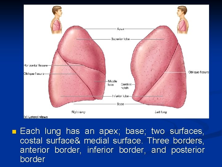
n Each lung has an apex; base; two surfaces, costal surface& medial surface. Three borders, anterior border, inferior border, and posterior border
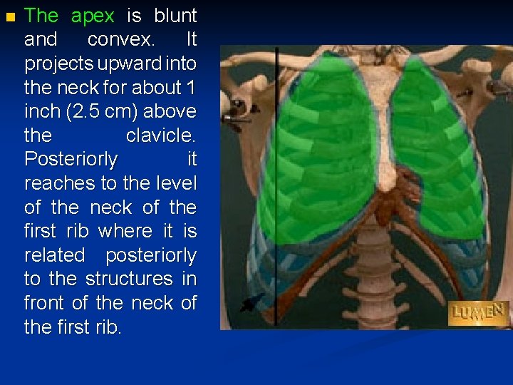
n The apex is blunt and convex. It projects upward into the neck for about 1 inch (2. 5 cm) above the clavicle. Posteriorly it reaches to the level of the neck of the first rib where it is related posteriorly to the structures in front of the neck of the first rib.

The following structures are related to the neck of the first rib from medial to lateral: 1 -Cervicothoracic ganglion 2 -Superior intercostal artery. 3 - Ventral ramus of the first thoracic nerve.
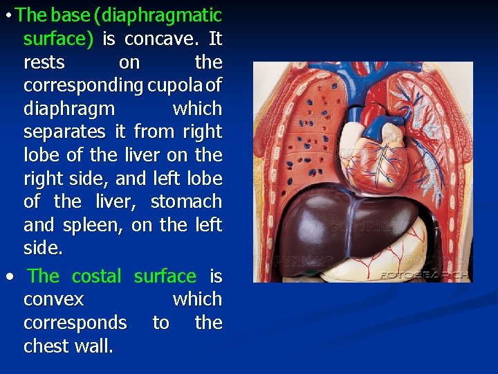
• The base (diaphragmatic surface) is concave. It rests on the corresponding cupola of diaphragm which separates it from right lobe of the liver on the right side, and left lobe of the liver, stomach and spleen, on the left side. • The costal surface is convex which corresponds to the chest wall.
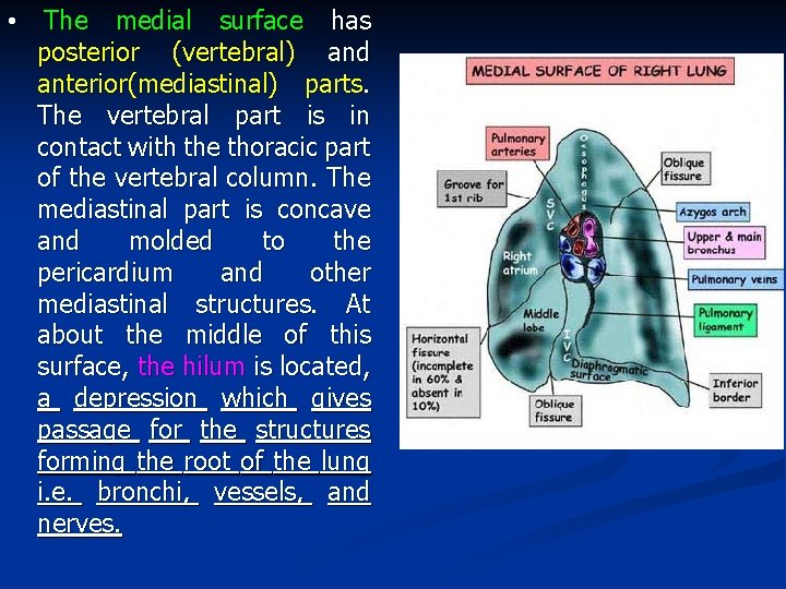
• The medial surface has posterior (vertebral) and anterior(mediastinal) parts. The vertebral part is in contact with the thoracic part of the vertebral column. The mediastinal part is concave and molded to the pericardium and other mediastinal structures. At about the middle of this surface, the hilum is located, a depression which gives passage for the structures forming the root of the lung i. e. bronchi, vessels, and nerves.
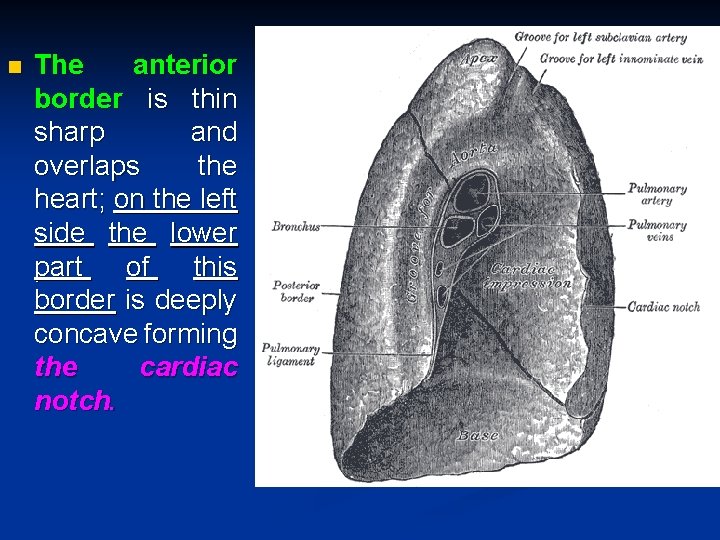
n The anterior border is thin sharp and overlaps the heart; on the left side the lower part of this border is deeply concave forming the cardiac notch.

The posterior border is thick, and broad to fit the paravertebral gutter i. e. on each side of the vertebral column. It is the rounded junction of costal and vertebral surfaces.
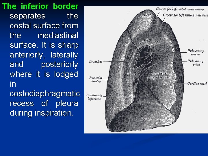
The inferior border separates the costal surface from the mediastinal surface. It is sharp anteriorly, laterally and posteriorly where it is lodged in costodiaphragmatic recess of pleura during inspiration.
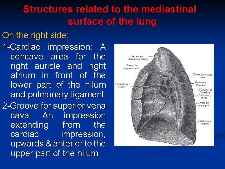
Structures related to the mediastinal surface of the lung On the right side: 1 -Cardiac impression: A concave area for the right auricle and right atrium in front of the lower part of the hilum and pulmonary ligament. 2 -Groove for superior vena cava: An impression extending from the cardiac impression, upwards & anterior to the upper part of the hilum.

3 -Groove for right brachiocephalic vein: It is the upward continuation of the superior vena cava impression. 4 -Groove for arch of azygos vein: It is a deep sulcus arching above the hilum. From its posterior part, a faint impression for azygos vein extends behind the oesephageal groove posterior to the hilum.

5 -Groove for oesophagus: It is a shallow vertical groove (made by the right side of the oesophagus) behind the hilum and the pulmonary ligament. It ascends upwards in contact with the lung (except at the area of azygos arch) till reaches just below the apex. It does not reach the lower limit of this surface.
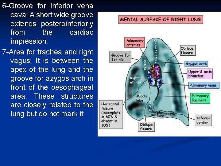
6 -Groove for inferior vena cava: A short wide groove extends posteroinferiorly from the cardiac impression. 7 -Area for trachea and right vagus: It is between the apex of the lung and the groove for azygos arch in front of the oesophageal area. These structures are closely related to the lung but do not mark it.
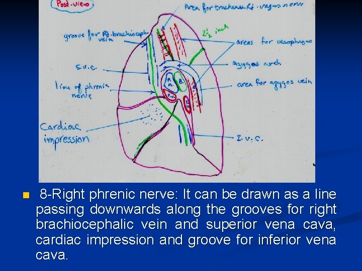
n 8 -Right phrenic nerve: It can be drawn as a line passing downwards along the grooves for right brachiocephalic vein and superior vena cava, cardiac impression and groove for inferior vena cava.
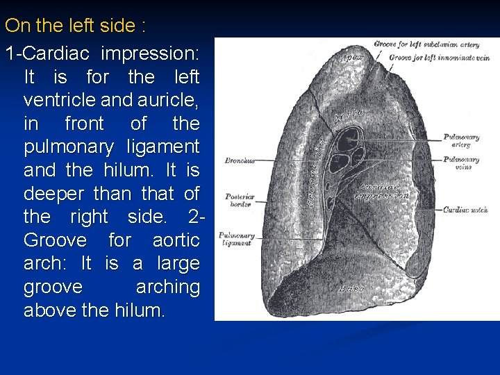
On the left side : 1 -Cardiac impression: It is for the left ventricle and auricle, in front of the pulmonary ligament and the hilum. It is deeper than that of the right side. 2 Groove for aortic arch: It is a large groove arching above the hilum.

3 -Groove for descending aorta: It is large and extends from the groove of the aortic arch, descending behind the hilum and pulmonary ligament. 4 -Groove for left subclavian artery: It is a vertical groove ascending from the summit of the groove for aortic arch to the apex of the lung. In front of it, there is a faint linear depression for the left brachiocephalic vein.
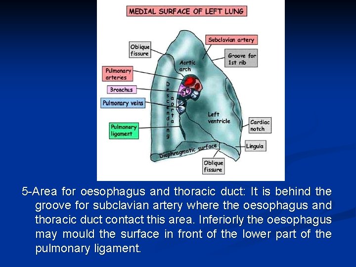
5 -Area for oesophagus and thoracic duct: It is behind the groove for subclavian artery where the oesophagus and thoracic duct contact this area. Inferiorly the oesophagus may mould the surface in front of the lower part of the pulmonary ligament.
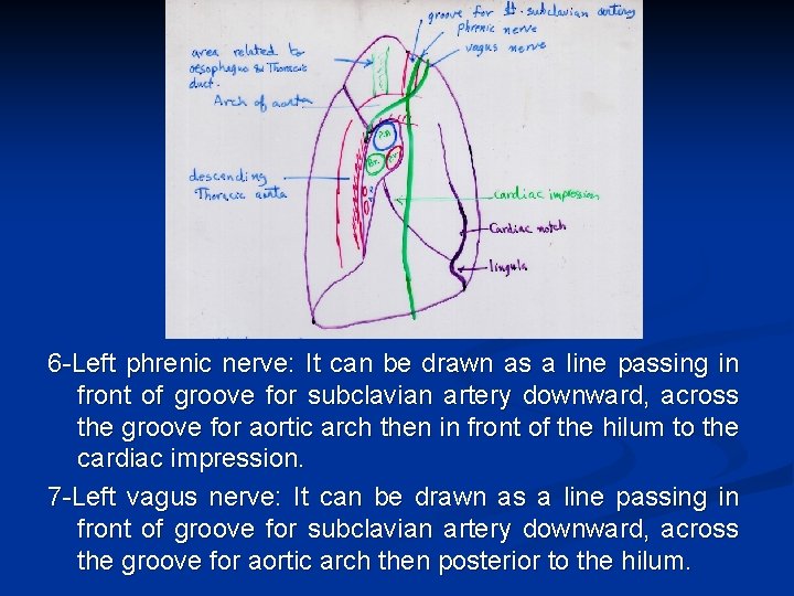
6 -Left phrenic nerve: It can be drawn as a line passing in front of groove for subclavian artery downward, across the groove for aortic arch then in front of the hilum to the cardiac impression. 7 -Left vagus nerve: It can be drawn as a line passing in front of groove for subclavian artery downward, across the groove for aortic arch then posterior to the hilum.
- Slides: 38