The term hemostasis prevention of blood loss Whenever

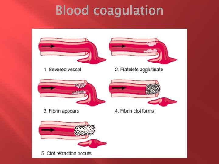
The term hemostasis = prevention of blood loss. Whenever a vessel is severed or ruptured, hemostasis is achieved by several mechanisms: (1) vascular constriction, (2) formation of a platelet plug, (3) formation of a blood clot as a result of blood coagulation, and (4) eventual growth of fibrous tissue into the blood clot to close the hole in the vessel permanently.

Immediately after a blood vessel has been cut or ruptured, the trauma to the vessel wall itself causes the smooth muscle in the wall to contract; this instantaneously reduces the flow of blood from the ruptured vessel. � The contraction results from (1) local myogenic spasm, (2) local factors from the traumatized tissues and blood platelets, and (3) nervous reflexes- are initiated by pain nerve impulses or other sensory impulses that originate from the traumatized vessel or nearby tissues. �

� � For the smaller vessels, the platelets are responsible for much of the vasoconstriction by releasing a vasoconstrictor substance, thromboxane A 2. The more severely a vessel is traumatized, the greater the degree of vascular spasm. The spasm can last for many minutes or even hours, during which time the processes of platelet plugging and blood coagulation can take place.

� If the cut in the blood vessel is very small - many very small vascular holes do develop throughout the body each day—the cut is often sealed by a platelet plug, rather than by a blood clot.
 are minute discs (1 to 4 micrometers)")
� � � Platelets (also called thrombocytes) are minute discs (1 to 4 micrometers) Are formed in the bone marrow from megakaryocytes, which are extremely large cells of the hematopoietic series in the marrow; the megakaryocytes fragment into the minute platelets either in the bone marrow or soon after entering the blood, especially as they squeeze through capillaries. The normal concentration of platelets in the blood is between 150, 000 and 300, 000 per microliter
 actin and myosin molecules, which")
In their cytoplasm are such active factors as (1) actin and myosin molecules, which are contractile proteins similar to those found in muscle cells, and still another contractile protein, thrombosthenin (cause the platelets to contract); (2) residuals of both the endoplasmic reticulum and the Golgi apparatus that synthesize various enzymes and especially store large quantities of calcium ions; (3) mitochondria and enzyme systems that are capable of forming adenosine triphosphate (ATP) and adenosine diphosphate (ADP); �
 enzyme systems that synthesize prostaglandins, which are local hormones that cause many vascular")
(4) enzyme systems that synthesize prostaglandins, which are local hormones that cause many vascular and other local tissue reactions; (5) an important protein called fibrin-stabilizing factor; and (6) a growth factor that causes vascular endothelial cells, vascular smooth muscle cells, and fibroblasts to multiply and grow, thus causing cellular growth that eventually helps repair damaged vascular walls.

When platelets come in contact with a damaged vascular surface (collagen fibers in the vascular wall), the platelets themselves immediately change their own characteristics : § They begin to swell; they assume irregular forms with numerous irradiating pseudopods protruding from their surfaces; § their contractile proteins contract forcefully and cause the release of granules that contain multiple active factors; § they become sticky so that they adhere to collagen in the tissues and to a protein called von Willebrand factor that leaks into the traumatized tissue from the plasma; § they secrete large quantities of ADP; and their enzymes form thromboxane A 2

The ADP and thromboxane in turn act on nearby platelets to activate them as well, and the stickiness of these additional platelets causes them to adhere to the original activated platelets. � At the site of any opening in a blood vessel wall, the damaged vascular wall activates successively increasing numbers of platelets that themselves attract more and more additional platelets, thus forming a platelet plug. This is at first a loose plug, but it is usually successful in blocking blood loss if the vascular opening is small. Then, during the subsequent process of blood coagulation, fibrin threads form. These attach tightly to the platelets, thus constructing an unyielding plug. �

� � The clot begins to develop in 15 to 20 seconds if the trauma to the vascular wall has been severe, and in 1 to 2 minutes if the trauma has been minor. Activator substances from the traumatized vascular wall, from platelets, and from blood proteins adhering to the traumatized vascular wall initiate the clotting process.

� � Within 3 to 6 minutes after rupture of a vessel, if the vessel opening is not too large, the entire opening or broken end of the vessel is filled with clot. After 20 minutes to an hour, the clot retracts; this closes the vessel still further. Platelets also play an important role in this clot retraction.
")
Once a blood clot has formed, it can follow one of two courses: (1) It can become invaded by fibroblasts, which subsequently form connective tissue all through the clot, or (2) it can dissolve. The usual course for a clot that forms in a small hole of a vessel wall is invasion by fibroblasts, beginning within a few hours after the clot is formed (which is promoted at least partially by growth factor secreted by platelets). This continues to complete organization of the clot into fibrous tissue within about 1 to 2 weeks. Conversely, when excess blood has leaked into the tissues and tissue clots have occurred where they are not needed, special substances within the clot itself usually become activated.

� � More than 50 important substances that cause or affect blood coagulation have been found in the blood and in the tissues—some that promote coagulation-procoagulants, and others that inhibit coagulation- anticoagulants. Whether blood will coagulate depends on the balance between these two groups of substances. In the blood stream, the anticoagulants normally predominate, so that the blood does not coagulate while it is circulating in the blood vessels. But when a vessel is ruptured, procoagulants from the area of tissue damage become “activated” and override the anticoagulants, and then a clot does develop.
 In response to rupture of the")
Clotting takes place in three essential steps: (1) In response to rupture of the vessel or damage to the blood itself, a complex cascade of chemical reactions occurs in the blood involving more than a dozen blood coagulation factors. The net result is formation of a complex of activated substances collectively called prothrombin activator. (2) The prothrombin activator catalyzes conversion of prothrombin into thrombin. (3) The thrombin acts as an enzyme to convert fibrinogen into fibrin fibers that enmesh platelets, blood cells, and plasma to form the clot.
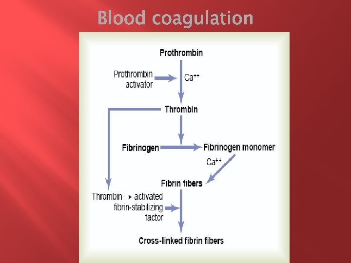

First, prothrombin activator is formed as a result of rupture of a blood vessel or as a result of damage to special substances in the blood. � Second, the prothrombin activator, in the presence of sufficient amounts of ionic Ca++, causes conversion of prothrombin to thrombin. � Third, the thrombin causes polymerization of fibrinogen molecules into fibrin fibers within another 10 to 15 seconds. The rate-limiting factor in causing blood coagulation is usually the formation of prothrombin activator and not the subsequent reactions beyond that point, because these terminal steps normally occur rapidly to form the clot itself. Platelets also play an important role in the conversion of prothrombin to thrombin. �

Prothrombin � is formed continually by the liver, and it is continually being used throughout the body for blood clotting. If the liver fails to produce prothrombin, in a day or so prothrombin concentration in the plasma falls too low to provide normal blood coagulation. � Vitamin K is required by the liver for normal formation of prothrombin as well as formation of a few other clotting factors. Therefore, either lack of vitamin K or the presence of liver disease that prevents normal prothrombin formation can decrease the prothrombin level so low that a bleeding tendency results. � is a plasma alpha 2 -globulin. It is present in normal plasma in a concentration of about 15 mg/dl. It is an unstable protein that can split easily into smaller compounds, one of which is thrombin, which has a molecular weight of 33, 700, almost exactly one half that of prothrombin.

� � Fibrinogen is a high-molecular-weight protein that occurs in the plasma in quantities of 100 to 700 mg/dl is formed in the liver (large molecular size!) little fibrinogen normally leaks from the blood vessels into the interstitial fluids, and because fibrinogen is one of the essential factors in the coagulation process, interstitial fluids ordinarily do not coagulate! when the permeability of the capillaries becomes pathologically increased, fibrinogen does then leak into the tissue fluids in sufficient quantities to allow clotting of these fluids in much the same way that plasma and whole blood can clot.

Thrombin is a protein enzyme with weak proteolytic capabilities. � It acts on fibrinogen to remove four low-molecular- weight peptides from each molecule of fibrinogen, forming one molecule of fibrin monomer that has the automatic capability to polymerize with other fibrin monomer molecules to form fibrin fibers. � many fibrin monomer molecules polymerize within seconds into long fibrin fibers that constitute the reticulum of the blood clot. In the early stages of polymerization, the fibrin monomer molecules are held together by weak noncovalent hydrogen bonding, and the newly forming fibers are not cross-linked with one another; therefore, the resultant clot is weak and can be broken apart with ease

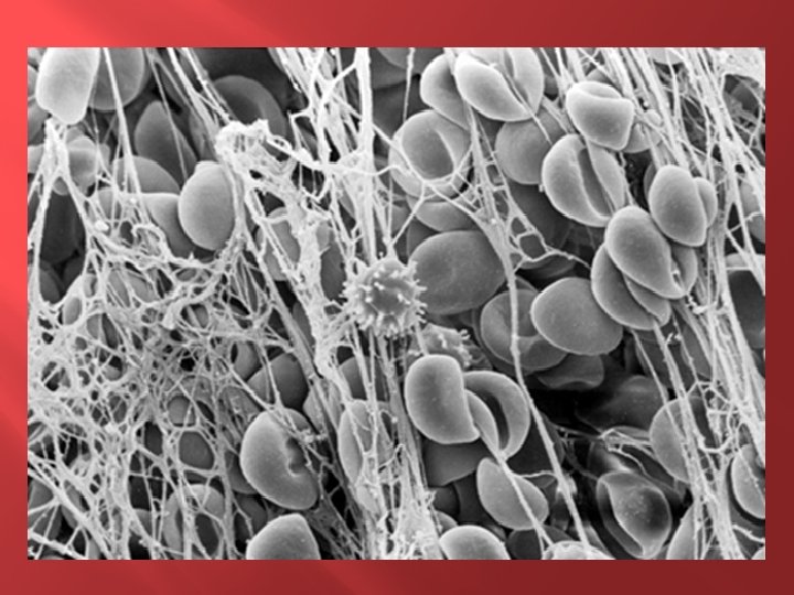
� � Before fibrin-stabilizing factor can have an effect on the fibrin fibers, it must itself be activated. The same thrombin that causes fibrin formation also activates the fibrin-stabilizing factor. Then this activated substance operates as an enzyme to cause covalent bonds between more and more of the fibrin monomer molecules, as well as multiple crosslinkages between adjacent fibrin fibers, thus adding tremendously to the three-dimensional strength of the fibrin meshwork. The clot is composed of a meshwork of fibrin fibers running in all directions and entrapping blood cells, platelets, and plasma. The fibrin fibers also adhere to damaged surfaces of blood vessels; therefore, the blood clot becomes adherent to any vascular opening and thereby prevents further blood loss.

� � � Within a few minutes after a clot is formed, it begins to contract and usually expresses most of the fluid from the clot within 20 to 60 minutes. The fluid expressed is called serum because all its fibrinogen and most of the other clotting factors have been removed; in this way, serum differs from plasma. Serum cannot clot because it lacks these factors. Platelets are necessary for clot retraction to occur. Therefore, failure of clot retraction is an indication that the number of platelets in the circulating blood might be low. Electron micrographs of platelets in blood clots show that they become attached to the fibrin fibers in such a way that they actually bond different fibers together. Furthermore, platelets entrapped in the clot continue to release procoagulant substances, one of the most important of which is fibrinstabilizing factor, which causes more and more cross-linking bonds between adjacent fibrin fibers.

� � In addition, the platelets themselves contribute directly to clot contraction by activating platelet thrombosthenin, actin, and myosin molecules, which are all contractile proteins in the platelets and cause strong contraction of the platelet spicules attached to the fibrin. The contraction is activated and accelerated by thrombin as well as by calcium ions released from calcium stores in the mitochondria, endoplasmic reticulum, and Golgi apparatus of the platelets.

� � Once a blood clot has started to develop, it normally extends within minutes into the surrounding blood. That is, the clot itself initiates a vicious circle (positive feedback) to promote more clotting. One of the most important causes of this is the fact that the proteolytic action of thrombin allows it to act on many of the other blood-clotting factors in addition to fibrinogen. For instance, thrombin has a direct proteolytic effect on prothrombin itself, tending to convert this into still more thrombin, and it acts on some of the blood-clotting factors responsible formation of prothrombin activator.
 trauma to the vascular wall and")
These mechanisms are set into play by (1) trauma to the vascular wall and adjacent tissues (2) trauma to the blood, or (3) contact of the blood with damaged endothelial cells or with collagen and other tissue elements outside the blood vessel. In each instance, this leads to the formation of prothrombin activator, which then causes prothrombin conversion to thrombin and all the subsequent clotting steps. �

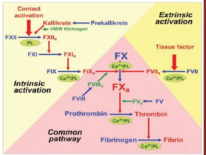
Prothrombin activator is generally considered to be formed in two ways, although, in reality, the two ways interact constantly with each other: (1) by the extrinsic pathway that begins with trauma to the vascular wall and surrounding tissues and (2) by the intrinsic pathway that begins in the blood itself. In both the extrinsic and the intrinsic pathways, a series of different plasma proteins called blood-clotting factors play major roles. Most of these are inactive forms of proteolytic enzymes. When converted to the active forms, their enzymatic actions cause the successive, cascading reactions of the clotting process. �

Fibrinogen Factor I Prothrombin Tissue factor Calcium Factor VII Factor III; tissue thromboplastin Factor IV Proaccelerin; labile factor; Ac-globulin (Ac-G) Serum prothrombin conversion accelerator (SPCA); proconvertin; stable factor Factor VIII Factor IX Antihemophilic factor (AHF) or globulin (AHG); antihemophilic factor A Plasma thromboplastin component (PTC); Christmas factor; antihemophilic factor B Factor XIII Prekallikrein HMWK kininogen (highmolecular-weight) kininogen Stuart factor; Stuart-Prower factor Plasma thromboplastin antecedent (PTA); antihemophilic factor C Hageman factor Fibrin-stabilizing factor Fletcher factor Fitzgerald factor

1. Release of tissue factor. Traumatized tissue releases a complex of several factors called tissue factor or tissue thromboplastin. This factor is composed especially of phospholipids from the membranes of the tissue plus a lipoprotein complex that functions mainly as a proteolytic enzyme. 2. Activation of Factor X—role of Factor VII and tissue factor. The lipoprotein complex of tissue factor further complexes with blood coagulation Factor VII and, in the presence of calcium ions, acts enzymatically on Factor X to form activated Factor X (Xa).

3. The activated Factor X combines immediately with tissue phospholipids that are part of tissue factor or with additional phospholipids released from platelets as well as with Factor V to form the complex called prothrombin activator. Within a few seconds, in the presence of calcium ions (Ca++), this splits prothrombin to form thrombin, and the clotting process proceeds as already explained. At first, the Factor V in the prothrombin activator complex is inactive, but once clotting begins and thrombin begins to form, the proteolytic action of thrombin activates Factor V.

� � This then becomes an additional strong accelerator of prothrombin activation. Thus, in the final prothrombin activator complex, activated Factor X is the actual protease that causes splitting of prothrombin to form thrombin; activated Factor V greatly accelerates this protease activity, and platelet phospholipids act as a vehicle that further accelerates the process. Note especially the positive feedback effect of thrombin, acting through Factor V, to accelerate the entire process once it begins.
 activation of Factor XII and (2) release of platelet")
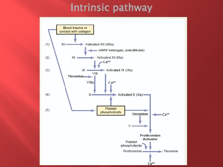
1. Blood trauma causes (1) activation of Factor XII and (2) release of platelet phospholipids. Blood trauma or contact with collagen wall collagen alters two important clotting factors in the blood: Factor XII and the platelets. When Factor XII is disturbed, such as by coming into contact with collagen or with a wettable surface such as glass, it takes on a new molecular configuration that converts it into a proteolytic enzyme called “activated Factor XII. ” Simultaneously, the blood trauma also damages the platelets because of adherence to either collagen or a wettable surface (or by damage in other ways), and this releases platelet phospholipids that contain the lipoprotein called platelet factor 3, which also plays a role in subsequent clotting reactions.

2. Activation of factor XI. The activated Factor XII acts enzymatically on factor XI to activate this factor as well, which is the second step in the intrinsic pathway. This reaction also requires HMW (high-molecularweight) kininogen and is accelerated by prekallikrein. 3. Activation of factor IX by activated factor XI. The activated factor XI then acts enzymatically on factor IX to activate this factor also.

4. Activation of factor X—role of factor VIII. The activated factor IX, acting in concert with activated factor VIII and with the platelet phospholipids and factor 3 from the traumatized platelets, activates factor X. It is clear that when either factor VIII or platelets are in short supply, this step is deficient. Factor VIII is the factor that is missing in a person who has classic hemophilia, for which reason it is called antihemophilic factor. Platelets are the clotting factor that is lacking in the bleeding disease called thrombocytopenia.

5. Action of activated factor X to form prothrombin activator—role of factor V. This step in the intrinsic pathway is the same as the last step in the extrinsic pathway. That is, activated factor X combines with factor V and platelet or tissue phospholipids to form the complex called prothrombin activator. The prothrombin activator in turn initiates within seconds the cleavage of prothrombin to form thrombin, thereby setting into motion the final clotting process, as described earlier.

� � Except for the first two steps in the intrinsic pathway, calcium ions are required for promotion or acceleration of all the blood-clotting reactions. Therefore, in the absence of calcium ions, blood clotting by either pathway does not occur. In the living body, the calcium ion concentration seldom falls low enough to significantly affect the kinetics of blood clotting. But, when blood is removed from a person, it can be prevented from clotting by reducing the calcium ion concentration below the threshold level for clotting, either by deionizing the calcium by causing it to react with substances such as citrate ion or by precipitating the calcium with substances such as oxalate ion.

� � Clotting occurs by both pathways simultaneously. Tissue factor initiates the extrinsic pathway, whereas contact of factor XII and platelets with collagen in the vascular wall initiates the intrinsic pathway. An especially important difference between the extrinsic and intrinsic pathways is that the extrinsic pathway can be explosive; once initiated, its speed of completion to the final clot is limited only by the amount of tissue factor released from the traumatized tissues and by the quantities of factors X, VII, and V in the blood. With severe tissue trauma, clotting can occur in as little as 15 seconds. The intrinsic pathway is much slower to proceed, usually requiring 1 to 6 minutes to cause clotting.
")
The most important factors for preventing clotting in the normal vascular system are (1) the smoothness of the endothelial cell surface, which prevents contact activation of the intrinsic clotting system; (2) a layer of glycocalyx on the endothelium (glycocalyx is a mucopolysaccharide adsorbed to the surfaces of the endothelial cells), which repels clotting factors and platelets, thereby preventing activation of clotting; and (3) a protein bound with the endothelial membrane, thrombomodulin, which binds thrombin �

� � the thrombomodulin-thrombin complex also activates a plasma protein, protein C, that acts as an anticoagulant by inactivating activated factors V and VIII. When the endothelial wall is damaged, its smoothness and its glycocalyx-thrombomodulin layer are lost, which activates both factor XII and the platelets, thus setting off the intrinsic pathway of clotting. If factor XII and platelets come in contact with the subendothelial collagen, the activation is even more powerful.

Among the most important anticoagulants in the blood itself are those that remove thrombin from the blood. The most powerful of these are (1) the fibrin fibers that themselves are formed during the process of clotting and (2) an alpha-globulin called antithrombin III or antithrombin-heparin cofactor. While a clot is forming, about 85 -90% of the thrombin formed from the prothrombin becomes adsorbed to the fibrin fibers as they develop. This helps prevent the spread of thrombin into the remaining blood and, therefore, prevents excessive spread of the clot. �

� � � Heparin is another powerful anticoagulant, but its concentration in the blood is normally low, so that only under special physiologic conditions does it have significant anticoagulant effects heparin is used widely as a pharmacological agent in medical practice in much higher concentrations to prevent intravascular clotting. The heparin molecule is a highly negatively charged conjugated polysaccharide. it has little or no anticoagulant properties, but when it combines with antithrombin III, the effectiveness of antithrombin III for removing thrombin increases by a hundredfold to a thousandfold, and thus it acts as an anticoagulant.

� � � The complex of heparin and antithrombin III removes several other activated coagulation factors in addition to thrombin, further enhancing the effectiveness of anticoagulation: activated Factors XII, X, and IX. H. is produced by many different cells of the body, but especially large quantities are formed by the basophilic mast cells located in the pericapillary connective tissue throughout the body. The basophil cells of the blood, which are functionally almost identical to the mast cells, release small quantities of heparin into the plasma. large quantities of heparin might be needed in these areas because the capillaries of the lungs and liver receive many embolic clots formed in slowly flowing venous blood; sufficient formation of heparin prevents further growth of the clots.
 that,")
� � � The plasma proteins contain a globulin called plasminogen (or profibrinolysin) that, when activated, becomes a substance called plasmin (or fibrinolysin). Plasmin is a proteolytic enzyme that resembles trypsin, the most important proteolytic digestive enzyme of pancreatic secretion. Plasmin digests fibrin fibers and some other protein coagulants such as fibrinogen, factor V, factor VIII, prothrombin, and factor XII. Therefore, whenever plasmin is formed, it can cause lysis of a clot by destroying many of the clotting factors, thereby sometimes even causing hypocoagulability of the blood.

� � � When a clot is formed, a large amount of plasminogen is trapped in the clot along with other plasma proteins. This will not become plasmin or cause lysis of the clot until it is activated. The injured tissues and vascular endothelium very slowly release a powerful activator called tissue plasminogen activator (t-PA) that a few days later, after the clot has stopped the bleeding, eventually converts plasminogen to plasmin, which in turn removes the remaining unnecessary blood clot. many small blood vessels in which blood flow has been blocked by clots are reopened by this mechanism. Thus, an especially important function of the plasmin system is to remove minute clots from millions of tiny peripheral vessels that eventually would become occluded were there no way to clear them.

Conditions that cause excessive bleeding in human beings Excessive bleeding can result from deficiency of any one of the many blood-clotting factors. Three particular types of bleeding tendencies that have been studied to the greatest extent are discussed here: bleeding caused by (1) vitamin K deficiency, (2) hemophilia, and (3) thrombocytopenia (platelet deficiency). �

� � � With few exceptions, almost all the blood-clotting factors are formed by the liver. Therefore, diseases of the liver (hepatitis, cirrhosis, and acute yellow atrophy) can sometimes depress the clotting system so greatly that the patient develops a severe tendency to bleed. Another cause of depressed formation of clotting factors by the liver is vitamin K deficiency. Vitamin K is necessary for liver formation of five of the important clotting factors: prothrombin, Factor VII, Factor IX, Factor X, and protein C. In the absence of vitamin K, subsequent insufficiency of these coagulation factors in the blood can lead to serious bleeding tendencies. Vitamin K is continually synthesized in the intestinal tract by bacteria, so that vitamin K deficiency seldom occurs in the normal person as a result of vitamin K absence from the diet (except in neonates before they establish their intestinal bacterial flora).

� � Hemophilia is a bleeding disease that occurs almost exclusively in males. In 85% it is caused by an abnormality or deficiency of factor VIII; this type of hemophilia is called hemophilia A or classic hemophilia. About 1 of every 10, 000 males in the United States has classic hemophilia. In the other 15 % of hemophilia patients, the bleeding tendency is caused by deficiency of factor IX. Both of these factors are transmitted genetically by way of the female chromosome. Therefore, almost never will a woman have hemophilia because at least one of her two X chromosomes will have the appropriate genes. If one of her X chromosomes is deficient, she will be a hemophilia carrier, transmitting the disease to half of her male offspring and transmitting the carrier state to half of her female offspring.

� � � Thrombocytopenia means the presence of very low numbers of platelets in the circulating blood. People with thrombocytopenia have a tendency to bleed, as do hemophiliacs, except that the bleeding is usually from many small venules or capillaries, rather than from larger vessels as in hemophilia. As a result, small punctate hemorrhages occur throughout all the body tissues. The skin of such a person displays many small, purplish blotches, giving the disease the name thrombocytopenic purpura. As stated earlier, platelets are especially important for repair of minute breaks in capillaries and other small vessels. Ordinarily, bleeding will not occur until the number of platelets in the blood falls below 50, 000/ml, rather than the normal 150, 000 to 300, 000. Levels as low as 10, 000/ml are frequently lethal.

� An abnormal clot that develops in a blood vessel is called a thrombus. Once a clot has developed, continued flow of blood past the clot is likely to break it away from its attachment and cause the clot to flow with the blood; such freely flowing clots are known as emboli. Also, emboli that originate in large arteries or in the left side of the heart can flow peripherally and plug arteries or arterioles in the brain, kidneys, or elsewhere. Emboli that originate in the venous system or in the right side of the heart generally flow into the lungs to cause pulmonary arterial embolism.
 Any")
The causes of thromboembolic conditions in the human being are usually twofold: (1) Any roughened endothelial surface of a vessel —as may be caused by arteriosclerosis, infection, or trauma—is likely to initiate the clotting process. (2) Blood often clots when it flows very slowly through blood vessels, where small quantities of thrombin and other procoagulants are always being formed.

� � Although blood removed from the body and held in a glass test tube normally clots in about 6 minutes, blood collected in siliconized containers often does not clot for 1 hour or more. The reason for this delay is that preparing the surfaces of the containers with silicone prevents contact activation of platelets and Factor XII, the two principal factors that initiate the intrinsic clotting mechanism.

� � � Conversely, untreated glass containers allow contact activation of the platelets and Factor XII, with rapid development of clots. Heparin can be used for preventing coagulation of blood outside the body as well as in the body. Heparin is especially used in surgical procedures in which the blood must be passed through a heart-lung machine or artificial kidney machine and then back into the person. Various substances that decrease the concentration of calcium ions in the blood can also be used for preventing blood coagulation outside the body: a soluble oxalate compound mixed in a very small quantity with a sample of blood causes precipitation of calcium oxalate from the plasma and thereby decreases the ionic calcium level so much that blood coagulation is blocked. Any substance that deionizes the blood calcium will prevent coagulation.

� � The negatively charged citrate ion is especially valuable for this purpose, mixed with blood usually in the form of sodium, ammonium, or potassium citrate. The citrate ion combines with calcium in the blood to cause an unionized calcium compound, and the lack of ionic calcium prevents coagulation. Citrate anticoagulants have an important advantage over the oxalate anticoagulants because oxalate is toxic to the body, whereas moderate quantities of citrate can be injected intravenously.
- Slides: 57