the system Types of Muscle LOCATION FEATURES Cardiac


 beyond")
 beyond")


. Muscle")

")


 = a neurotransmitter in neurons that carries action")

- Slides: 34
the system
Types of Muscle LOCATION FEATURES Cardiac heart only striated, one nucleus Skeletal voluntary muscles, connected to bones striated, many nuclei Smooth involuntary muscles non-striated, one nucleus TYPE
of muscle tissue Muscles are like coils: Contractility = ability to shorten (contract) beyond resting length
of muscle tissue Muscles are like coils: Extensibility = ability to stretch (extend) beyond resting length
of muscle tissue Muscles are like coils: Elasticity = ability to return to normal, resting state (recoil) after being contracted or extended
of muscle tissue t n e m e v Mo Breath in g • Moving body parts • Pumping blood throughout body • Pushing body contents through digestive system • Expelling body contents through the urinary & reproductive tracts All of these functions help your body maintain homeostasis! Posture Sitting, standing & maintaining stable body positions Body Temperature Because muscles are highly active, they are responsible for most of the body’s heat production
in muscles Cellular respiration = your cells turn the food you eat into energy. Cellular respiration happens in mitochondria (the “powerhouse” of cells). Muscle fibers have many mitochondria…they need to make a lot of energy to power muscle movement!
of skeletal muscle The smallest units of muscle are called MYOFILAMENTS. Thin myofilaments = ACTIN. Thick myofilaments = MYOSIN. is why is h T. in e t o r p f o e made r a in s o y m d n they a ; t ie d Actin r u o y f o t r a rtant p o p im n a h c u s is tissue! protein le c s u m r u o y f o s lock are the building b
of skeletal muscle Bundles of myofilaments are called MYOFIBRILS. Bundles of myofibrils make up muscle cells (also called muscle FIBERS).
of skeletal muscle FASCIA are sheets of connective tissue that are wrapped around sections of muscle to separate them. Individual muscle fibers are separated by fascia called ENDOMYSIUM.
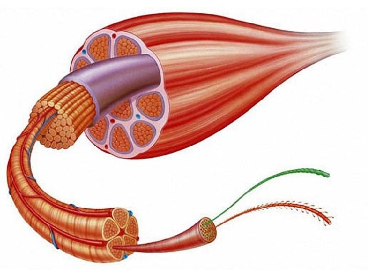
of skeletal muscle Muscles are made of many muscle FIBERS (aka muscle cells). Muscle fibers are arranged in bundles called FASCICLES. Individual fascicles are separated by fascia called PERIMYSIUM. Blood vessels and nerves are embedded between fascicles of muscle.
of skeletal muscle Fascicles are arranged in bundles called MUSCLES. Individual muscles are separated by fascia called EPIMYSIUM.
of skeletal muscle Epimysium Perimysium Tendon Fascicle Actin filament Muscle fiber (wrapped in endomysium) Blood vessels Myofibril Myosin filament
Label the following: Muscle fibers Fascicle Endomysium Perimysium
BELLRINGER Place the following terms in order from smallest/most deep to largest/most superficial part of skeletal muscle: Endomysium Epimysium Perimysium Muscle Myofilaments Muscle Fiber Muscle Fascicle Myofibril ANSWER: Myofilaments Myofibril Muscle Fiber Endomysium Muscle Fascic le Perimysium Muscle Epimysium
A closer look at By now, you should know that… Muscle fibers (are made of) Myofibrils (are made of) Myofilaments and that… Actin (thin) and Myosin (thick) are myofilaments made of protein.
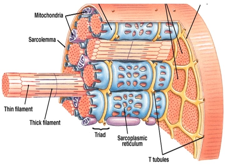
A closer look at t n e r e f if d e v a h s e ll Muscle cell organe … s ll e c r e h t o n a h t names Sarcolemma = cell membrane of a muscle fiber. Controls what enters and exits the cell. Sarcoplasm = cytoplasm of a muscle fiber. Jelly-like fluid that holds other organelles in place. Sarcoplasmic Reticulum (SR) = endoplasmic reticulum (ER) of a muscle fiber. Stores calcium.
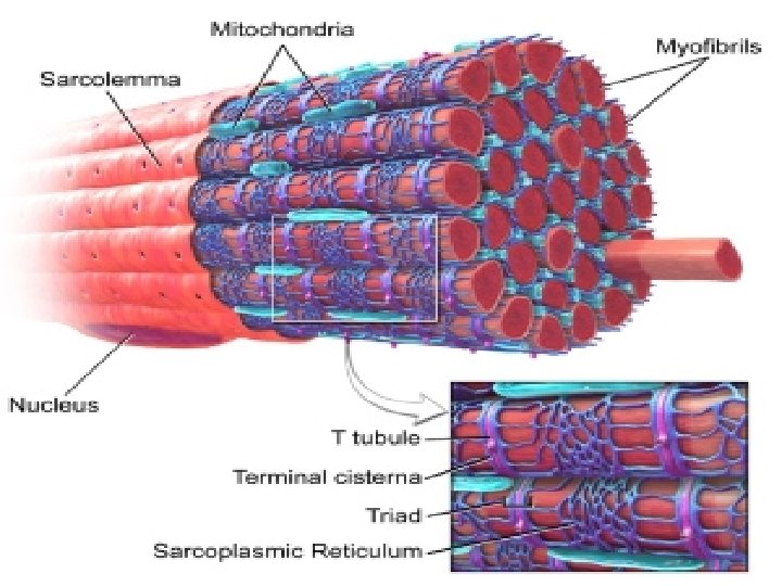
A closer look at Mitochondria are embedded between the myofibrils Myofibrils are made of protein filaments (myosin and actin) Sarcoplasmic Reticulum (SR) surrounds each myofibril inside a muscle cell the t c e n n o c s le u b u T T SR of one myofibril o it with the one next t
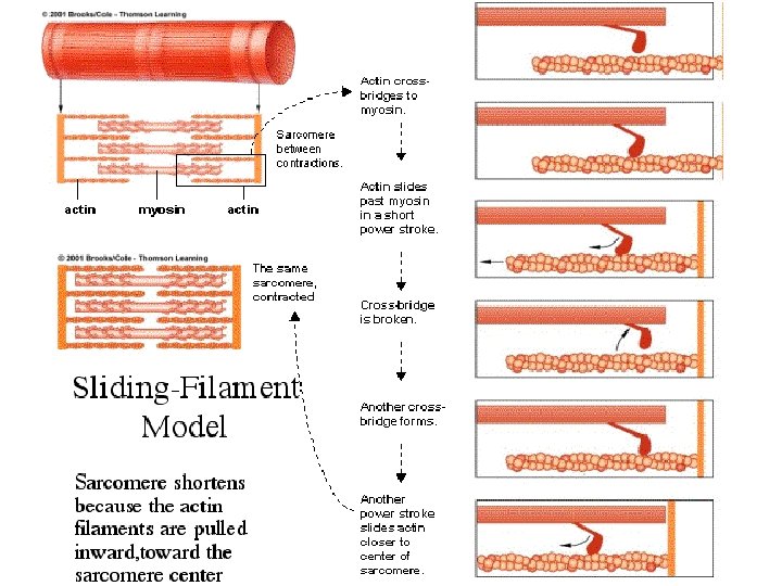
A closer look at e il t c a r t n o c ic s a b = Sarcomere unit of muscle cell Sarcomere Myosin and actin filaments are overlapped in a specific pattern to form each sarcomere…
structure Actin is shown in pink Myosin is shown in purple • Z line = place where actin fibers in one sarcomere attach to actin in next sarcomere (stabilizes actin filaments) • M line = stabilizes myosin • A band = thick zone (appears dark because it contains both myosin and actin) • H zone = part of the A band with only myosin (actin does not overlap) • I band = thin zone (appears light because it only contains thin actin filaments, no myosin)
abel l & Draw tures c the pi OR! L IN CO H Zone Each Z-Line is where one sarcomere ends and the next one begins
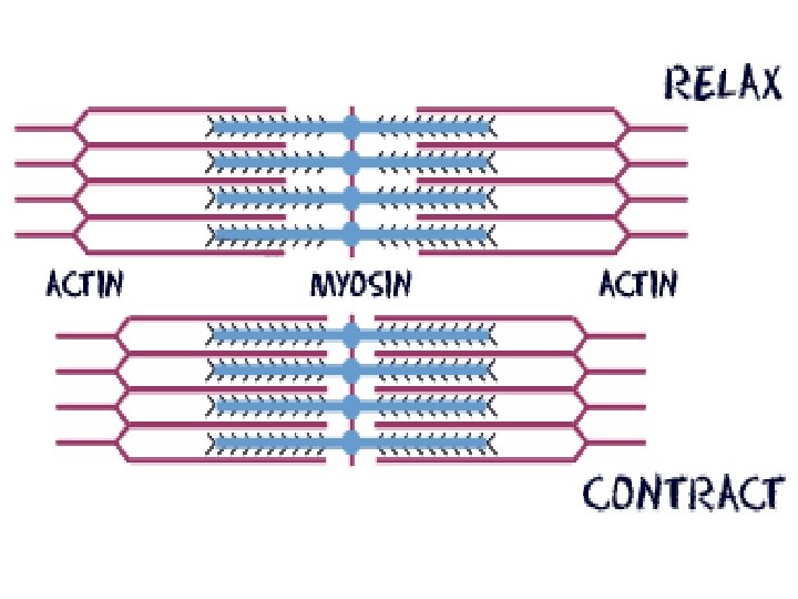
THE SLIDING FILAMENT THEORY Sliding filament theory = theory of how muscle tissue contracts (shortens). The contraction of a muscle occurs as the thin filaments slide past the thick filaments, shortening each sarcomere. The sliding filament theory involves 6 different molecules plus calcium ions. The 6 molecules are: Acetylcholine (Ach) myosin actin tropomyosin troponin ATP
Sarcomere Structure H Zone Shortening H zone, bringing z lines closer, shortening I bands
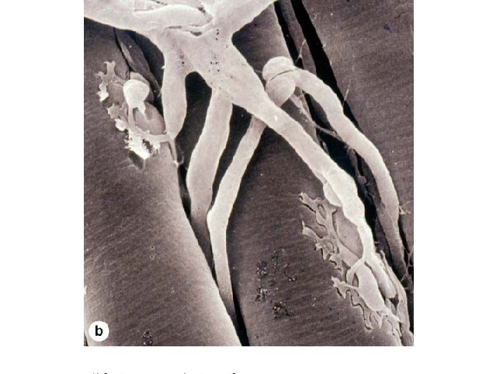
Motor Unit The muscle fiber and the motor neuron that controls it.
Motor Unit Vocabulary • Neuromuscular Junction= the site of connection between a nerve cell and a muscle fiber • Neuron= nerve cell that transmits brain signal to other cells in the body (muscle fibers!) • Axon= branch of a neuron that attaches to muscle fibers • Axon Terminal= the end of an axon (where it terminates); the part that attaches to the muscle
Motor Unit Vocabulary • Motor Endplate= the place on a muscle fiber where the axon attaches • Synaptic cleft= the very small space between an axon terminal and a motor endplate
Motor Unit Vocabulary • Acetylcholine (ACh) = a neurotransmitter in neurons that carries action potential (the potential energy to do work) from the brain • Tropomyosin = thread-like protein that wraps around actin filaments, blocking the binding sites where myosin wants to attach • Troponin = protein that is attached to tropomyosin and can move it
➊Signal to move a muscle is sent from brain to neuromuscular junctions as an action potential. ➋ACh is released from the axon terminal of a motor neuron and binds to ACh receptors on the motor end plate. ➌The action potential is transferred along the muscle’s sarcolemma and down the T-tubules. ➑Calcium is sent back to the SR. ➒Troponin and ➐When brain signal stops, myosin detaches from actin. ➍The action potential triggers tropomyosin rebinds to actin ➓The muscle fiber relaxes. Ca 2+ to be released from the sarcoplasmic reticulum (SR). ➎The Ca 2+ binds to troponin, which pulls ➏Myosin filaments can now attach to actin and pull it inward to contract the muscle fiber. tropomyosin away from the actin filaments, exposing their binding sites.