The Submandibular Region ANYANWU GE Introduction The submandibular

The Submandibular Region ANYANWU GE

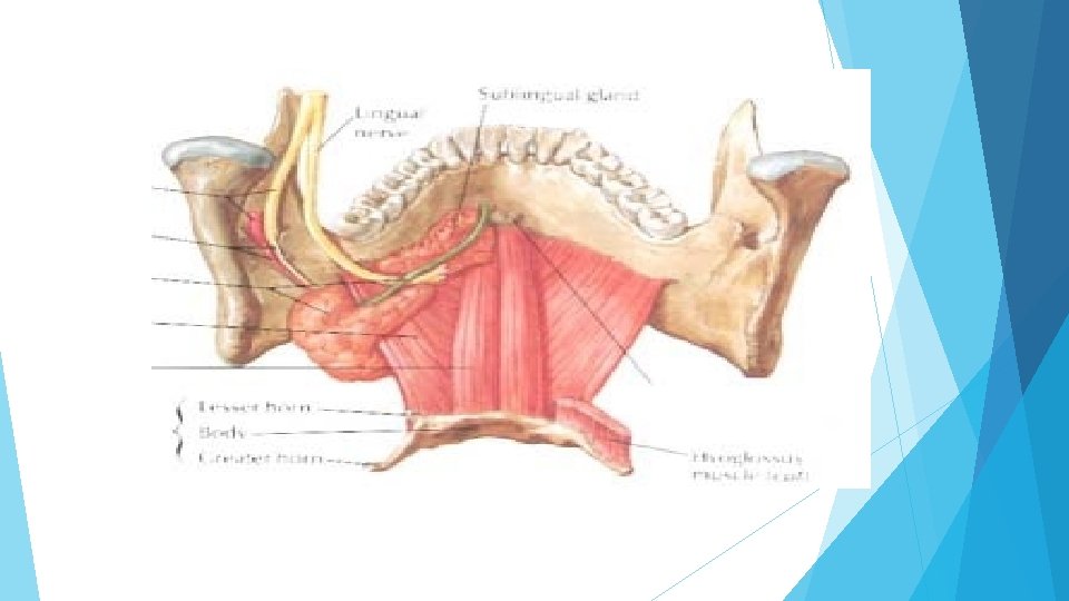
Introduction The submandibular region is the region under the cover of the body of the mandible, and between the mandible and the hyoid bone. It contains the suprahyoid muscles submandibular and sublingual glands, submandibular ganglion, and lingual artery
 Mylohyoid Geniohyoid Stylohyoid Digastric")
Suprahyoid Muscles (My Gravy Spoon, Darling) Mylohyoid Geniohyoid Stylohyoid Digastric

Suprahyoid muscles q The suprahyoid muscles which connect the hyoid bone to the skull, are: Ø Digastric. Ø Stylohyoid Ø Mylohyoid. Ø Geniohyoid
 united by an")
Digastric Ø It consist of two fleshy bellies (Posterior and Anterior) united by an intermediate rounded tendon. Ø It lies below the body of the mandible, and extends, in a curved form, from the mastoid process to the chin. q Origin & Insertion: Ø Posterior belly arises from the medial surface of the mastoid process, then it passes downward and forward crossing the carotid sheath, and ends in the intermediate tendon Ø Anterior belly runs and is attached to the lower border mandible, near the forward and medially the digastric fossa in of the body of the median plane

Digastric q The two bellies end in an intermediate tendon which perforates the stylohyoid q The intermediate tendon is held in connexion with the body and greater cornu of hyoid bone by a fibrous loop q Nerve supply: posterior belly: facial nerve( 2 nd) anterior belly: nerve to the mylohyoid (branch of the mandibular division of the trigeminal nerve)(1 st pharyngeal arch)

Digastric- Superficial relations Its superficial surface is in relation with 5 muscles, 2 glands, one vein & one bone 1. Platysma. 2. Sternomastoid. 3. Splenius capitis. 4. Longissimus capitis. 5. Parotid gland. 6. Submandibular gland. 7. Stylohyoid. 8. Retromandibular vein. 9. Mastoid process.

Digastric- Deep relations q Deep to the anterior belly of the digatric is the mylohyoid. q Deep to the posterior belly of the digastric are: Ø Ø Ø Ø Ø Obliques superiorius, occipital artery, rectus capitis lateralis, transverse process of atlas, accessory nerve, internal jugular vein, hypoglossal nerve, internal and external carotid arteries. facial and lingual arteries hyoglossus

Digastric q Action Ø The digastric depresses the mandible and can elevate the hyoid bone

Stylohyoid Muscle q It lies along the upper border of the posterior belly of the digastric muscle q Origin Ø It arises by a small delicate tendon from the posterior surface of the styloid process q Insertion Ø It is inserted into the body of hyoid bone q It is perforated near its insertion by the tendon of digastric q Innervation Ø It is supplied by the facial nerve q Actions Ø It can elevate and draw backwards the hyoid bone

Mylohyoid Muscle q It is a flat, triangular muscle q It is situated deep to the anterior belly of the digastric q With its fellow on of the opposite side, it forms the muscular floor for the mouth cavity q Origin: Ø q q Mylohyoid line of the mandible. Insertion: Ø Posterior fibers into the body of the hyoid bone. Ø Anterior fibers into the mylohyoid raphe which extends from the symphysis menti to the body of the hyoid bone. Nerve supply: Ø mylohyoid branch of the inferior alveolar nerve.

Mylohyoid Muscle

Mylohyoid Muscle q Action The mylohyoid elevates the floor of the mouth in the first stage of deglutition Ø It elevates the hyoid bone Ø It depresses the mandible Ø

Geniohyoid Muscle The geniohyoid muscle is a slender muscle, situated superior to the mylohyoid. q Origin: from the inferior mental spine, behind the symphysis menti. q Insertion: anterior surface of the body of the hyoid bone. q Action: elevates the hyoid bone and draws it forward; or it depresses the mandible. q Nerve supply: first cervical nerve through the hypoglossal nerve

Submandibular gland q a major salivary gland q situated in the anterior part of digastric triangle in submandibular region q about the size of walnut q roughly ‘J’- shaped - being indented by posterior border of mylohyoid muscle q It has two parts a larger superficial part (body) and a smaller deep process q The two parts are continuous with each other, forming a "U" shape around the posterior border of the mylohyoid muscle. The body of the gland is in and inferior to the digastric triangle and also partly under cover of the mandible q composed of a mixture of serous and mucous acini

Submandibular gland- superficial part q q q this part fills the digastric triangle extends upwards deep to the mandible up to the mylohyoid muscle It has three surfaces: inferior (covered by skin and platysma), lateral (related to the medial surface of the mandible), and medial (related to the mylohyoid, hyoglossus, and digastric muscles). partially enclosed between two layers of deep cervical fascia superficial layer of fascia covers the inferior surface of the gland is attached to the base of mandible deep layer covers the medial surface of the gland is attached to the mylohyoid line of mandible

Submandibular gland- Superficial part Relations q inferior surface is covered by Ø Ø Ø q skin platysma muscle cervical branch of facial nerve deep fascia facial vein submandibular lymph nodes lateral surface is related to submandibular fossa on the mandible Ø insertion of pterygoid Ø facial artery Ø

Submandibular gland- Superficial part q medial surface maybe divided into three parts Ø anterior part - related to mylohyoid muscles, nerve and vessels Ø middle part - related to hyoglossus, styloglossus, lingual nerve, submandibular ganglion and hypoglossal nerve Ø posterior part - related to styloglossus, stylohyoid ligament, the ninth nerve, and the wall of pharynx. Inferiorly it overlaps stylohyoid and posterior belly of digastric

Submandibular gland – Deep part q small in size q lies in the intermuscular interval between mylohyoid, below and laterally, and medial to hyoglossus and styloglossus q posteriorly continuous with superficial part around the posterior border of mylohyoid q anteriorly extends up to posterior end of sublingual gland

Submandibular gland Blood supply and lymphatic drainage q supplied by facial artery q veins drain into common facial or lingual vein q lymph passes to submandibular lymph nodes Innervation q supplied by branches from the submandibular ganglion q these branches convey: q Ø secretomotor fibres Ø sensory fibres from lingual nerve Ø vasomotor fibres from plexus on facial artery secretomotor pathway (parasympathetic) - begins in the superior salivary nucleus Ø preganglionic fibres pass through sensory root of facial nerve, geniculate ganglion, facial nerve, chorda tympani, lingual nerve, to reach submandibular ganglion Ø post ganglionic fibres emerges from the ganglion and enter submandibular gland
 Ø regulates submandibular secretions through vasoconstriction of the arteries that")
q vasomotor function (sympathetic) Ø regulates submandibular secretions through vasoconstriction of the arteries that supply it Ø increased sympathetic activity reduces glandular bloodflow decreasing salivary secretions producing an enzyme rich serous saliva. The submandibular duct Ø is about 5 cm long Ø Its wall is thinner than that of parotid duct Ø It opens by one to three orifices into the oral cavity on the sublingual papilla, at the side of the frenulum linguae

LINGUAL ARTERY BEGINNING: from anterior aspect of external carotid artery, in the carotid triangle, opposite the tip of greater cornu of hyoid bone COURSE: has a tortuous course, divided into 3 parts: FIRST PART: forms a loop crossed by hypoglossal nerve SECOND PART: runs along upper border of greater cornu of hyoid bone, deep to hyoglossus THIRD PART: ascends along anterior border of hyoglossus & runs along the under surface of tongue to reach its tip & anastomoses with artery of opposite side

BRANCHES OF LINGUAL ARTERY FROM FIRST PART: Suprahyoid artery: runs along upper border of greater cornu of hyoid bone, superficial to hyoglossus, supplying adjacent muscles FROM SECOND PART: Two dorsal lingual arteries: supply dorsum of tongue FROM THIRD PART: Sublingual artery: supplies sublingual gland & mucous membrane of floor of mouth N. B. : VEINS CORRESPONDING TO BRANCHES OF LINGUAL ARTERY UNITE TO FORM A SINGLE LINGUAL VEIN THAT DRAINS INTO INTERNAL JUGULAR VEIN

Submandibular ganglion q It is a small parasympathetic ganglion lying superficial to hyoglossus & is connected to lingual nerve by 2 roots (anterior & posterior q The submandibular ganglion lies on the lateral surface of the hyoglossus muscle, medial to the mylohyoid muscle, superior to the submandibular duct and hypoglossal nerve, and inferior to the lingual nerve, from which it is suspended by several branches. q Origin of fibers: superior salivary nucleus in pons q q

Submandibular ganglion q Preganglionic parasympathetic fibers derived from the chorda tympani travel in the lingual nerve and synapse in the submandibular ganglion. q Some of the postganglionic secretory fibers enter the submandibular gland; others, by entering the lingual nerve, reach the sublingual gland. q Postganglionic sympathetic fibers (from the superior cervical ganglion) pass through the submandibular ganglion and are distributed with the parasympathetic fibers

SUBMANDIBULAR GANGLION Postganglionic fibers: 1. To submandibular gland: fibers are distributed directly to the gland 2. To sublingual gland: Fibers pass along anterior root to lingual nerve again Lingual nerve transmits fibers to sublingual gland

Thank You 28
- Slides: 28