
The Study of Cells Light microscope LMs visible
The Study of Cells § Light microscope (LMs) visible light passes through the specimen and then through glass lenses. § The lenses refract light such that the image is magnified into the eye or a video screen. § Low magnification and resolution. § Does not kill the specimen. ( A problem with EM’s) § Magnification is the ratio of an object’s image to its real size. (ie: 4 X, 100 X) § Resolution is a measure of image clarity.

Copyright © 2002 Pearson Education, Inc. , publishing as Benjamin Cummings
Electron Microscopes § Transmission electron microscopes (TEM) § study the internal ultrastructure of cells. § A TEM aims an electron beam through a thin section of the specimen. § Electromagnets focus and magnify specimen. § Specimens are stained with heavy metals to enhance contrast.
Electron Microscopes § Scanning electron microscopes (SEM) § studying surface structures. § The sample surface is covered with a thin film of gold. § The beam excites electrons on the surface. § These secondary electrons are collected and focused on a screen. § Image produced by SEM appears 3 -D

TEM SEM
Studying Organelles § Light microscopes allow the study of individual cells. § The small, functional structures of cells, organelles, require the use of EM’s. § Cell fractionation separates (based on relative density) the organelles using an ultracentrifuge.
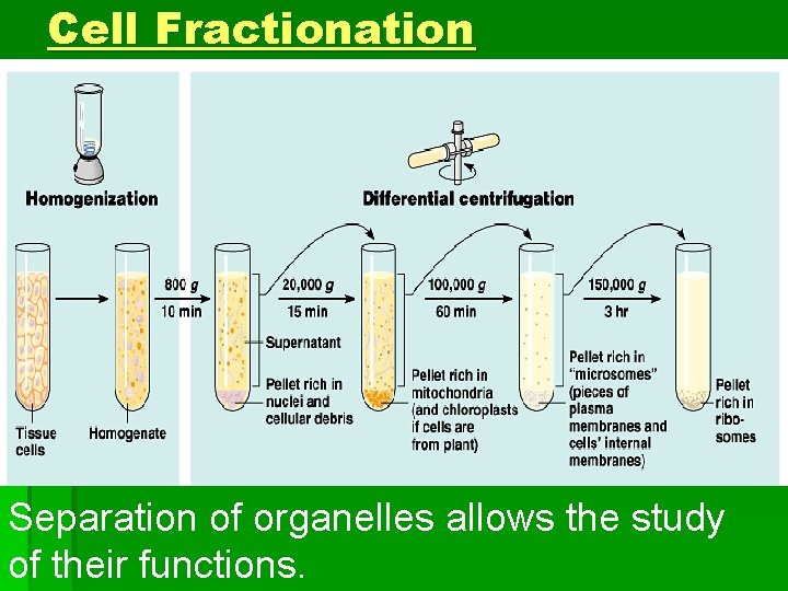
Cell Fractionation Separation of organelles allows the study of their functions.
Isolation/Study of Organelles § Step 1: Homogenization § Gentle, mechanical disruption of the cells. § Breaks open the membranes. § Step 2: Ultracentrifugation § Spinning of cells separates the denser organelles as pellet (solid) and less dense particles remain suspended in supernatant (liquid).
Prokaryotic and Eukaryotic Cells § Prokaryotic Cells (ie: bacteria) § Lack a membrane bound nucleus § DNA is found in nucleoid (no membrane) § Eukaryotic Cells (ie: yeast, humans) § “True”, membrane-bound nucleus contains chromosomes. § DNA separate from rest of the cell.
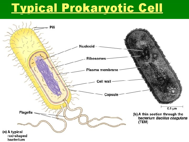
Typical Prokaryotic Cell
Eukaryotic Cells vs Prokaryotic Cells § Cytoplasm– Fluid region between the cell membrane and the nuclear membrane. (All of material in prok. is cytoplasm). § Membrane bound organelles are found suspended in the cytoplasm of eukaryotic cells (absent in prokaryotes). § Eukaryotic cells are generally much bigger than prokaryotic cells. (bacteria = 1 -10 microns, eukaryote = 10 -100 microns)
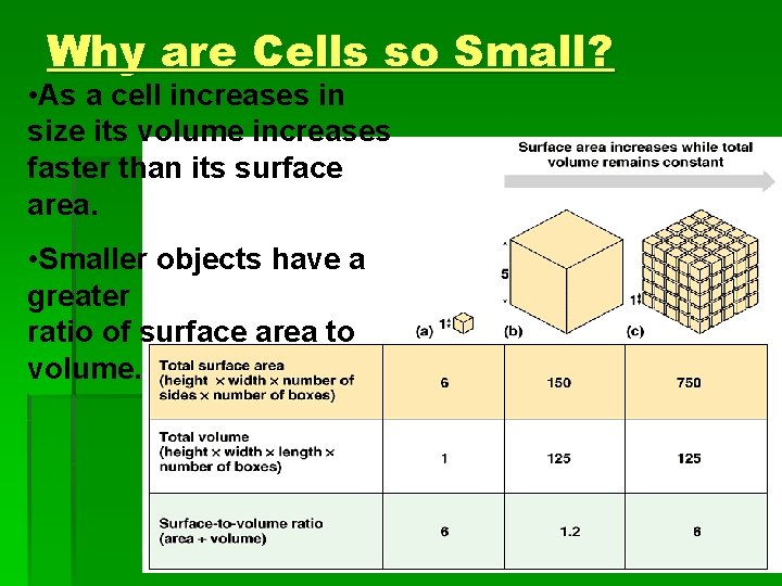
Why are Cells so Small? • As a cell increases in size its volume increases faster than its surface area. • Smaller objects have a greater ratio of surface area to volume.
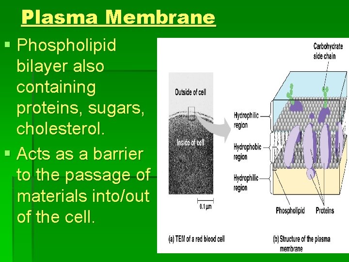
Plasma Membrane § Phospholipid bilayer also containing proteins, sugars, cholesterol. § Acts as a barrier to the passage of materials into/out of the cell.
Importance of Surface Area § The more cytoplasm a cell has, the more gas/material exchange it needs. § Since it is the membrane that allows this exchange, it does not make sense to have large volume cells (small surface area). Therefore, cells are microscopic (best surface to volume ratio). § Therefore, large organisms have more cells, not larger cells.
Internal Membranes § Eukaryotic organelles tend to have secondary internal membranes. § Separate the metabolic reactions spatially. § Provide microenvironments for metabolic reactions to take place. § Enzymes are often found stored inside membrane bound structures.

Typical Animal Cell

Typical Plant Cell
Nucleus § The nucleus contains most DNA in eukaryotic cell (mitochondria and chloroplasts). § Approx. 5 micron diameter. § Separated from cytoplasm via double membrane. § Nuclear pore allows passage of large molecules into and out of the nucleus (ie: m. RNA)
Nuclear Structure § Nuclear lamina (filament proteins) line the inside of the nuclear envelope and maintain shape. § DNA and associated proteins are organized into fibrous material, chromatin. § In a normal cell they appear as diffuse mass. § Cell Division- chromatin fibers coil up to be seen as separate structures- chromosomes. § All eukaryotic cells have characteristic # of chromosomes (e. g. , human = 46 (23 pairs))
The Nucleolus § Ribosomal subunits are assembled. § Subunits pass through nuclear pore to unite and form functional ribosomes in cytoplasm.
Nuclear Control § The nucleus controls the cell by determining which proteins are synthesized (depends on DNA). § Nucleus synthesizes m. RNA and this molecule passes through nuclear pore. § m. RNA works along side ribosomes to create the cell’s functional proteins.

Ribosomes § Assemble amino acid subunits into proteins. § Found in higher numbers in cells that make lots of proteins. (ie: pancreas) § Free Ribosomes – function unattached in the cytoplasm. § Proteins made tend to function in cytoplasm. § Bound Ribosomes – Function bound to endoplasmic reticulum. § Proteins made tend to be packaged inside organelles.
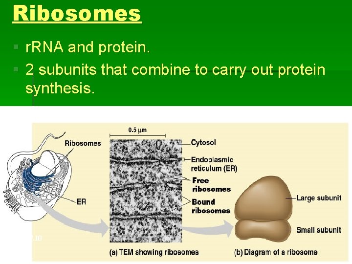
Ribosomes § r. RNA and protein. § 2 subunits that combine to carry out protein synthesis. Fig. 7. 10 Copyright © 2002 Pearson Education, Inc. , publishing as Benjamin Cummings
Endomembrane System § System of internal membranes in a eukaryotic cell. § Either in direct contact or connected via transfer of vesicles (sacs of membrane) § Membranes have diverse functions and structures. § Nuclear envelope, endoplasmic reticulum, Golgi apparatus, lysosomes, vacuoles, and the plasma membrane.
Endoplasmic Reticulum (ER) § Network of fluid filled tubules (cisternae) § Roughly ½ of eukaryotic membrane tissue § Continuous with the nuclear envelope § Smooth ER = lacks ribsosomes § Rough ER = ribosomes bound to cytoplasmic side of ER membrane
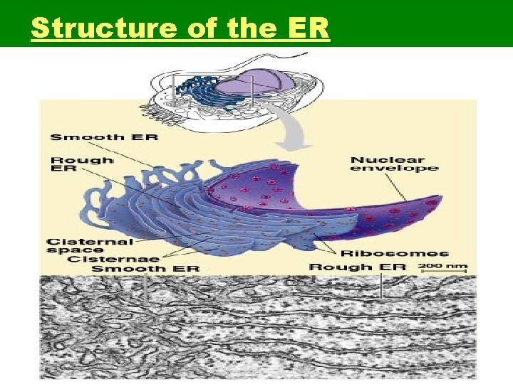
Structure of the ER
The Smooth ER § Synthesis of lipids, steroids (ie: sex hormones) § Carb metabolism (ie: liver cells hydrolysis glycogen into glucose utilizes enzymes in smooth ER) § Detoxification of drugs/poisons (ie: smooth ER enzymes make drugs more soluble add –OH). Tolerance = more Smooth ER § Involved in Ca ion movement during muscle contraction.
Functions of Rough ER § Attached ribosomes = protein synthesis § Abundant in cells that make proteins § Manufactured proteins are “threaded” through pore into the cisternal space of ER. § Glycoproteins – covalently bonded to carb. § Secretory proteins are packaged into transport vescicles and sent to various locations in the cell.
Rough ER § Also manufactures phospholipids from precursors in cytoplasm. § Assembles phospholipids and proteins into new membrane sections. § ER membrane can expand or transfer new membrane via vescicles to other parts of endomembrane system.
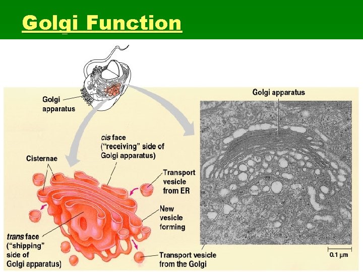
Golgi Apparatus § Responsible for modifying, packaging, and transporting products of the ER throughout the cell. § System of flattened membranous sacs (cisternae) § Has two poles; cis and trans faces. § The cis and trans faces of the golgi act as the “receiving” and “shipping” departments.
Golgi Function
Golgi Function § Products of ER are modified from cis to trans faces through golgi § Manufactures some polysaccharides (pectin) § Tags, sorts, and packages materials into transport vescicles
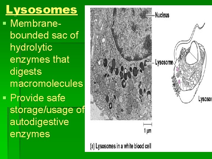
Lysosomes § Membranebounded sac of hydrolytic enzymes that digests macromolecules § Provide safe storage/usage of autodigestive enzymes

Lysosomal Enzymes § Lysosomal enzymes can hydrolyze proteins, fats, polysaccharides, and nucleic acids. § Enzymes work best at p. H 5 § Proteins in the lysosomal membrane pump hydrogen ions from the cytosol to the lumen of the lysosomes.
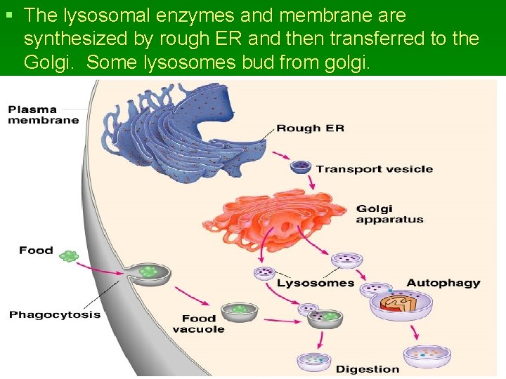
§ The lysosomal enzymes and membrane are synthesized by rough ER and then transferred to the Golgi. Some lysosomes bud from golgi.

Phagocytosis § Lysosomes can fuse with food vacuoles § As the polymers are digested, monomers pass out to the cytosol to become nutrients of the cell. § Autophagy-- Lysosomes fuse with/digests another organelle or part of the cytosol. Renews cellular material.
Lysosome Function § Pre-programmed cell death during development and throughout cell lifetime § Pompe’s disease (liver) and Tay-Sachs disease (brain) § Serious metabolic disorders in which sufferers lack a (or many) functional hydrolytic enzyme (s). § Cells accumulate undigestible substrates in lysosomes.
Vacuoles § Membrane bound sacs. Similar to vesicles only larger. § Three main types of vacuoles: § Food vacuoles, formed by phagocytosis, fuse with lysosomes. § Contractile vacuoles (freshwater protists)-- pump excess water out of the cell. § Central vacuoles are found in many mature plant cells.
Central Vacuole § Tonoplast– membrane that regulates passage into/out of the central vacoule. § Stores proteins or inorganic ions § Depositing metabolic byproducts § Storing pigments § Storing defensive compounds against herbivores § Increases surface to volume ratio for the whole cell
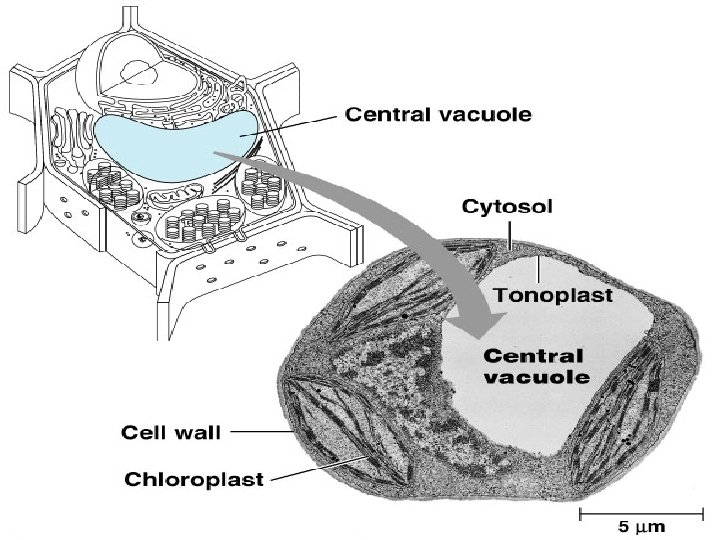
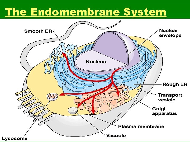
The Endomembrane System

Mitochondria and Chloroplasts § Convert energy into usable forms for cells. § Mitochondria are the sites of cellular respiration § Chloroplasts (plants, eukaryotic algae) are the site of photosynthesis § Have own DNA (mitochondrial used in forensics § Operate and reproduce autonomously § Mobile -- move around the cell along tracks in the cytoskeleton
Mitochondria § The number of mitochondria is correlated with aerobic metabolic activity § Mitochondria have a smooth outer membrane and a highly folded inner membrane, the cristae § Fluid-filled space between membranes § Cristae present ample surface area for the enzymes that synthesize ATP
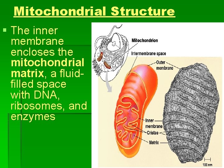
Mitochondrial Structure § The inner membrane encloses the mitochondrial matrix, a fluidfilled space with DNA, ribosomes, and enzymes
Plastids § Amyloplasts store starch in roots and tubers § Chromoplasts store pigments for fruits and flowers § The chloroplast produces sugar via photosynthesis § Green pigment chlorophyll § Concentrated in photosynthetic cells (ie: leaf cells)
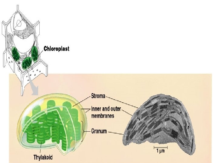
Chloroplast Structure § The processes in the chloroplast are separated from the cytosol by two membranes. § Inside the innermost membrane is a fluid-filled space, the stroma, in which float membranous sacs, the thylakoids.
Chloroplast Structure § Stroma contain DNA, ribosomes, and enzymes for photosynthesis. § Thylakoids (flattened sacs) are stacked into grana and are critical for converting light to chemical energy.
Peroxisomes § Peroxisomes contain enzymes that transfer hydrogen from various substrates to oxygen § Produce H 2 O 2 (poison) but also convert it to water § Break fatty acids down to smaller molecules that are transported to mitochondria for fuel § Detoxify alcohol and other harmful compounds
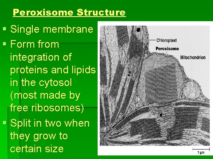
Peroxisome Structure § Single membrane § Form from integration of proteins and lipids in the cytosol (most made by free ribosomes) § Split in two when they grow to certain size
Glyoxisomes § Specialized peroxisomes § Convert fatty acids in seeds to sugars § easier energy and carbon source to transport
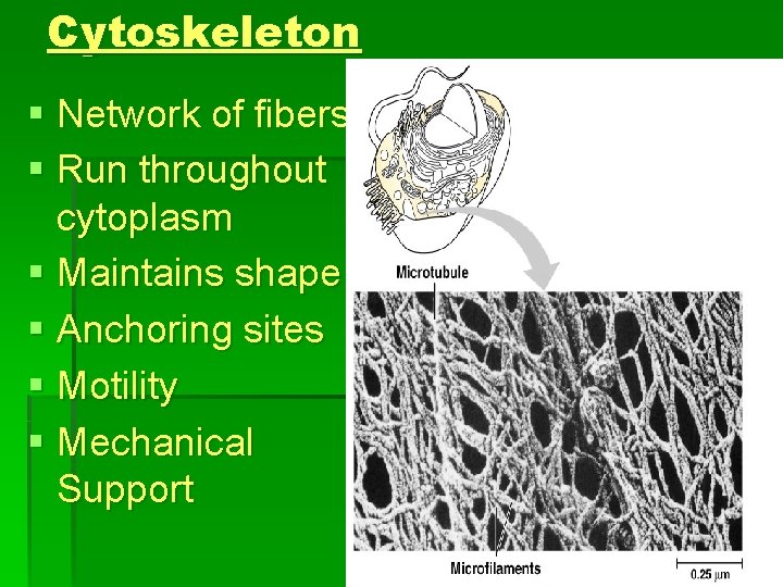
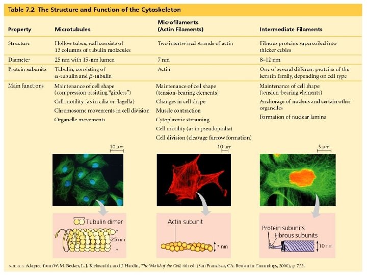
Cytoskeleton § Network of fibers § Run throughout cytoplasm § Maintains shape § Anchoring sites § Motility § Mechanical Support
Cytoskeleton and Motility § Motor molecules – specialized proteins that interact with cytoskeleton § Allow movement of cell (cilia and flagella) and movement of materials around cell (organelles, vesicles, etc. ) § Muscle contractions § Cytoplasmic streaming
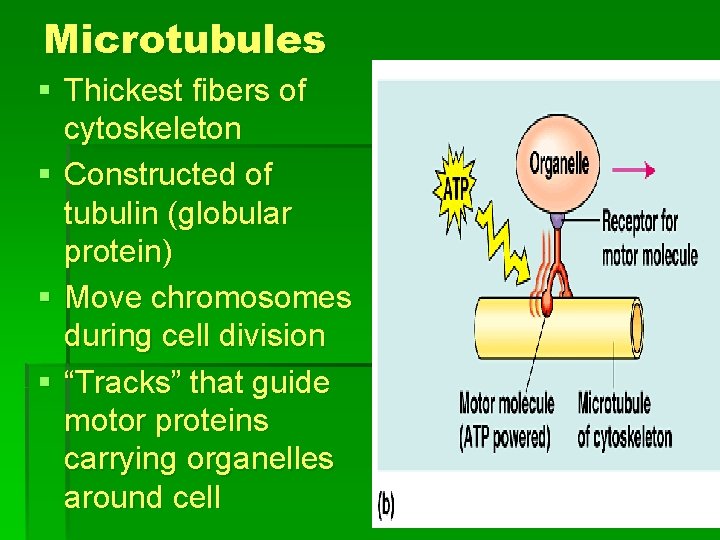
Microtubules § Thickest fibers of cytoskeleton § Constructed of tubulin (globular protein) § Move chromosomes during cell division § “Tracks” that guide motor proteins carrying organelles around cell

Centrioles § Some microtubules are arranged into a pair of centrioles in animal cells. § During cell division, centrioles replicate.
Cilia and Flagella § Microtubules are the central structural supports in cilia and flagella § Propel unicellular organisms through water § Move fluid across large surfaces in multicellular organisms (ie: mucus in lungs) § Cilia occur in large numbers per cell (ie: paramecia) § Flagella are only one or two per cell (ie: sperm cell)
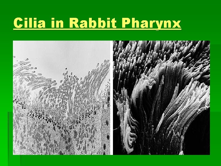
Cilia in Rabbit Pharynx
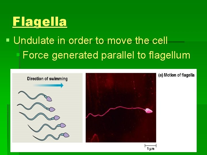
Flagella § Undulate in order to move the cell § Force generated parallel to flagellum
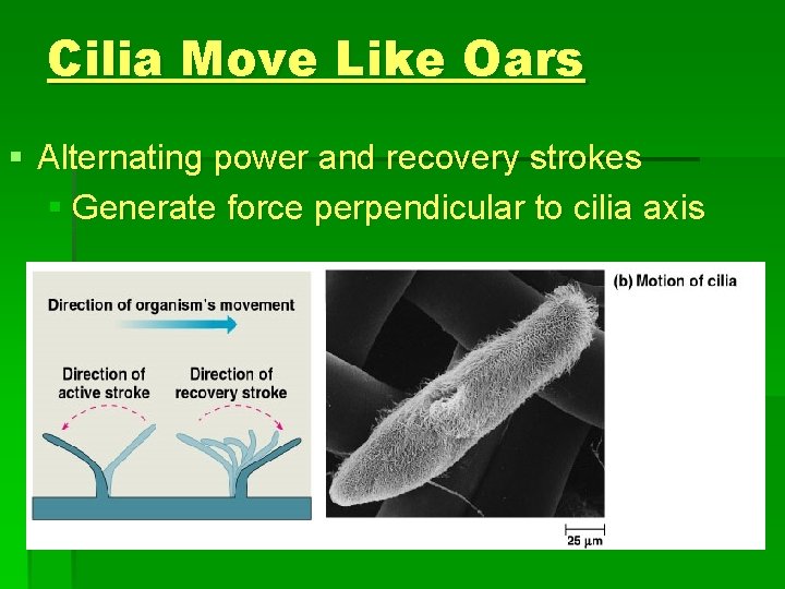
Cilia Move Like Oars § Alternating power and recovery strokes § Generate force perpendicular to cilia axis
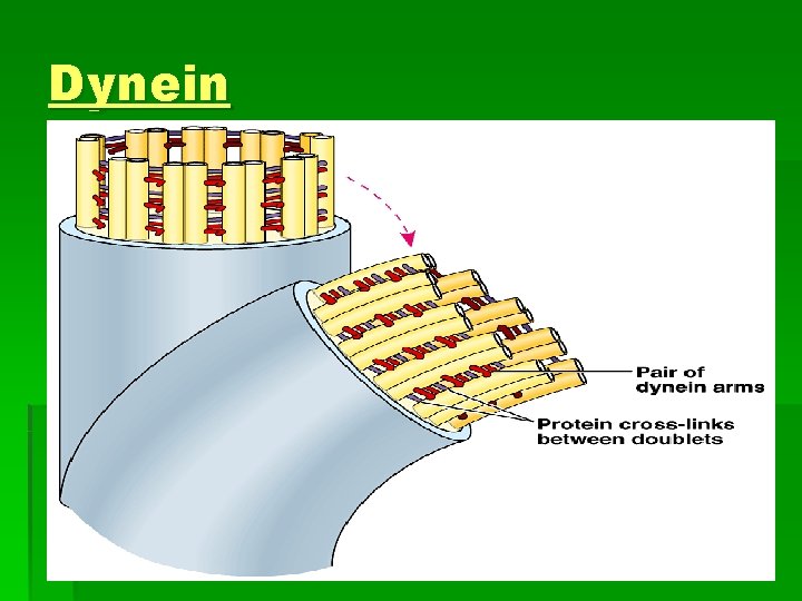
Structure of Cilia and Flagella § Core of microtubules sheathed by a plasma membrane § Nine doublets of microtubules arranged around a pair at the center, the “ 9 + 2” pattern. § Flexible “wheels” of proteins connect outer doublets to each other and to the core. § Outer doublets connected by motor proteins.

Cilia and Flagella Structure § Anchored in the cell by a basal body, whose structure is identical to a centriole.
Dynein § Motor protein § Provides power to drive cilia and flagella § Addition/removal of P from ATP causes the movement of dynein § Alternately grab, move, and release microtubules of cilia/flagella § Protein cross links provide rigidity
Dynein

Microfilaments § The thinnest cytoskeletal fibers § Resist tension § Solid rods of globular protein actin (muscle cells) § Form 3 -D network just inside plasma membrane
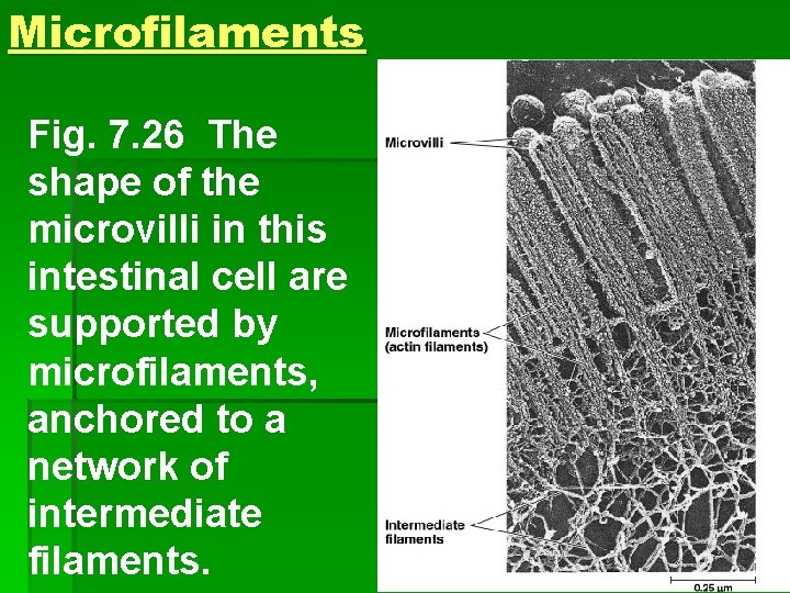
Microfilaments Fig. 7. 26 The shape of the microvilli in this intestinal cell are supported by microfilaments, anchored to a network of intermediate filaments.
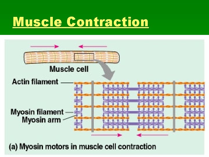
Actin in Muscle Cells § Thousands of actin filaments are arranged parallel to one another. § Thicker filaments, composed of a motor protein, myosin, interdigitate with the thinner actin fibers. § Myosin walks along the actin filament, pulling stacks of actin fibers together and shortening the cell. (muscle contraction)
Muscle Contraction
Other Microfilaments § Microfilaments separate the cytoplasm in animal cells during cell division § Amoeba (localized contraction) § Pseudopodia -- cellular extensions § extend and contract through the reversible assembly and contraction of actin subunits into microfilaments
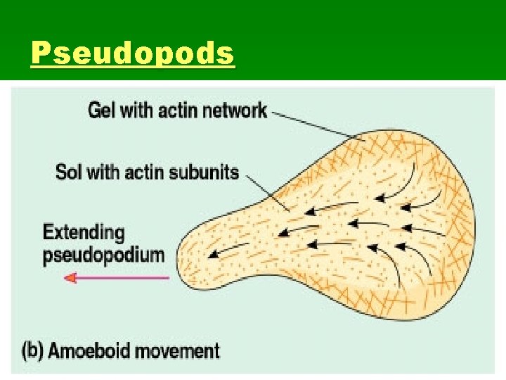
Pseudopods

Cytoplasmic Streaming § In plant cells (and others), actinmyosin interactions drive cytoplasmic streaming § Circular flow that speeds distribution of materials through the cell
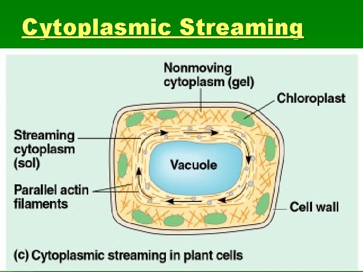
Cytoplasmic Streaming
Intermediate Filaments § In between microtubule and microfilament diameter § Tension bearing filaments § Composed of keratins (family of proteins) § More permanent structures than other 2 § Reinforce cell shape and provide strength § Fix organelle location within the cytoplasm
Cell Walls § Surround plant cells, many prokaryotes, fungi, and protists § Protect cell § Maintain shape § Limit excessive water loss § Supports plant against gravity
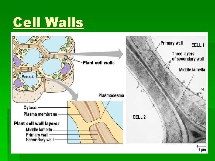
Cell Wall Structure § Cellulose microfibrils embedded in protein, polysaccharide matrix § Primary Cell Wall § Middle Lamella § Secondary Cell Wall
Cell Walls
Extracellular Matrix § The ECM provides structure to cell wall lacking animal cells. § The ECM is a network of fibrous proteins and sugars (glycoproteins). Mostly collagen. § The ECM is connected to the cytoskeleton via integrins (intermembrane proteins) § This connection allows for coordination of changes inside and outside of cell.
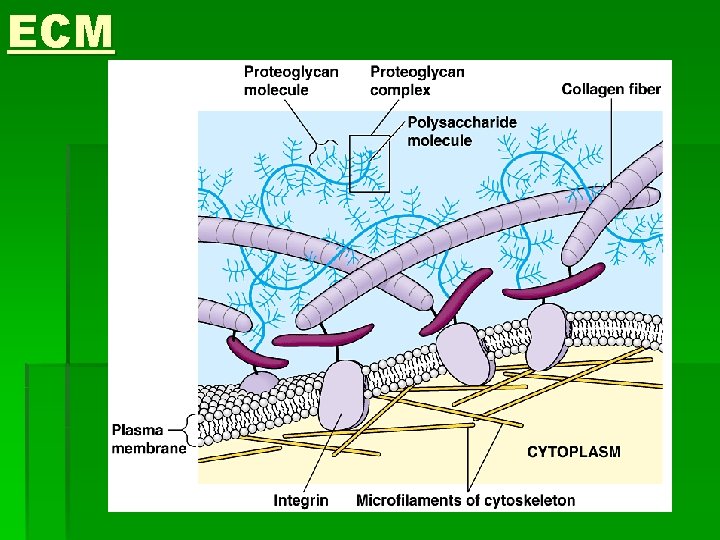
ECM
ECM § ECM helps regulate cell activity § Embryonic cell migration follows “grain” of ECM § Can communicate with the nucleus to influence gene expression § May help coordinate cells of a tissue with one another § Receptor molecules
Intracellular Junctions § Neighboring cells in tissues, organs, or organ systems adhere, interact, and communicate through direct physical contact. § Plasmodesmata (plants) -- channels that allow cytosol to pass between adjacent cells
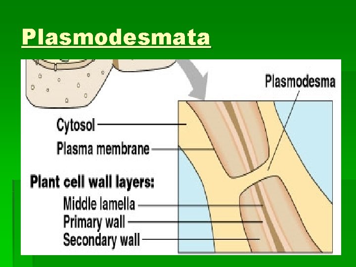
Plasmodesmata
Intercellular Junctions in Animals § Tight Junctions– Membranes of adjacent cells fused. § Desmosomes (anchoring junctions) fasten cells together into strong sheets § Gap junctions (communicating junctions) provide cytoplasmic channels between adjacent cells
- Slides: 82