THE STRUCTURE OF THE GENETIC MATERIAL DNA is


, Cytosine (C), Guanine")
 Cytosine (C) Pyrimidines Guanine (G) Adenine (A) Purines")
, Cytosine (C),")


")


 molecules")


- Slides: 46
THE STRUCTURE OF THE GENETIC MATERIAL
DNA is a Double-Stranded Helix § In 1953, James D. Watson and Francis Crick deduced the structure of DNA, using X-ray crystallography data of DNA from the work of Rosalind Franklin and Maurice Wilkins and Chargaff’s observation that in DNA the amount of adenine was equal to the amount of thymine and the amount of guanine was equal to that of cytosine.
DNA is a Double-Stranded Helix § Watson and Crick reported that DNA consisted of two polynucleotide strands wrapped into a double helix. – The sugar-phosphate backbone is on the outside. – The nitrogenous bases are perpendicular to the backbone in the interior. – Specific pairs of bases give the helix a uniform shape. – A pairs with T, with two hydrogen bonds – G pairs with C, with three hydrogen bonds
DNA and RNA are Polymers of Nucleotides § DNA and RNA are nucleic acids composed of long chains of nucleotides § A nucleotide = nitrogenous base, five-carbon sugar, and phosphate group § Nucleotides are joined to one another by a sugar-phosphate backbone. © 2012 Pearson Educaton, Inc.
DNA and RNA are Polymers of Nucleotides § The polynucleotide strands of DNA run in opposite directions or, are, antiparallel § There are 5’ (prime) and 3’ (prime) ends
Phosphate group on 5’ end Hydrogen bonds G T C A A C T G Partial chemical structure Hydroxyl group on 3’ end
§ Each DNA nucleotide has a different nitrogenous base: Adenine (A), Cytosine (C), Guanine (G) or Thymine (T) Nitrogenous base (can be A, G, C, or T) Thymine (T) Phosphate group Sugar (deoxyribose) DNA nucleotide
Nitrogenous Bases of DNA Thymine (T) Cytosine (C) Pyrimidines Guanine (G) Adenine (A) Purines
§ Each RNA nucleotide also has a different nitrogenous base: Adenine (A), Cytosine (C), Guanine (G) or Uracil (U) Nitrogenous base (can be A, C, G, or U) Sugar (ribose)
DNA REPLICATION
DNA Replication Depends on Specific Base Pairing Rules § DNA replication follows a Semiconservative Model § The two DNA strands separate so that they can serve as a template § Enzymes will read the parental strands and link nucleotides according to base pairing rules forming the new complementary strand § Semiconservative Replication means that each new DNA helix has one old strand with one new strand § DNA replication ensures that all somatic cells in a multicellular organism carry the same genetic information
A T G A A T Parental DNA molecule T A G C Daughter strand T C G T C A G C C Parental strand G C G G T C C T A G T A C C T A T G A T A A C A T G T Daughter DNA molecules
DNA Replication Proceeds in Two Directions at Many Sites Simultaneously § DNA replication begins at the origins of replication where, – DNA unwinds producing a replication bubble – Replication proceeds in both directions from the origin – Replication ends when products from the bubbles merge with each other
Parental DNA molecule Origin of replication “Bubble” Two daughter DNA molecules Parental strand Daughter strand
Daughter Strand Synthesis in DNA Replication § Both daughter strands are synthesized 5’ to 3’; meaning new nucleotides are added only to the 3’ end of a growing strand – The daughter strand that is synthesized in one continuous piece, toward replication fork = leading strand – The other daughter strand synthesized in (Okazaki) fragments = lagging strand – Lagging strand synthesis is due to the antiparallel nature of DNA
Daughter Strand Synthesis in DNA Replication
DNA Replication Requires a Host of Enzymes § Four key Enzymes are involved in DNA replication 1. Helicase-breaks H-bonds, unwinding (separating) the DNA double helix 2. DNA polymerase reads template strand adds nucleotides to a growing chain, proofreads and corrects improper base pairings 3. DNA ligase joins Okazaki fragments into a continuous DNA strand 4. RNA Primase- synthesizes a small RNA nucleotide sequence (primer) to provide a free 3’ end for DNA pol. to add nucleotides to *DNA polymerases and DNA ligase also repair damaged DNA*
THE FLOW OF GENETIC INFORMATION FROM DNA TO RNA TO PROTEIN
The DNA Genotype is Expressed as Proteins, Which Provide the Molecular Basis for Phenotypic Traits § Connections between genes and proteins: – In the 1940’s Beadle and Tatum suggested a one gene –one enzyme hypothesis based on studies of inherited metabolic diseases – Their hypothesis is still accepted but with important changes: – The hypothesis includes all proteins not just enzymes and that one gene codes for one polypeptide
The DNA Genotype is Expressed as Proteins, Which Provide the Molecular Basis for Phenotypic Traits § DNA specifies traits by dictating protein synthesis (genotype determines phenotype) § The molecular chain of command is from DNA to RNA to protein § Transcription is the synthesis of RNA using DNA as a template. § Translation is the synthesis of proteins using RNA as a template.
Figure 10. 6 A_s 3 DNA Transcription RNA NUCLEUS Translation Protein CYTOPLASM
Genetic Information in DNA is written in m. RNA as Codons § The genetic code is written in DNA as three base sequences called triplets § During transcription the genetic code in DNA is copied into complementary three base sequences in m. RNA called codons – Each codon determines the amino acid that will be added to a growing polypeptide chain – 64 codons are possible A A A C C G G C A A Transcription U U U G G C C G U U Translation Polypeptide amino acid 1 amino acid 2 amino acid 3 amino acid 4
Third base First base Second base
Transcription § RNA polymerase oversees transcription by – Unwinding DNA, reading the bases, and joining the appropriate RNA nucleotides together – RNA pol. enzymes synthesize (all) RNA molecules – There are three steps in transcription. – Initiation, elongation, termination
Transcription § Initiation - RNA pol. Binds the promoter § Elongation - m. RNA grows longer § Termination –RNA pol. reaches the terminator sequence and detaches from m. RNA and the gene being transcribed
RNA polymerase DNA of gene Promoter DNA 1 Initiation Terminator DNA
2 Elongation Area shown in Figure 10. 9 A Growing RNA
3 Completed RNA Termination Growing RNA polymerase
Transcription Produces Genetic Messages in the Form of RNA § Messenger RNA (m. RNA) – The RNA formed from transcription, carrying the genetic code is called m. RNA – m. RNA carries the message from DNA (nucleus) to ribosomes (cytoplasm) – In prokaryotes transcription and translation occur at the same place and time – In eukaryotes, m. RNA must exit nucleus via nuclear pores to enter cytoplasm – Eukaryotic m. RNA has introns or interrupting sequences that separate exons or coding regions
Eukaryotic RNA is Processed Before Leaving the Nucleus § m. RNA processing involves: – The addition of extra nucleotides to the ends of m. RNA in the form of a 5’ cap and 3’ tail. This helps to: – facilitate export of m. RNA from nucleus – protect m. RNA from attack by cellular enzymes – help ribosomes bind m. RNA § RNA splicing removes introns and joins exons to produce a continuous coding sequence.
Exon Intron Exon DNA Cap RNA transcript with cap and tail Transcription Addition of cap and tail Introns removed Tail Exons spliced together m. RNA Coding sequence NUCLEUS CYTOPLASM
The Genetic Code Dictates How Codons are Translated into Amino Acids § Translation involves switching from the nucleotide “language” to the amino acid “language” and requires t. RNA’s to act as translators § Characteristics of the Genetic Code § Three RNA nucleotides (one codon) specify one amino acid. § AUG = the start codon signals the ribosome to start translating; AUG also codes for methionine § There are 3 “stop” codons that signal the ribosome to stop translating
Strand to be transcribed DNA T A C T T C A A T A T G A A G T T T C T A G Transcription RNA A U G A A G U U A G Translation Start codon Polypeptide Met Stop codon Lys Phe
The Genetic Code Dictates How Codons are Translated into Amino Acids § The Genetic Code is: – Redundant, more than one codon for the same amino acid – Unambiguous, in that any codon for one amino acid does not code for any other amino acid, – Nearly universal, the genetic code is shared by organisms from the simplest bacteria to the most complex plants and animals
Transfer RNA Molecules Serve as Interpreters During Translation § Transfer RNA (t. RNA) molecules read codons in m. RNA to help synthesize a polypeptide chain Amino acid attachment site § A t. RNA has a three base sequence or anticodon to read an m. RNA codon Anticodon
Ribosomes Build Polypeptides With the Help of t. RNA’s § Translation occurs on the surface of a Ribosome. – Ribosomes coordinate the functioning of m. RNA , t. RNA & therefore protein synthesis Growing polypeptide – Ribosomes have binding sites for m. RNA and t. RNA – Ribosomes have small and large subunits composed of ribosomal RNA (r. RNA) and m. RNA proteins – Subunits come together during translation The next amino acid to be added to the polypeptide t. RNA Codons
Translation § Initiation occurs in two steps. 1. An m. RNA molecule binds to a small ribosomal subunit and the first t. RNA binds to m. RNA at the start codon – The first t. RNA has the anticodon UAC. 2. A large ribosomal subunit joins the small subunit, forming a functional ribosome – The first t. RNA occupies the P site, which will hold the growing peptide chain – The A site is available to receive the next t. RNA
Met Large ribosomal subunit Initiator t. RNA P site m. RNA U A C A U G Start codon 1 Small ribosomal subunit 2 U A C A U G A site
Translation Elongation is the addition of amino acids to the polypeptide chain and each cycle of elongation has three steps. 1. Codon recognition: The anticodon of an incoming t. RNA molecule, carrying its amino acid, pairs with the m. RNA codon in the A site of the ribosome. 2. Peptide bond formation: The new amino acid is joined to the chain. 3. Translocation: t. RNA is released from the P site and the ribosome moves t. RNA from the A site into the P site.
Polypeptide P site m. RNA Amino acid A site Anticodon Codons 1 Codon recognition m. RNA movement Stop codon 2 New peptide bond 3 Translocation Peptide bond formation
Translation § Termination 1. Ribosome reaches a stop codon 2. Completed polypeptide is freed from the last t. RNA 3. The ribosome splits into separate subunits
Mutations Can Change the Meaning of Genes § A mutation is any change in the nucleotide sequence of DNA. § Mutations can be caused by – spontaneous errors that occur during DNA replication or crossing over – mutagens, like high-energy radiation (X-rays), UV light and chemicals, viruses
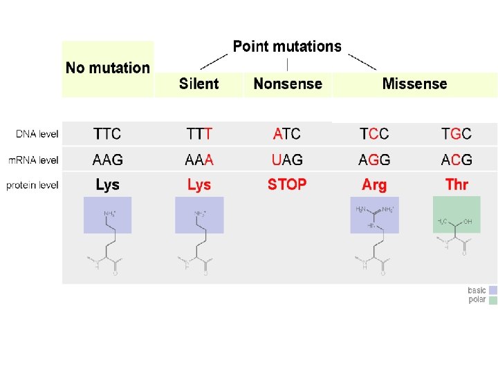
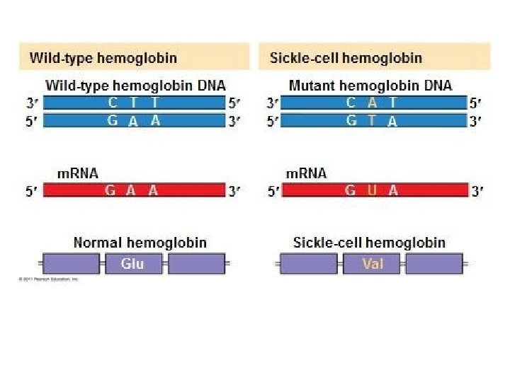
Mutations Can Change the Meaning of Genes § Mutations within a gene can be divided into two general categories. 1. Base substitutions involve the replacement of one nucleotide with another. Base substitutions may – Produce a silent mutation where the mutation has no effect on the polypeptide because the right amino acid is still added – Produce a missense mutation where the mutation causes the wrong amino acid to be added to the polypeptide – Produce a nonsense mutation where the mutation produces a stop codon in the m. RNA instead of an amino acid.
Mutations Can Change the Meaning of Genes 2. Mutations can result in deletions or insertions that may – Alter the reading frame (triplet grouping) of the m. RNA, so that nucleotides are grouped into different codons= frame shift mutation. – This leads to significant changes in amino acid sequence downstream of the mutation, and produces a nonfunctional polypeptide.