The Structure and Function of Macromolecules Overview The


 HO 1 2 3")


 (C")

 Dehydration reaction")


")


 A ribbon")


 � Stores DNA information for the synthesis of specific proteins –")


- Slides: 63
- The Structure and Function of Macromolecules � Overview: The Molecules of Life � Another level in the hierarchy of biological organization is reached when small organic molecules are joined together � Macromolecules � Are large molecules composed of smaller molecules � Are complex in their structures � Include proteins, carboydrates, lipids, and nucleic acids like DNA
Most macromolecules are polymers, built from monomers Three of the classes of life’s organic molecules are polymers � Carbohydrates � Proteins � Nucleic � A acids polymer � Is a long molecule consisting of many similar building blocks called monomers
The Synthesis and Breakdown of Polymers � Monomers form larger molecules by condensation reactions also called dehydration reactions Short polymer Unlinked monomer HO 1 2 3 OH H HO Dehydration removes a water molecule, forming a new bond H 2 O HO 1 2 3 4 H Longer polymer (a) Dehydration reaction in the synthesis of a polymer
� Polymers can disassemble by � Hydrolysis (also called digestion) HO 1 2 3 4 Hydrolysis adds a water molecule, breaking a bond H H 2 O HO (b) Hydrolysis of a polymer 1 2 3 OH HO H
The Diversity of Polymers � Each class of polymer � Is � � � formed from a specific set of monomers All living organisms are composed of the same types of polymers made up of the same monomer types – proteins, carbohydrates and nucleic acids. However, each organism is composed of many unique polymers (unique proteins, carbohydrates and nucleic acids) based on the arrangement of monomers An immense variety of polymers can be built from a small set of monomers
Carbohydrates serve as fuel and building material � Carbohydrates � Include � both simple sugars and their polymers Monosaccharides (simple sugars) � Are the simplest sugars � Can be used for fuel - glucose � Can be converted into other organic molecules Nucleotides include a 5 carbon sugar, ribose or deoxyribose � Can be combined into polymers
Examples of monosaccharides Triose sugars Pentose sugars (C 3 H 6 O 3) (C 5 H 10 O 5) H O H Aldoses C O Hexose sugars (C 6 H 12 O 6) H C H O C C OH H C OH HO C H C OH H H Ribose H C OH H HO C H C OH HO C H H C OH H H Glucose H H H C OH C O Galactose H C OH C O O C OH HO H H C OH Dihydroxyacetone H C OH H Ribulose O C H Glyceraldehyde Ketoses � C H H Fructose
� Monosaccharides Notice the carbons are numbered and this numbering system remains when they form a ring in water. � May be linear � Can form rings H C O 1 H 2 C OH HO 3 C H H 4 C 5 OH C 6 C OH OH 6 CH OH 2 5 C H H OH 4 C OH 3 C H 6 CH OH 2 O H H H 2 C OH H 4 C 1 C O OH 5 C H OH 3 C H CH 2 OH O H H 6 H 1 C 2 C OH OH 4 HO O 5 H OH 3 H H 1 OH 2 OH H (a) Linear and ring forms. Chemical equilibrium between the linear and ring structures greatly favors the formation of rings. To form the glucose ring, carbon 1 bonds to the oxygen attached to carbon 5.
Disaccharides Consist of two monosaccharides Are joined by a glycosidic linkage (a) Dehydration reaction in the synthesis of maltose. The bonding of two glucose units forms maltose. The glycosidic link joins the number 1 carbon of one glucose to the number 4 carbon of the second glucose. Joining the glucose monomers in a different way would result in a different disaccharide. CH 2 OH H HO O H OH HO O H OH H CH 2 OH H H OHOH HO O H OH H H 1– 4 glycosidic 1 linkage H 4 O H H OH O H OH CH 2 OH H OH OH H 2 O Glucose CH 2 OH H (b) Dehydration reaction in the synthesis of HO sucrose. Sucrose is a disaccharide formed from glucose and fructose. Notice that fructose, though a hexose like glucose, forms a five-sided ring. O H OH H H CH 2 OH HO CH 2 OH O H OH OH Maltose H H HO CH 2 OH HO H H O H 1– 2 glycosidic 1 linkage H O CH 2 OH O 2 H H HO CH 2 OH OH H 2 O Glucose Fructose In living systems, these reactions are always done by enzymes. Cellular enzymes are controlled Sucrose Notice that the chemical reactions take place at the functional groups
Polysaccharides � Are polymers of sugars � Serve many roles in organisms Storage �Starch is a polymer of glucose only �Glycogen is also a polymer of glucose Cell wall - structure �Cellulose is a polymer of glucose �Chitin
Starch Is a polymer consisting entirely of glucose monomers Chloroplast Starch � Is the major storage form of glucose in plants 1 m Amylose Amylopectin (a) Starch: a plant polysaccharide
� Glycogen � Consists of glucose monomers � Is the major storage form of glucose in animals Mitochondria Glycogen granules 0. 5 m Glycogen (b) Glycogen: an animal polysaccharide
Variety from monomers and the covalent bond type H H 4 CH 2 O H OH H Which type of bond H OH HO H OH glucose depends on the enzyme which is controlled by the cell. O CH 2 O H H O OH H 4 1 OH H HO H C OH HO C H H C OH H OH glucose (a) and glucose ring structures CH 2 O H O HO 4 1 OH OH OH CH 2 O H O O 4 1 OH OH (b) Starch: 1– 4 linkage of glucose monomers CH 2 O H O HO OH CH 2 O H O OH 1 O 4 OH O O CH 2 O OH OH H H (c) Cellulose: 1– 4 linkage of glucose monomers OH
� Is Cellulose a major component of the tough walls that enclose plant cells Cell walls Cellulose microfibrils in a plant cell wall About 80 cellulose molecules associate to form a microfibril, the main architectural unit Microfibrilof the plant cell wall. 0. 5 m Plant cells Parallel cellulose molecules are held together by hydrogen bonds between hydroxyl groups attached to carbon atoms 3 and 6. Figure 5. 8 OH CH 2 OH O O OH OH O O O CH OH OH CH 2 OH 2 H CH 2 OH OH OH CH 2 OH O O OH OH OH O O O O CH OH OH CH 2 OH 2 H Glucose monomer Cellulose molecules A cellulose molecule is an unbranched glucose polymer.
� Cellulose is difficult to digest � Cows have microbes in their stomachs to facilitate this process What do these microbes have that will allow them to break down cellulose?
� Chitin, another important structural polysaccharide � Is found in the exoskeleton of arthropods � Can be used as surgical thread CH 2 O H O OH H H NH C O CH 3 (a) The structure of the (b) Chitin forms the exoskeleton of arthropods. This cicada chitin monomer. is molting, shedding its old exoskeleton and emerging in adult form. (c) Chitin is used to make a strong and flexible surgical thread that decomposes after the wound or incision heals.
Lipids are a diverse group of hydrophobic molecules � Lipids � Are the one class of large biological molecules that do not consist of polymers � Share the common trait of being hydrophobic � Include Fats Phospholipids steroids
The synthesis and structure of a fat, or triglycerol H H C OH HO H C OH C H H H C C H H H C H H C H Fatty acid (palmitic acid) Glycerol (a) Dehydration reaction in the synthesis of a fat Ester linkage O H C O C H C H O C H H C H H C H H C H H C H (b) Fat molecule (triacylglycerol) H C H H C H H C H H C H H C H H Again, notice where the chemical reaction takes place. H H C H H C H H C H H
Fatty acids Vary in the length and number and locations of double bonds they contain Saturated fatty acids � Have the maximum number of hydrogen atoms possible (saturated with hydrogen) � Have no double bonds Stearic acid Oleic acid (b) Unsaturated fat and fatty acid (a) Saturated fat and fatty acid • Unsaturated fatty acids --Have one or more double bonds cis double bond causes bending
A single bond allows rotation, is longer and not a strong as a double bond A double bond is stronger, shorter, and more rigid. Bonds help to determine the 3 -D shape of a molecule.
Phospholipids have only two fatty acids plus a phosphate group instead of a third fatty acid Hydrophilic head Consists of a hydrophilic “head” and hydrophobic “tails” CH 2 O O P O– + N(CH ) 3 3 Choline Phosphate O CH 2 CH O O C CH 2 Glycerol O Hydrophobic tails � (a) Structural formula Fatty acids Hydrophilic head Hydrophobic tails (b) Space-filling model (c) Phospholipid symbol
� The structure of phospholipids � Results in a bilayer arrangement found in cell membranes WATER Hydrophilic head WATER Hydrophobic tail
Steroids - Are lipids characterized by a carbon skeleton consisting of four fused rings � One steroid, cholesterol � Is found in cell membranes � Is a precursor for some hormones H 3 C When written as a ring, all points are carbon unless written in otherwise. HO CH 3 Is this molecule polar or nonpolar?
Cholesterol fills in the spaces left by the kinks; stiffens the bilayer and makes it less fluid and less permeable. Do concept check 5. 3 How do you think bacteria, which do not use cholesterol, adjust the fluidity of their cell membrane?
Both saturated and trans fats correlate with heart problems and high levels or blood cholesterol. Atherosclerosis Animal fats found in meat, butter, and cream are usually saturated, and solid at room temperature. Plant oils like corn oil contain more unsaturated fatty acids. Peanut and olive oil contain monounsaturated fatty acids
Proteins have many structures, resulting in a wide range of functions
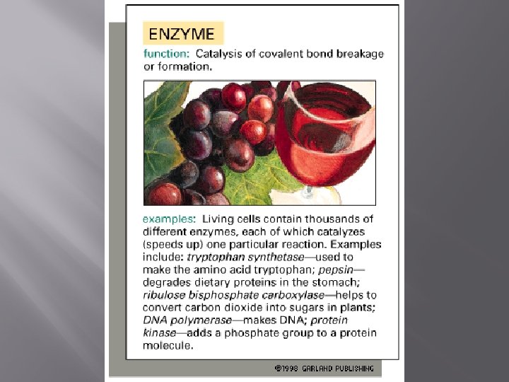
� Enzymes � Are often a type of protein that acts as a catalyst, speeding up chemical reactions 1 Active site is available for a molecule of substrate, the reactant on which the enzyme acts. Is this part of the protein polar or nonpolar? Substrate (sucrose) 2 Substrate binds to enzyme. Glucose OH Enzyme (sucrase) H 2 O Fructose Enzyme remains unchanged, ready to work again. H O 4 Products are released. 3 Substrate is converted to products.
Polypeptides � Are � polymers of amino acids A protein � Can consist of only one large polypeptide consists of more than one polypeptides (subunits) bound together by non-covalent interactions Hemoglobin � Some very small polypeptides are referred to as peptides � Amino acids � Are organic molecules possessing both carboxyl and amino groups � Differ in their properties due to differing side chains, called R groups
Proteins are composed of amino acid building blocks and are diverse in structure (shape) and function. Amino acids have an amino group and an acid group bound to a central carbon. Amino group Acid group This central carbon forms 4 single bonds. One with the amino group, one with the carboxylic acid, one with hydrogen, and the last with a variety of different chemical groups (R group).
� 20 different amino acids make up proteins CH 3 H H 3 N+ C CH 3 O H 3 N+ C H Glycine (Gly) O– C H 3 N+ C H Alanine (Ala) O– CH CH 3 O C CH 2 O H 3 N+ C H Valine (Val) CH 3 O– C O C H Leucine (Leu) H 3 C H 3 N+ O– CH C O C H Isoleucine (Ile) O– Nonpolar CH 3 CH 2 S NH CH 2 H 3 N+ C H CH 2 O H 3 N+ C O– Methionine (Met) C H CH 2 O C H 3 N+ O– Phenylalanine (Phe) C H O H 2 C CH 2 H 2 N C O C H O– C O– Tryptophan (Trp) Proline (Pro) Know the structure of an amino acid, not all the R groups.
OH OH Polar CH 2 H 3 N+ C CH O H 3 N+ C O– H Serine (Ser) C CH 2 O H 3 N+ C O– H C CH 2 O C H O– C H 3 N+ O H 3 N+ C O– H C Electrically charged CH 2 H 3 N+ C O H H 3 N+ NH 3+ O C C O– H Glutamine (Gln) NH 2 C CH 2 CH 2 C O CH 2 C H O– H 3 N+ C H Aspartic acid (Asp) C H 3 N+ – O C CH 2 C O– CH 2 Basic O– O O Asparagine (Asn) Acidic –O CH 2 H Tyrosine (Tyr) Cysteine (Cys) Threonine (Thr) C NH 2 O C SH CH 3 OH NH 2 O Glutamic acid (Glu) C Lysine (Lys) NH 2+ H 3 N+ CH 2 O O– NH+ CH 2 H 3 N+ C H NH CH 2 O C C O– H O C O– Arginine (Arg) Histidine (His) Know both the name and abbreviation of all amino acids along with their chemical nature – polar, nonpolar, charged, acidic, . . .
the amino group on one amino acid and the carboxyl group on another amino acid. The formation of a peptide bonds in a tetrapeptide
� Amino acids � Are linked by peptide bonds between the amino group of one amino acid and the acid group of the other amino acid Peptide bond OH OH CH 2 H H N The chemical reaction again takes place at the functional groups! 2 H C C N C C OH H H O (a) 2 OH DESMOSOMES SH OH Peptide CH 2 bond CH 2 H H N C C H O (b) Each peptide CH bond is in a H plane. This N C C OH H O DESMOSOMES contributes to HO the shape of the protein. SH Amino end (N-terminus) H H N C C H O N C C OH H O Carboxyl end (C-terminus) Side chains Backbone
Protein Conformation and Function � Two models of protein conformation Groove (a) A ribbon model A protein’s specific conformation (shape and chemical nature) Groove determines how it functions. (b) A space-filling model
Four Levels of Protein Structure � Primary structure � Is the unique sequence of amino acids in a polypeptide HN + Covalent bonds Peptide backbone imposes some restrictions on the folding of a protein. Why? Gly Pro. Thr Gly 3 Amino end Amino acid subunits Glu Cys. Lys. Seu Leu. Pro Met Val Lys Val Leu Asp Ala. Val Arg Gly Ser Pro Ala Glu Lle Asp Thr Lys Ser Lys Trp Tyr Leu Ala Gly lle Ser Pro. Phe. His Glu Ala Thr Phe. Val Asn His Ala Glu Val Asp Tyr Arg Ser Arg Gly Pro Thr Ser Tyr Thr lle Ala Leu Ser Pro Ser. Tyr Thr Ala Val Lys. Glu Thr Asn. Pro o c – o Carboxyl end
Secondary structure � � Is the folding or coiling of the polypeptide into a repeating configuration � Includes the helix and the pleated sheet Amino acid subunits O H H C C N H R R O H H C C N O H H R helix C H R O C N H N H O C H C R N H O C O C H C O N H N C C H R R H N C C N OH H R O C C H R O H H C C N OH H R O C H H H C N HC C C N HC N N H H C O C C O R R O H H C C N R R O C H H NH C N C H O C R R C C O R H C N H O C H All based on hydrogen bonds between the peptide bonds of different amino acids
� Tertiary structure � Is the overall three-dimensional shape of a polypeptide after it “folds” into a stable form. � Results from interactions between amino acids and R groups What are these? Hydrogen bond CH 2 O H 3 C CH CH 3 H 3 C CH 3 CH Hydrophobic interactions and van der Waals interactions Polypeptide backbone HO C CH 2 S S CH 2 Disulfide bridge O CH 2 NH 3+ -O C CH 2 Ionic bond
The distribution of polar and nonpolar amino acids is important in how a protein folds. The nonpolar side chains tend to cluster in the interior of a molecule, avoiding contact with water, while the polar side chains arrange themselves near the outside.
Hydrophobic areas also tend to be found spanning the lipid bilayer of membranes like the plasma membrane. Transmembrane proteins often cross the membrane in an alpha helix because the peptide bond itself is hydrophic unless all partial charges are equalized in an alpha helix or beta sheet.
Quaternary structure � Is the overall protein structure that results from the aggregation of two or more polypeptide subunits
Hemoglobin contains two alpha globin subunits and two beta globin subunits. Heme is the site where oxygen is carried There are many large multisubunit proteins in cells.
Larger protein molecules may contain more than one polypeptide chain or subunit. The region that interacts with another molecule through noncovalent bonds is the binding site.
Sickle-cell disease results from a single amino acid substitution in the protein hemoglobin Normal β hemoglobin Sickle-cell β Primary Val hemoglobin Val His Leu Thr Pro Glul Glu. . . His Leu Thr Pro Val Glu. . . structure 1 2 3 4 5 6 7 Primary structure Secondary and tertiary structures Secondary subunit and tertiary structures Quaternary Hemoglobin A structure Molecules do not associate with one another, each carries oxygen. Function Red blood cell shape Figure 5. 21 Normal cells are full of individual hemoglobin molecules, each carrying oxygen Quaternary structure subunit Function 10 m Red blood cell shape Exposed hydrophobic region Hemoglobin S Molecules interact with one another to crystallize into a fiber, capacity to carry oxygen is greatly reduced. Fibers of abnormal hemoglobin deform cell into sickle shape. The sickle-cell hemoglobin does not fold into the proper shape because the amino acid sequence (Primary structure) is incorrect.
� 20 different amino acids make up proteins CH 3 H H 3 N+ C CH 3 O H 3 N+ C H Glycine (Gly) O– C H 3 N+ C H Alanine (Ala) O– CH CH 3 O C CH 2 O H 3 N+ C H Valine (Val) CH 3 O– C O C H Leucine (Leu) H 3 C H 3 N+ O– CH C O C H Isoleucine (Ile) O– Nonpolar CH 3 CH 2 S NH CH 2 H 3 N+ C H CH 2 O H 3 N+ C O– Methionine (Met) C H CH 2 O C H 3 N+ O– Phenylalanine (Phe) C H O H 2 C CH 2 H 2 N C O C H O– C O– Tryptophan (Trp) Proline (Pro) Know the structure of an amino acid, not all the R groups.
OH OH Polar CH 2 H 3 N+ C CH O H 3 N+ C O– H Serine (Ser) C CH 2 O H 3 N+ C O– H Threonine (Thr) C CH 2 O C H O– C H 3 N+ O H 3 N+ C O– H Tyrosine (Tyr) Cysteine (Cys) C Electrically charged CH 2 H 3 N+ C O H C H 3 N+ – O C C O– H Glutamine (Gln) H 3 N+ NH 2 C CH 2 CH 2 C O CH 2 C H O– H 3 N+ C H Aspartic acid (Asp) CH 2 Asparagine (Asn) NH 3+ O CH 2 C O– O Basic O– O CH 2 H Acidic –O C NH 2 O C SH CH 3 OH NH 2 O Glutamic acid (Glu) C Lysine (Lys) NH 2+ H 3 N+ CH 2 O O– NH+ CH 2 H 3 N+ C H NH CH 2 O C C O– H O C O– Arginine (Arg) Histidine (His) Know both the name and abbreviation of all amino acids along with their chemical nature – polar, nonpolar, charged, acidic, . . .
Protein conformation � � Depends on � the sequence of amino acid side chains (with R groups) and � the physical and chemical conditions of the protein’s environment Denaturation is when a protein unravels and loses its native conformation Increased temperature What kinds of bonds are broken here? Denaturation Change in p. H Organic solvent (hydrophobic) Denatured protein Normal protein Renaturation What kinds of bonds are not broken here?
The Protein-Folding Problem � Most proteins � Probably go through several intermediate states on their way to a stable conformation. � Many proteins are being made in the cell all of the time. How do the fold correctly, how do they interact with their subunits correctly?
� Chaperonins � Are protein molecules that assist in the proper folding of other proteins Polypeptide Cap Correctly folded protein Hollow cylinder Chaperonin (fully assembled) 2 The cap attaches, causing 3 The cap comes Steps of Chaperonin the cylinder to change shape in off, and the properly Action: such a way that it creates a folded protein is 1 An unfolded polyhydrophilic environment for the released. peptide enters the cylinder from one end. folding of the polypeptide.
� X-ray crystallography � Is used to determine a protein’s three-dimensional structure X-ray diffraction pattern Photographic film Diffracted X-rays X-ray beam source Do concept check 5. 4 Figure 5. 24 Crystal Nucleic acid Protein (a) X-ray diffraction pattern (b) 3 D computer model
Nucleic acids store and transmit hereditary information � Genes � Are the units of inheritance Code for the amino acid sequence of polypeptides � Are � made of nucleic acids There are two types of nucleic acids � Deoxyribonucleic acid (DNA) � Ribonucleic acid (RNA)
Deoxyribonucleic acid (DNA) � Stores DNA information for the synthesis of specific proteins – DNA is the “ genetic material” inherited from parents 1 � Directs RNA synthesis Synthesis of m. RNA in the nucleus m. RNA � Directs protein synthesis NUCLEUS indirectly through messenger RNA CYTOPLASM 2 Movement of m. RNA into cytoplasm via nuclear pore m. RNA Ribosome 3 Synthesis of protein Polypeptide Amino acids
The Structure of Nucleic Acids � 5’ end 5’C Nucleic acids � Exist as polymers called polynucleotides �Each polynucleotide � Consists of monomers called nucleotides O 3’C O Nucleoside Nitrogenous base O O 5’C O P O CH 2 O 3’C OH O 5’C 3’ end (a) Polynucleotide, or nucleic acid O Phosphate group (b) Nucleotide 3’C Pentose sugar
Nucleotide Monomers Are made up of nucleosides and phosphate groups pyrimidines � Nitrogenous bases Pyrimidines NH 2 O O C C CH 3 C N CH C CH HN HN CH C C CH N N O O H H H Cytosine Thymine (in DNA) Uracil (in RNA) Uracil (in U C U T Nucleoside Purines Nitrogenous base O O P O O NH 2 C N CC NH N HC HC C CH N C N NH 2 N N H H Adenine Guanine A G 5’C CH 2 O O Phosphate group 3’C 5” Pentose sugars HOCH 2 O OH 4’ H H 1’ 5” HOCH 2 O OH 4’ H H 1’ H H H 3’ 2’ OH H OH OH Deoxyribose (in DNA) Ribose (in RNA) H (b) Nucleotide Figure 5. 26 (c) Nucleoside components
Nucleotide polymers are made up of nucleotides linked by the–OH group on the 3´ carbon of one nucleotide and the phosphate on the 5´ carbon on the next So they “grow” at the 3’ end.
� The sequence of bases along a nucleotide polymer � Is unique for each gene
The DNA Double Helix Anti-parallel complementary
Complementary base pairs Do concept check 5. 5
DNA and Proteins as Tape Measures of Evolution � Molecular comparisons � Help biologists sort out the evolutionary connections among species Ribosomal RNA gene sequence is conserved. Look for differences.