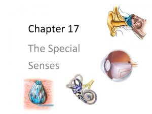
THE SPECIAL SENSES THE EYES AND EARS Chapter


- Slides: 51

THE SPECIAL SENSES THE EYES AND EARS Chapter 11

Function of the Eyes ◦ Receptor organs of sight ◦ Receive images and transmits them to the brain ◦ Oculus means eye Plural is oculi ◦ OD = Right eye (oculus dexter) ◦ OS = Left eye (Oculus sinister) ◦ OU = Both eyes or each eye ◦ *** Joint commission on accreditation of healthcare organizations (JCAHO) recommends NOT using the abbreviations

Structures of the eyes ◦ Adnexa – accessory or adjoining anatomical parts of an organ ◦ Adnexa of the eye structures outside the eyeball ◦ Orbit = eye socket – protects the eye ◦ 6 major eye muscles ◦ Work together to create binocular vision ◦ Allows depth perception (3 D vision)

More structures of the eye ◦ Canthus: angle where upper and lower eyelids meet ◦ Conjunctiva (kon-junk-TYE-vah): transparent mucous membrane on underside of the eyelids and over the eyelids (protective)

Lacrimal Apparatus ◦ AKA tear apparatus – structures that produce, store and remove tears ◦ Lacrimal glands – secrete tears ◦ Underside of the upper eyelid ◦ Lacrimal canal: duct at the corner of the eye. They collect tears and empty them into lacrimal sacs. Crying is an overflow ◦ Lacrimal duct: drains tears to the nose

Walls of the eye ◦ Sclera: Outer layer ◦ white of the eye ◦ Tough outer layer (except cornea) ◦ Choroid (KOH-roid): opaque middle layer ◦ Many blood vessels ◦ Retina: innermost layer ◦ Receives nerve impulses and transmits to the brain via the optic nerve

Eyeball ◦ Ocular: pertaining to the eye ◦ Extraocular: outside the eye ◦ Intraocular: within the eyeball ◦ Divided into 2 segments ◦ Anterior segment: front 1/3 of eyeball ◦ Filled with watery aqueous humor (fluid) ◦ Posterior segment: back 2/3 of eyeball ◦ Lined with retina ◦ Filled with jelly-like vitreous humor (gel)

Retina ◦ Sends images to the brain via the optic nerve ◦ Rods: black and white receptors ◦ Cones: color receptors ◦ Macula: light-sensitive area in the center of the retina responsible for sharp central vision ◦ Fovea centralis: pit in the middle of macula. Good color vision (many cones no rods) ◦ Optic disk: AKA blind spot – where retinal nerves enter optic nerve. No rods or cones

Uvea ◦ Pigmented layer of eye ◦ Ciliary body: Within the choroid ◦ Contains muscles/ligaments that allow the lens to focus near and far ◦ Produces aqueous humor ◦ Iris: colorful – surrounds the pupil ◦ Muscles dilate or constrict the pupil which ↑ or ↓ the amount of light that enters the eye.

More eye structures ◦ Cornea: transparent ◦ Covers iris and pupil ◦ Primary structure for focusing ◦ Pupil: black circular opening ◦ Allows light to enter the eye ◦ Lens: clear flexible curved ◦ Behind iris and pupil ◦ Focuses images on the retina

Normal action of the eyes ◦ Accommodation: process whereby eyes make adjustments for seeing at varying distances. ◦ Change in pupil size, movement of eyes, change in shape of lens ◦ Convergence: Simultaneous inward movement of the eyes toward each other. ◦ Singular binocular vision as an object comes closer

Normal vision ◦ Refraction: ability of the lens to bend light rays so they focus on the retina. ◦ Emmetropia: normal relationship between refractive power of the eye and the shape of the eye.

Medical specialties ◦ Ophthalmologist: physician (MD) Dx and Tx of the eye ◦ Vision correction and surgery ◦ Optometrist: Doctor of optometry ◦ Dx of eye diseases and conditions ◦ Vision (glasses? ? ? ) ◦ Optician: Designs, fits and dispenses lenses for vision correction Dr. Daniel Neely at Riley Pediatric ophthalmologist

Eyelid pathology ◦ Ptosis: drooping of upper eyelid often d/t paralysis ◦ Periorbital edema: swelling of tissues around the eye/s ◦ Chalazion: (kah-LAY-zee-on) nodule or cyst on upper eyelid ◦ d/t Clogged sebaceous gland ◦ Hordeolum: AKA stye ◦ Puss filled painful lesion. ◦ Usually d/t infected sebaceous gland

More eyelid pathology ◦ Entropion: Inversion of and eyelid ◦ Usually lower eyelid ◦ Eyelashes rub on cornea ◦ Ectropion: eversion (turning out) of the edge of an eyelid ◦ Usually affects lower lid ◦ Irritates eye and prevents tears from draining well.

Adnexa Pathology ◦ Conjunctivitis: AKA pinkeye – inflammation of the conjunctiva ◦ Allergy or infection (bacterial or viral) ◦ Dacryoadenitis: inflammation of the lacrimal gland ◦ Bacterial, viral or fungal

More pathology ◦ Subconjunctival hemorrhage (sub-kon-junk-TIH-val): bleeding between the conjunctiva and the sclera ◦ Usually d/t injury ◦ Xerophthalmia: AKA dry eye – dry eye surfaces including the conjunctiva

Uvea, cornea, iris and sclera pathology ◦ Uveitis: inflammation of the uvea (choroid, ciliary body and iris). Can lead to blindness ◦ Iritis: (eye-RYE-tis)most common form of uveitis affecting the structures in the front of the eye (like the iris) ◦ Keratitis: inflammation of the cornea (infection) ◦ Scleritis: inflammation of the sclera (infections, chemical injuries or autoimmune disease)

Eye pathology ◦ Anisocoria (an-ih-so-KOH-ree-ah): unequal pupil size ◦ Congenital or d/t head injury, aneurysm or other pathology ◦ Cataract: Progressive loss of lens transparency and visual clarity ◦ Retinitis pigmentosa: progressive degeneration of the retina ◦ Affects night and peripheral vision ◦ Detected by dark pigmented spots on retina.

More eye pathology ◦ Miosis: contraction of pupil ◦ drugs ◦ Mydriasis (mih-DRY-ah-sis): dilation of pupil ◦ drugs, diseases, or trauma ◦ Nystagmus: involuntary constant rhythmic movement of the eyeball ◦ Congenital or d/t neurological injury or drugs ◦ Papilledema: Swelling and inflammation of the optic nerve where it enters the eye (at the optic disk) ◦ D/t ↑ intracranial pressure or a tumor

Glaucoma and Macular Degeneration ◦ Glaucoma: Increased intraocular pressure d/t blocked flow of fluid out of eye ◦ Damages retinal and optic nerves ◦ Can cause loss of peripheral vision or blindness ◦ Macular Degeneration: Progressive condition ◦ Macula damaged ◦ Loss of central vision, but not total blindness

Types of Glaucoma (Links to videos)

Open-angle glaucoma ◦ AKA chronic glaucoma ◦ Most common ◦ Trabecular meshwork blocked ◦ Pressure increases ◦ Can be detected early with regular eye exams (tonometry and visual field testing) ◦ Treatment: Laser Trabeculoplasty ◦ Creates openings in the meshwork to allow fluid to drain properly

Closed-angle glaucoma ◦ Opening between the cornea and iris narrows ◦ Fluid cannot reach the trabecular meshwork (the drain) ◦ Sudden increase in pressure ◦ Sx: Severe pain, nausea, red eye, blurred vision ◦ Blindness in as little as 2 days ◦ Treatment: Iridectomy (ir-ih-DECK-tohmee): surgical removal of a portion of the tissue of the iris ◦ laser iridotomy (small hole created) is used more often ◦ Fewer side effects

Functional Defects ◦ Diplopia: Double vision ◦ Hemianopia: blindness in ½ of visual field ◦ Nyctalopia (nick-tah-LOH-pee -ah): difficulty seeing at night (AKA night blindness)

More functional defects ◦ Photophobia: excessive sensitivity to light ◦ Can be d/t migraines, too long in contacts, drug use or inflammation ◦ Presbyopia: common changes w/ aging ◦ Decreased near vision ◦ Lens less flexible, muscles weaker

Strabismus ◦ Is when the eyes point in different directions ◦ Eye muscles are unable to focus together ◦ Esotropia: AKA crosseyes ◦ Eyes deviated inward ◦ Exotropia: Aka walleye ◦ Eyes deviated outward

Refractive Disorders ◦ Are focusing problems ◦ Lens and cornea do not bend light so that it focuses properly on the retina ◦ Ametropia: Any error where images are not focused properly on the retina

Examples of Ametropia ◦ Astigmatism ◦ Poor focusing d/t uneven curvatures of the cornea ◦ Hyperopia: AKA farsightedness ◦ Light focuses beyond the retina ◦ Childhood and 40+ years ◦ Myopia: AKA Nearsightedness ◦ Light focuses in front of the retina ◦ Around puberty

Blindness ◦ Inability to see, although some sight can remain ◦ 20/200 or less = legally blind ◦ Amblyopia: diminished vision or partial loss of sight ◦ Often in 1 eye ◦ No detectable disease of the eye ◦ Scotoma: AKA blind spot

Diagnostics for vision ◦ Snellen chart: measure of visual acuity ◦ 1 st number – distance from chart (20 feet) ◦ 2 nd number – deviation from normal ◦ Visual field testing: AKA perimetry ◦ Looks for loss of peripheral (side) vision ◦ Can indicate glaucoma or optic nerve disorder

Diagnostics for eyes ◦ Ophthalmoscopy (of-thal-mos-kuh-pee) AKA funduscopy – use of an ophthalmoscope to visually examine the fundus (back part) of the eye. ◦ Tonometry: measurement of intraocular pressure ◦ High can indicate glaucoma ◦ Several ways to test: air puff or instrument right on cornea

More diagnostics for the eye ◦ Fluorescein staining (floo-res-ee-in): using fluorescent dye on the surface of the eye. ◦ Drops or a strip applicator ◦ Makes a corneal abrasion appear bright green ◦ Fluorescein angiography: study of the blood vessels in the retina of the eye. ◦ IV injection of fluorescein dye as a contrast medium

PERRLA ◦ Pupils ◦ Equal ◦ Round ◦ Responsive (Reactive) ◦ Light and ◦ Direct and consensual ◦ Accommodation ◦ Pupils constrict when object brought closer Performing the Exam When performing a pupillary exam, it sometimes helps to illuminate pupils indirectly from the side, so you can actually see what is happening. 1. Observe the pupil size and shape at rest, looking for anisocoria (one pupil larger than the other) 2. Observe the direct response (constriction of the illuminated pupil) 3. Observe the consensual response (constriction of the opposite pupil) 4. Repeat with the opposite pupil 5. Check for accommodation (constriction of pupil when viewing a close object)

Treatments ◦ Tarsorrhaphy: suturing the eyelids together. ◦ Performed to protect eye if pt. cannot close eye normally (paralysis) ◦ Enucleation: Removal of the eyeball, but leaves the eye muscles intact

Treatments for retinal detachment ◦ * when the retina separates from the choroid ◦ Retinopexy: surgery to reattach the retina ◦ Vitrectomy (vih-TRECK-tohmee): removal/replacement of vitreous humor ◦ Treat retinal detachment or when vitreous humor is bloody or cloudy (diabetic retinopathy)

Vision Treatments ◦ Radial Keratotomy ◦ Treats myopia ◦ Incisions made in cornea ◦ Flattens ◦ Brings focal point of the eye closer to the retina to improve distance vision ◦ LASIK (Laser Assisted in Situ Keratomileusis (ker·a·to·mi·leu·sis)) “carving the cornea using a laser” ◦ Corneal flap created, then the shape changed using a laser

Function of the Ears ◦ Receptors for hearing ◦ Receives sound impulses ◦ Transmits them to the brain ◦ Inner ear maintains balance ◦ Auditory: pertaining to hearing ◦ Acoustic: pertaining to sound/hearing

Structures of the ears: Outer ear ◦ Pinna: Aka auricle – outside part of the ear ◦ Captures sounds ◦ External auditory canal: transmits sound waves to the tympanic membrane (eardrum) of the middle ear ◦ Cerumen: AKA earwax ◦ Secreted by glands in the auditory canal. ◦ Protective – traps insects, dust and debris and bacteria

The Middle Ear ◦ Transmits sound from outer ear to inner ear ◦ Tympanic membrane: AKA eardrum ◦ Sound waves are received by the eardrum and transmitted via vibrations ◦ Mastoid process: Temporal bone with hollow air space ◦ Surrounds the middle ear

More middle ear ◦ Bone conduction: when eardrum vibrates and causes the tiny bones of the middle ear to vibrate which transmits the sound on to the inner ear. ◦ Auditory Ossicles: 3 tiny bones of the middle ear ◦ Malleus: AKA the hammer ◦ Incus: AKA anvil ◦ Stapes: AKA stirrup ◦ Eustachian Tubes: Tubes that lead from the middle ear to the nose/throat ◦ Equalize air pressure in the middle ear with that of outside the ear

Inner Ear ◦ Sensory receptors for hearing and balance ◦ Labyrinth: All the structures of the inner ear ◦ Cochlea: snail-shaped structure where sound vibrations are converted into nerve impulses. ◦ Semicircular canals: contain liquid and sensitive hair-like cells that sense when the liquid has moved. (the liquid moves when your head moves) ◦ Acoustic nerves: transmits info to brain

The ear

Pathology: outer ear ◦ Otalgia: AKA earache ◦ Otomycosis: Fungal infection of external auditory canal ◦ *** Your book says this is AKA “swimmer’s ear”, but “swimmer’s ear” is usually bacterial ◦ Otorrhea: any discharge from the ear ◦ Otopyorrhea: flow of puss from the ear

Middle Ear Pathology ◦ Otitis Media: inflammation of the middle ear ◦ Acute OM: r/t URI (upper respiratory infection) common in young children ◦ Serous OM: fluid buildup in middle ear w/o symptoms or infection ◦ Can follow acute OM, or result from obstructed Eustachian tubes ◦ Infectious Myringitis: contagious inflammation of the eardrum (blisters) ◦ Related to otitis media ◦ Otosclerosis: Ankylosis (fusing)of the middle ear bones. ◦ Results in conductive hearing loss

Inner ear pathology ◦ Vertigo: sense of whirling, dizziness, and loss of balance. ◦ Can be accompanied by n/v ◦ Tinnitus: ringing in the ears. ◦ Associated with hearing loss; more common with prolonged exposure to loud noises ◦ Meniere’s disease: rare chronic disorder ◦ Amount of fluid in the inner ear increases intermittently ◦ Results in attacks of vertigo, fluctuating hearing loss and tinnitus

Hearing Loss ◦ Deafness: complete or partial loss of the ability to hear ◦ Presbycusis (pres-beh-KOO-sis): gradual loss of senorineural hearing that occurs as the body ages ◦ Conductive hearing loss: sound waves are prevented from passing from the air to the fluid-filled inner ear ◦ Earwax, infection, fluid in middle ear, punctured eardrum, otosclerosis, scaring ◦ Can often be treated ◦ Sensorineural hearing loss: AKA nerve deafness – auditory nerve or hair cells in the inner ear are damaged. ◦ Usually d/t age, noise exposure or a tumor near the cranial nerve

Diagnostic procedures ◦ Audiometry: use of an audiometer to measure hearing acuity ◦ Audiometer: electronic device that produces acoustic stimuli at a set frequency and intensity ◦ Monaural vs binaural testing: testing one or 2 ears ◦ Tympanometry: Use of air pressure in the ear canal to test for disorders of the middle ear. ◦ Middle ear fluid, Eustachian tube obstruction or evaluation of conductive hearing loss

◦ Mastoidectomy: surgical Treatments: Middle Ear removal of the mastoid cells. ◦ Mastoiditis not controlled by antibiotics, or in preparation for a cochlear implant ◦ Myringotomy: small surgical incision into the eardrum to relieve pressure from excess pus or fluid, or to allow placement of ear tubes ◦ Ear tubes: tiny ventilating tubes that provide ongoing drainage and relieve pressure after ear infections

More middle ear treatments ◦ Stapedectomy: surgical removal of the top portion of the stapes bone and the insertion of a prosthetic device (piston) ◦ Tympanoplasty: surgical correction of a damaged middle ear

Inner ear treatments LINK ◦ Labyrinthectomy: surgical removal of all or a portion of the labyrinth ◦ Alleviates uncontrolled vertigo, but causes complete hearing loss in that ear ◦ Cochlear implant: electronic device that bypasses the damaged portion of the ear and directly stimulates the auditory nerve
 Chapter 11 medical terminology learning exercises
 Special senses the eyes and ears
 What is the difference between somatic and special senses
 Messiners
 Thermoreceptors
 Anatomy and physiology chapter 8 special senses
 The general and special senses chapter 9
 Down syndrome ears
 Chapter 17 special senses answer key
 Cranucle
 Chapter 10 special senses
 Taste bud
 Chapter 15 special senses
 Let him who has ears
 Eyes that see and ears that hear
 Impaired eyes and ears
 Cranial nerve acronym
 The general senses
 Somatic and special senses
 Eyes are to spectacles as ears are to
 Hear us from heaven jared anderson
 Special senses physiology
 5 special senses
 Special senses physiology
 Special senses quiz
 Physiology
 Conclusion of special senses
 Building vocabulary activity: the special senses
 Building vocabulary activity: the special senses
 Houses the receptors for hearing
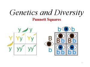
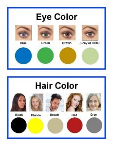
 Eye color punnett square
 What is the genotype of the man? ii i a i i b i i a i a
 Pf
Pf Punnett square for eye color
 Green eyes vs hazel
 A chapter on ears
 5 senses and 5 elements
 Facts about taste
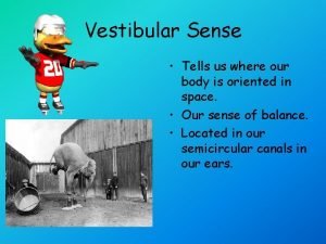
 Vestibular sense vs kinesthesis
 In your notebook identify
 Receptor cells for the vestibular sense are located in the
 7 senses of the body
 Epiglottis taste buds
 Super senses of cow
 Five senses at the beach
 Classification of sensory system
 Descriptive essay beach using five senses
 Memery
 Sense responsibility
 Is imagery language or structure
 Section 35-1 human body systems answer key
 Visual channel in hci
