
The Special Senses Objective Describe the structure of
The Special Senses
Objective • Describe the structure of vertebrate sensory organs and relate structure to function in vertebrate sensory systems.

The 5 Special Senses 1. 2. 3. 4. 5. Olfaction Gustation Vision Equilibrium Hearing
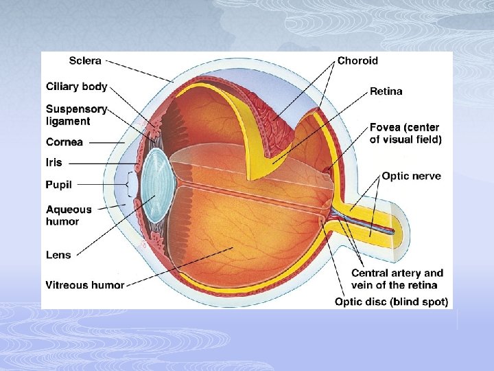
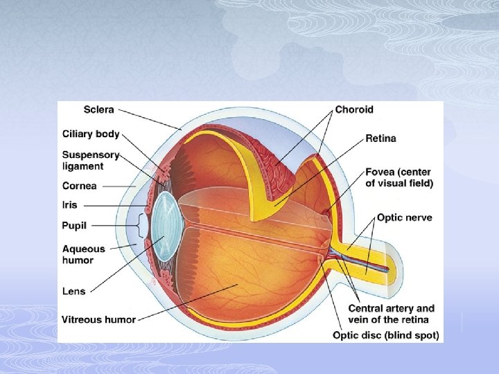
Vision - Eye Anatomy • Cornea – clear protective layer of eye (also refracts light) • no blood vessels: good for transplants • Iris – colored muscle of eye which protects retina by changing pupil size • Pupil – allows light into eye • Aqueous humor – watery fluid filled substance that gives the front of eye its shape • Lens – refracts light onto retina • Vitreous humor – jelly like fluid that gives eye it’s shape
Accessory Structures of the Eye • Protect the eye and aid eye function • • • Eyebrows Eyelids (palpebrae) Conjunctiva Lacrimal apparatus Extrinsic eye muscles
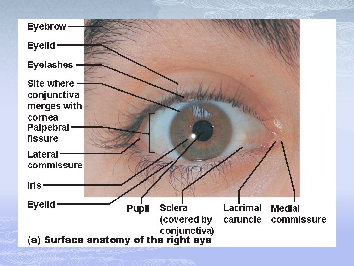
Eyebrow Eyelid Eyelashes Site where conjunctiva merges with cornea Palpebral fissure Lateral commissure Iris Eyelid Sclera Lacrimal (covered by caruncle conjunctiva) (a) Surface anatomy of the right eye Pupil Medial commissure
Eyebrows • Overlie the supraorbital margins • Function in • Shading the eye • Preventing perspiration from reaching the eye

Eyelids • Protect the eye anteriorly • Palpebral fissure—separates eyelids • Lacrimal caruncle—elevation at medial commissure; contains oil and sweat glands • Tarsal plates—internal supporting connective tissue sheet • Levator palpebrae superioris—gives the upper eyelid mobility
Eyelids • Eyelashes • Nerve endings of follicles initiate reflex blinking • Lubricating glands associated with the eyelids • Tarsal (Meibomian) glands • Sebaceous glands associated with follicles • Ciliary glands between the hair follicles
Conjunctiva • Transparent membrane • Palpebral conjunctiva lines the eyelids • Bulbar conjunctiva covers the white of the eyes • Produces a lubricating mucous secretion
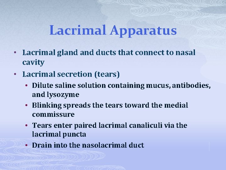
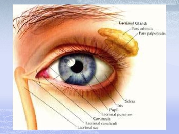
Lacrimal Apparatus • Lacrimal gland ducts that connect to nasal cavity • Lacrimal secretion (tears) • Dilute saline solution containing mucus, antibodies, and lysozyme • Blinking spreads the tears toward the medial commissure • Tears enter paired lacrimal canaliculi via the lacrimal puncta • Drain into the nasolacrimal duct

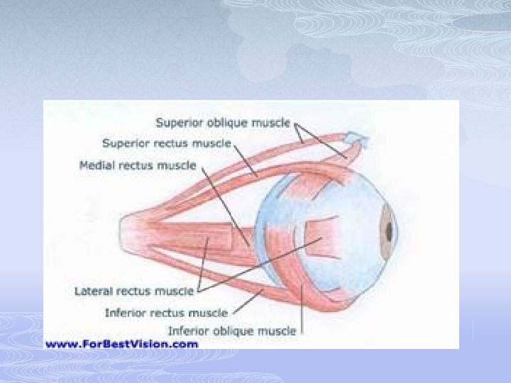
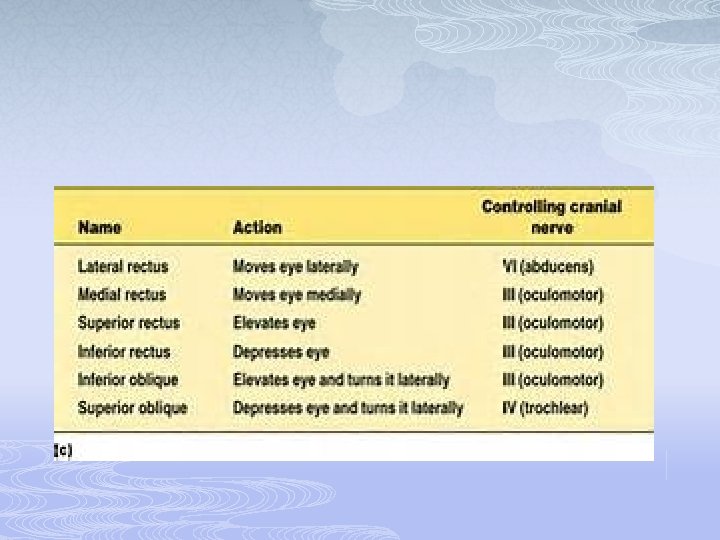
Extrinsic Eye Muscles • Six straplike extrinsic eye muscles • Originate from the bony orbit • Enable the eye to follow moving objects • Maintain the shape of the eyeball • Four rectus muscles originate from the common tendinous ring; names indicate the movements they promote • Two oblique muscles move the eye in the vertical plane and rotate the eyeball
Structure of the Eyeball • Wall of eyeball contains three layers • Fibrous • Vascular • Sensory • Internal cavity is filled with fluids called humors • The lens separates the internal cavity into anterior and posterior segments (cavities)
Fibrous Layer • Outermost layer; dense avascular connective tissue • Two regions: sclera and cornea 1. Sclera • • • Opaque posterior region Protects and shapes eyeball Anchors extrinsic eye muscles White part of external eye Outer most protective layer
Fibrous Layer 2. Cornea: • Transparent anterior 1/6 of fibrous layer • Bends light as it enters the eye • Numerous pain receptors contribute to blinking and tearing reflexes
Vascular Layer (Uvea) • Middle pigmented layer • Three regions: choroid, ciliary body, and iris 1. Choroid region • Posterior portion of the uvea • Supplies blood to all layers of the eyeball; blood vessels are located here • Brown pigment absorbs light to prevent visual confusion
Vascular Layer 2. Ciliary body • Ring of tissue surrounding the lens • Smooth muscle bundles (ciliary muscles) control lens shape • Capillaries of ciliary processes secrete fluid • Ciliary zonule (suspensory ligament) holds lens in position
Vascular Layer 3. Iris • The colored part of the eye • Pupil—central opening that regulates the amount of light entering the eye n Close vision and bright light—sphincter papillae (circular muscles) contract; pupils constrict n Distant vision and dim light—dilator papillae (radial muscles) contract; pupils dilate n Changes in emotional state—pupils dilate when the subject matter is appealing or requires problem-solving skills
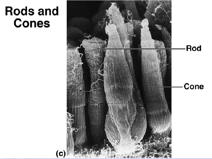
Retina • Retina – back part of eye that contains rods & cones for detecting light • Ganglion cell axons • Run along the inner surface of the retina • Leave the eye as the optic nerve • Optic disc (blind spot) • Site where the optic nerve leaves the eye • Lacks photoreceptors
Photoreceptors • Rods • More numerous at peripheral region of retina, away from the macula lutea • Operate in dim light • Provide indistinct, fuzzy, non color peripheral vision
Photoreceptors • Cones • Found in the macula lutea; concentrated in the fovea centralis • Operate in bright light • Provide high-acuity color vision
Internal Chambers and Fluids • Posterior segment contains vitreous humor that: • Transmits light • Supports the posterior surface of the lens • Holds the neural retina firmly against the pigmented layer • Contributes to intraocular pressure
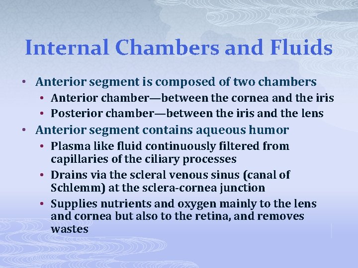
Internal Chambers and Fluids • Anterior segment is composed of two chambers • Anterior chamber—between the cornea and the iris • Posterior chamber—between the iris and the lens • Anterior segment contains aqueous humor • Plasma like fluid continuously filtered from capillaries of the ciliary processes • Drains via the scleral venous sinus (canal of Schlemm) at the sclera-cornea junction • Supplies nutrients and oxygen mainly to the lens and cornea but also to the retina, and removes wastes
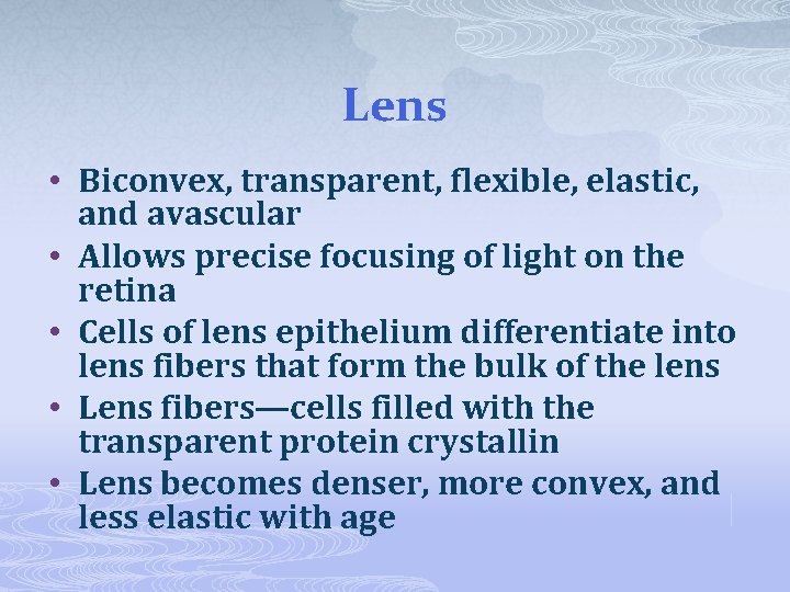
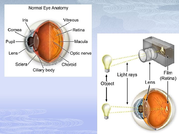
Lens • Biconvex, transparent, flexible, elastic, and avascular • Allows precise focusing of light on the retina • Cells of lens epithelium differentiate into lens fibers that form the bulk of the lens • Lens fibers—cells filled with the transparent protein crystallin • Lens becomes denser, more convex, and less elastic with age
Problems of Refraction • Myopia (nearsightedness)—focal point is in front of the retina, e. g. in a longer than normal eyeball • Corrected with a concave lens • Hyperopia (farsightedness)—focal point is behind the retina, e. g. in a shorter than normal eyeball • Corrected with a convex lens • Astigmatism—caused by unequal curvatures in different parts of the cornea or lens • Corrected with cylindrically ground lenses, corneal implants, or laser procedures
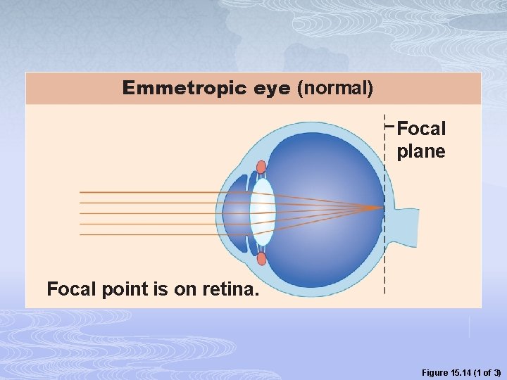
Emmetropic eye (normal) Focal plane Focal point is on retina. Figure 15. 14 (1 of 3)
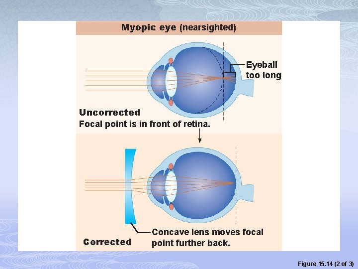
Myopic eye (nearsighted) Eyeball too long Uncorrected Focal point is in front of retina. Corrected Concave lens moves focal point further back. Figure 15. 14 (2 of 3)
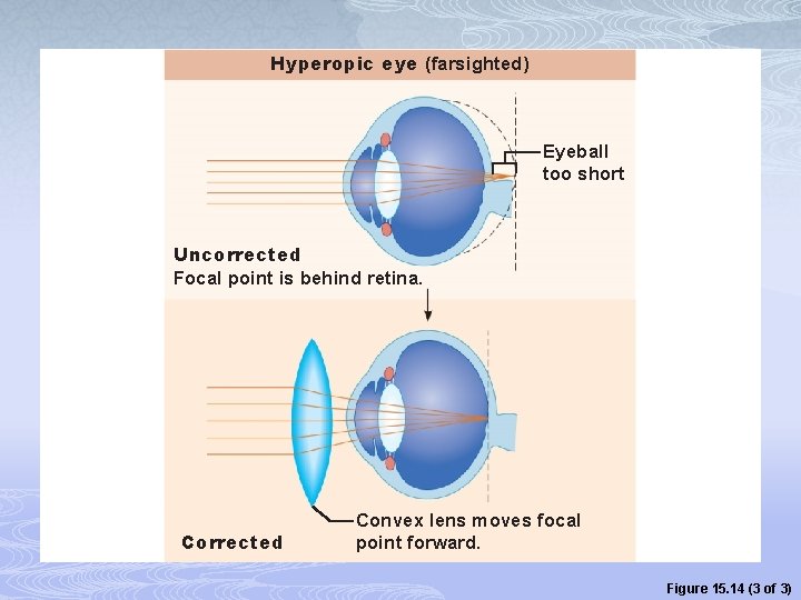
Hyperopic eye (farsighted) Eyeball too short Uncorrected Focal point is behind retina. Corrected Convex lens moves focal point forward. Figure 15. 14 (3 of 3)
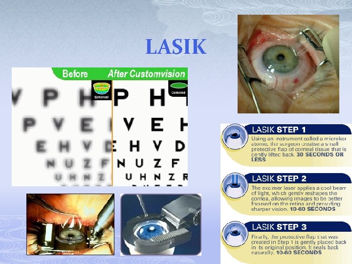
LASIK

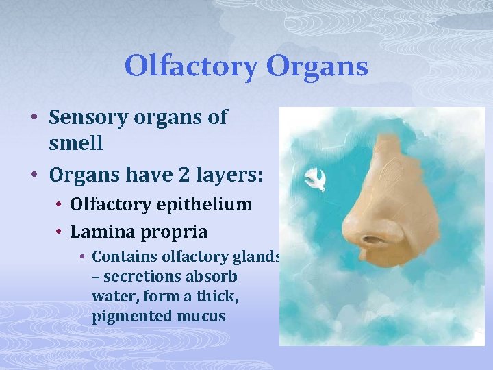
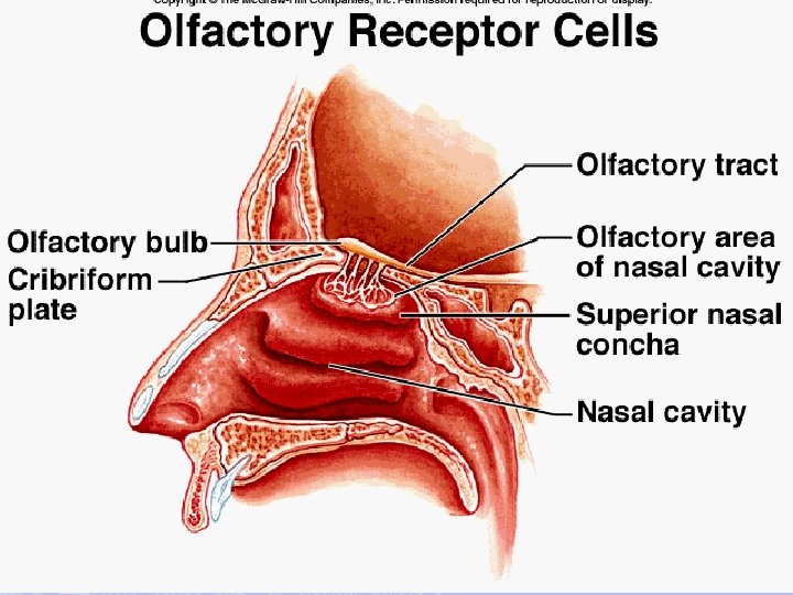
Olfactory Organs • Sensory organs of smell • Organs have 2 layers: • Olfactory epithelium • Lamina propria • Contains olfactory glands – secretions absorb water, form a thick, pigmented mucus
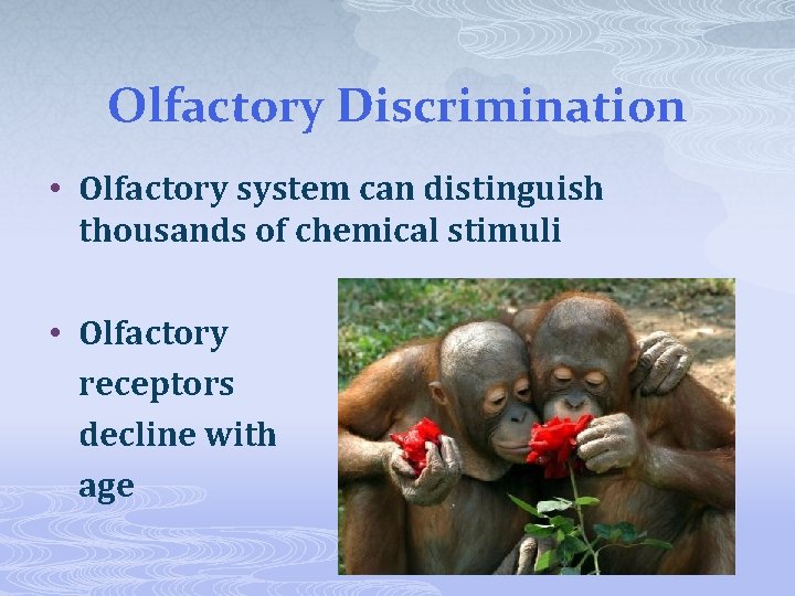
Olfactory Discrimination • Olfactory system can distinguish thousands of chemical stimuli • Olfactory receptors decline with age
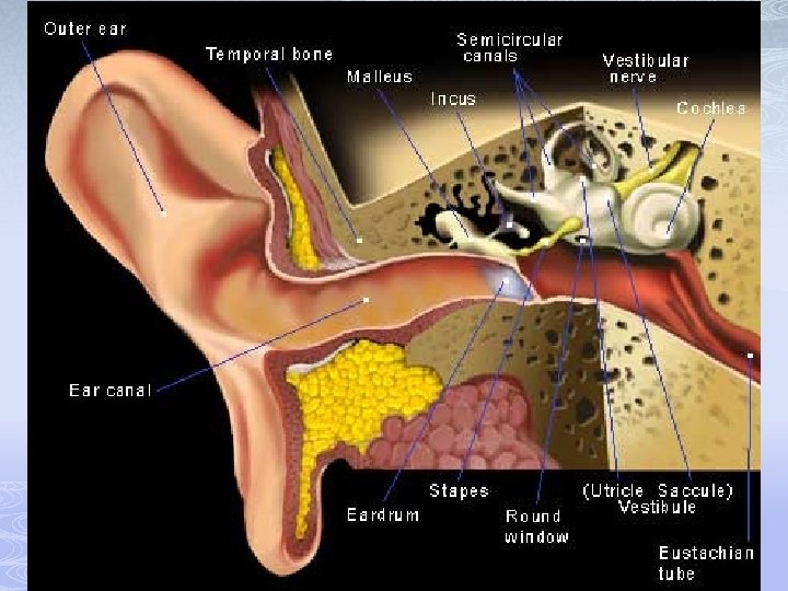
Special Senses of the Ear • Special senses of equilibrium and hearing are provided by the inner ear • Equilibrium • Position of the head in space • Hearing • Detect and determine sound waves
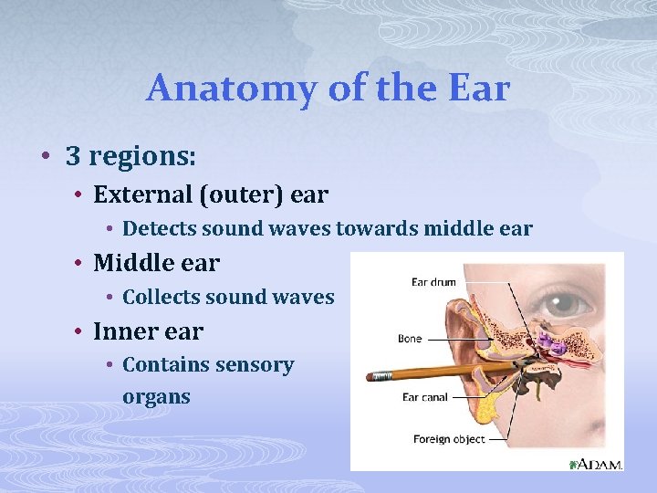
Anatomy of the Ear • 3 regions: • External (outer) ear • Detects sound waves towards middle ear • Middle ear • Collects sound waves • Inner ear • Contains sensory organs
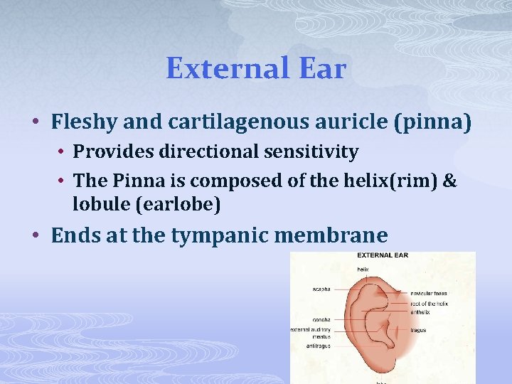
External Ear • Fleshy and cartilagenous auricle (pinna) • Provides directional sensitivity • The Pinna is composed of the helix(rim) & lobule (earlobe) • Ends at the tympanic membrane
• Ear drum • Thin, semitransparent sheet • Delicate • Protected by cerumen • Boundary between external and middle ears • Connective tissue membrane that vibrates in response to sound • Transfers sound energy to the bones of the middle ear
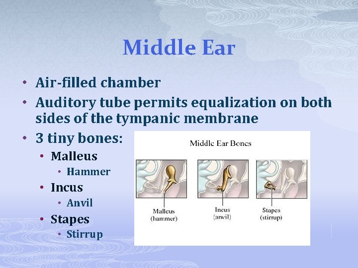
Middle Ear • Air-filled chamber • Auditory tube permits equalization on both sides of the tympanic membrane • 3 tiny bones: • Malleus • Hammer • Incus • Anvil • Stapes • Stirrup
Middle Ear • Epitympanic recess—superior portion of the middle ear • Pharyngotympanic (auditory) tube— connects the middle ear to the nasopharynx • Equalizes pressure in the middle ear cavity with the external air pressure
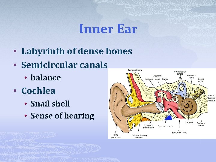
Inner Ear • Labyrinth of dense bones • Semicircular canals • balance • Cochlea • Snail shell • Sense of hearing
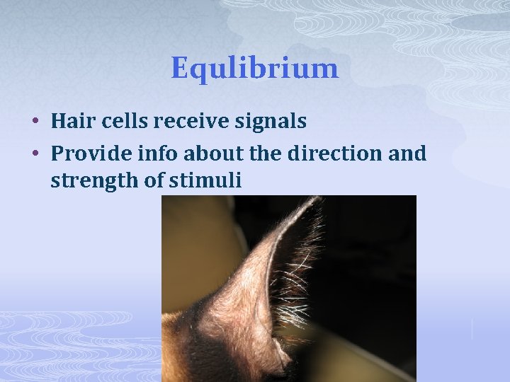
Equlibrium • Hair cells receive signals • Provide info about the direction and strength of stimuli
Hearing • Receptors can detect frequency and intensity of sounds • Intensity is measured in decibels (d. B) • Normal conversation 60 -70 d. B • Telephone dial tone 80 d. B • Sustained exposure resulting in hearing loss: 90 d. B • Rock concert 115 d. B • Loudest recommended with protection: 140 d. B • Gun blast 140 d. B

Gustatory Receptors • Taste receptors • Aka taste buds • Found on tongue • An adult has ~3000 taste buds • 3 types of projections: • Filiform • Fungiform • Circumvallate
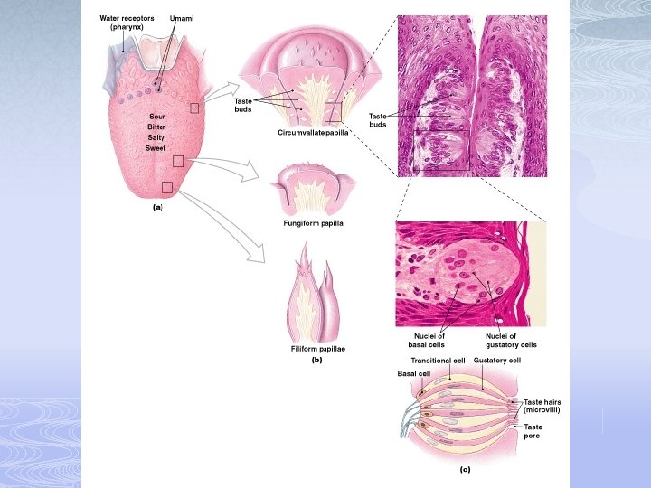
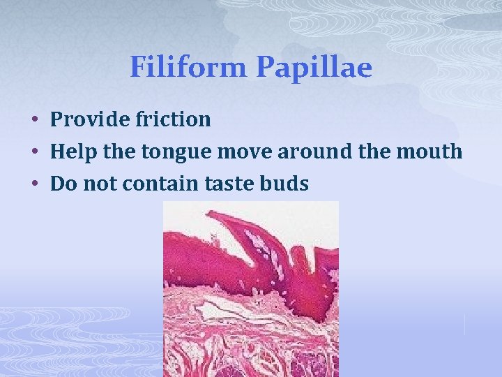
Filiform Papillae • Provide friction • Help the tongue move around the mouth • Do not contain taste buds
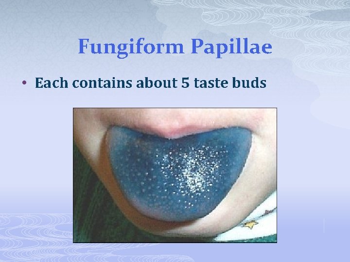
Fungiform Papillae • Each contains about 5 taste buds
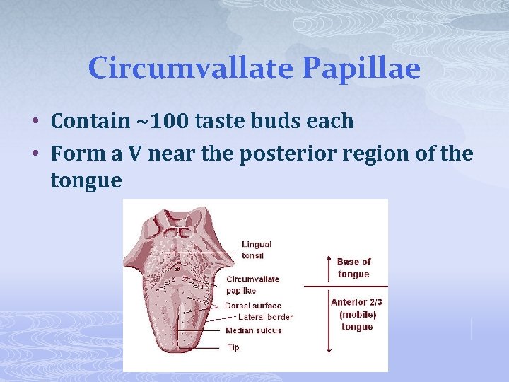
Circumvallate Papillae • Contain ~100 taste buds each • Form a V near the posterior region of the tongue
Aging and Taste • 10, 000 taste buds when born • # declines dramatically by age 50 • *if we cannot smell the food, we believe it is more bland
Physiology of Taste • In order to be tasted, a chemical: • Must be dissolved in saliva • Must contact gustatory hairs • Binding of the food chemical (tastant) • Depolarizes the taste cell membrane, causing release of neurotransmitter • Initiates a generator potential that elicits an action potential
Influence of Other Sensations on Taste • Taste is 80% smell • Thermoreceptors, mechanoreceptors, nociceptors in the mouth also influence tastes • Temperature and texture enhance or detract from taste
- Slides: 60