The Sliding Filament Theory Microstructure of Skeletal Muscle


- Slides: 25

The Sliding Filament Theory

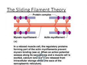
Microstructure of Skeletal Muscle (recap) Actin-Myosin Orientation: • Myosin filament (thick). • Have long rod-shaped tails with 2 globular heads. • The heads form cross bridges.

Microstructure of Skeletal Muscle (recap) • Actin filament (thin). • Tropomyosin is a rigid, rod-shaped protein which lies in grooves on either side of actin. • Troponin is complex of 3 globular proteins.


Structures involved • Myofibril: A cylindrical organelle running the length of the muscle fiber, containing Actin and Myosin filaments. • Sarcomere: The functional unit of the Myofibril, divided into: I, A & H bands. • Actin: A thin, contractile protein filament, containing 'active' or 'binding' sites. • Myosin: A thick, contractile protein filament, with protrusions known as Myosin Heads. • Tropomyosin: An actin-binding protein which regulates muscle contraction. • Troponin: A complex of three proteins, attached to Tropomyosin.

Structure of Skeletal Muscle Microstructure of Skeletal Muscle

Structure of Skeletal Muscle The Sarcoplasmic Reticulum and Transverse Tubules

Muscular Contraction Excitation-Contraction Coupling • Depolarization of motor end plate (excitation) is coupled to muscular contraction • Overview: – Action potential travels down transverse tubules and causes release of Ca+2 from SR – Ca+2 binds to troponin and causes position change in tropomyosin =Exposing active sites on actin – Strong binding state formed between actin and myosin – Contraction occurs

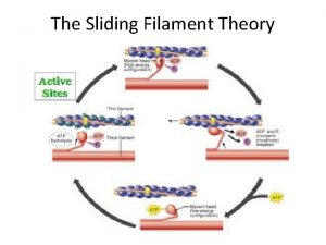
Muscle Contraction: • The process of a muscle contracting can be divided into 5 sections: • Excitation: 1. A nerve impulse (AP) arrives at the neuromuscular junction, which causes a release of a chemical called Acetylcholine (Ach) into the synaptic cleft. - The presence of Acetylcholine causes Calcium (Ca+) to be released from the sarcoplasmic reticulum.

2. In the presence of high concentrations of Ca+, the Ca+ binds to Troponin changing its shape. - This moves Tropomyosin from the active site of the Actin to expose uncovered sites. - Therefore, Myosin filaments can now attach to the Actin forming a cross-bridge.

Tropomyosin, Ca+ & ATP (cont) • Ca+ causes tropomyosin to be displaced. • So it no longer blocks the myosin binding site • So myosin and actin can bind together allowing cross bridge cycling

3. The myosin head binds to the actin, tilts, forcing the actin to move toward the myosin this requires ATP (energy)

4. When an ATP molecule binds to the Myosin head, the Myosin detaches from the Actin and the cross-bridge is broken - When the ATP is then broken down the Myosin head can again attach to an Actin binding site further along the Actin filament and repeat the 'power stroke'.

• Energy released produces crossbridge movement (power stroke). • New ATP attaches to myosin crossbridge to dissociate from actin.


5. This process of muscular contraction can last for as long as there is enough ATP and Ca+ stores. - Once the impulse stops the Ca+ is pumped back to the Sarcoplasmic Reticulum and the Actin returns to its resting position causing the muscle to lengthen and relax.

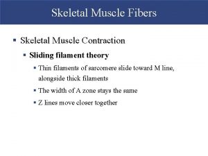
• During contraction, neither myosin nor actin filaments change in length. • They slide past each other. • A band remains same, I band decreases. • H zone decreases on contraction.

Sarcomere Relaxed

Sarcomere Partially Contracted

Sarcomere Completely Contracted

The Sliding Filament Theory… • Therefore, when the muscle contracts sarcomeres become smaller • However, the filaments do not change in length. • Instead they slide past each other (overlap) • So actin filaments slide between myosin filaments = the zone of overlap is larger

Muscular Contraction The Sliding Filament Model • Also called the swinging lever-arm model • Muscle shortening occurs due to the movement of the actin filament over the myosin filament • Formation of cross-bridges between actin and myosin filaments • Power stroke • Reduction in the distance between Z lines of the sarcomere

Therefore… sliding-filament theory: Muscle fibers shorten or lengthen because thick and thin myofilaments slide past each other without the filaments changing length. • See http: //thepoint. lww. com Animation: Sliding Filament Theory.
 Ratchet theory of muscle contraction
 Crash course sliding filament theory
 Sliding filament theory
 Winding filament theory
 Sliding filament theory
 Source
 Sliding filament theory notes
 Pic of smooth muscle
 Sliding filament
 Bio 137
 Structure of dictionary
 Hypereutectoid microstructure
 Development of microstructure in isomorphous alloys
 Market microstructure trading strategies
 Microstructure of ferrous metals
 High frequency market microstructure
 Roll's model bid ask spread
 Pharynx wall layers
 The five golden rules of skeletal muscle activity
 Centrally acting skeletal muscle relaxants
 Skeletal muscle relaxants classification
 Non depolarizing muscle relaxant classification
 Antispasmodic
 Twizzler muscle model
 Comparison of skeletal cardiac and smooth muscle
 Skeletal muscle relaxants classification
