THE SKULL 2 Divisions Cranium Face Most complex


 1 frontal bone 2 parietal bones 2 temporal bones 1 occipital")


- Slides: 27
THE SKULL 2 Divisions Cranium Face
• Most complex bony structure • 22 bones in all • Mostly flat bones, but not all!
Cranial Bones: 8 • • Friday Find and describe: 1. frontal bone 2. occipital bone 3. sphenoid bone 4. ethmoid bone 5. parietal bones (2) 6. temporal bones (2)
Functions of Cranial Bones • Enclose and protect the brain • Attachment sites for head and neck muscles
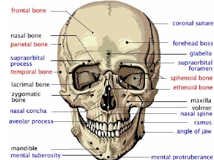
Functions of Facial Bones • 1. form framework of face • 2. contain cavities for special sense organs • 3. openings for food/air passage • 4. secure the teeth • 5. anchor the facial muscles of expression
• ALL BONES OF THE SKULL ARE FIRMLY LOCKED IN PLACE BY JOINTS CALLED SUTURES • Four major sutures
THE CRANIUM (8) 1 frontal bone 2 parietal bones 2 temporal bones 1 occipital bone 1 sphenoid bone 1 ethmoid bone
THE FRONTAL BONE
Parietal Bones: Form most of the superior and lateral aspects of the skull Figure 7. 3 a
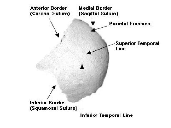
Parietal Bones • Curved, rectangular bones forming majority of the cranium • All four major sutures articulate with this bone
Look up the 4 major sutures of the skull
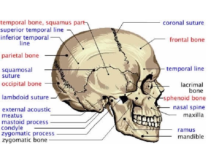
Parietal Bones and Major Associated Sutures • Four sutures mark the articulations of the parietal bones – Coronal suture – articulation between parietal bones and frontal bone anteriorly – Sagittal suture – where right and left parietal bones meet superiorly – Lambdoid suture – where parietal bones meet the occipital bone posteriorly – Squamosal or squamous suture – where parietal and temporal bones meet
Occipital Bone and Its Major Markings • Forms most of skull’s posterior wall and base • Major markings include the posterior cranial fossa, foramen magnum, occipital condyles, and the hypoglossal canal Figure 7. 2 b
Temporal Bones • Lateral sides of the skull • Zygomatic process connects with zygomatic arch • External auditory meatus (external ear) • Styloid process muscle attachment for tongue and neck • Mastoid process neck muscles
Temporal Bones Figure 7. 5
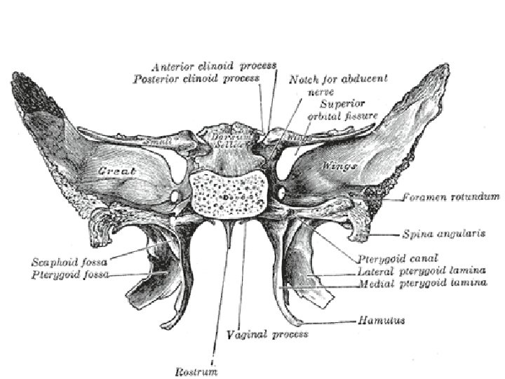
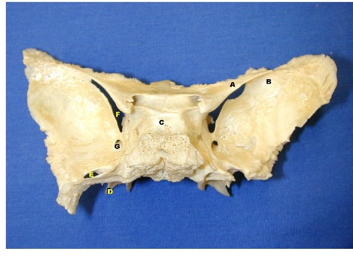
Sphenoid Bone • Spans width of middle cranial fossa • Articulates with all other cranial bones • Three parts greater wing, lesser wing and pterygoid processes • Sella turcica enclosure for pituitary gland
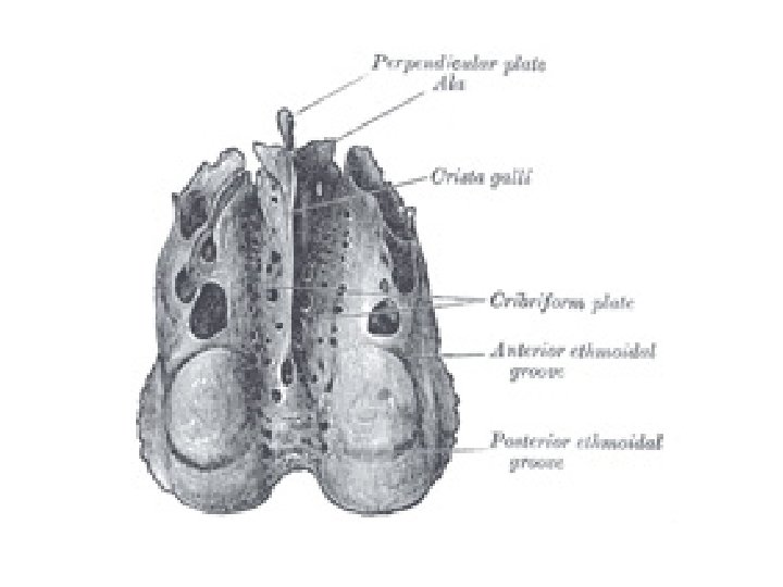
Ethmoid Bone • • Complex shape Nasal cavity and orbital cavity Cribriform plate roof of the nasal cavity Crista galli- outermost covering of the brain cover connects here
Maxillary Bones • Medially fused bones that make up the upper jaw and the central portion of the facial skeleton • Facial keystone bones that articulate with all other facial bones except the mandible • Their major markings include palatine, frontal, and zygomatic processes, the alveolar margins, inferior orbital fissure, and the maxillary sinuses
Maxillary Bone Figure 7. 8 b