The Skeletal System Structure The Skeletal System Parts

 · Joints")


- Slides: 26
The Skeletal System: Structure
The Skeletal System · Parts of the skeletal system · Bones (skeleton) · Joints · Cartilages · Ligaments (bone to bone)(tendon=bone to muscle) · Divided into two divisions · Axial skeleton- skull, spinal column · Appendicular skeleton – limbs and girdle Copyright © 2003 Pearson Education, Inc. publishing as Benjamin Cummings
Bones are classified by their shape: 1. Long- bones are longer than they are wide (arms, legs) 2. Short- usually square in shape, cube like (wrist, ankle) 3. Flat- flat , curved (skull, Sternum) 4. Irregular- odd shapes (vertebrae, pelvis)
Classification of Bones on the Basis of Shape Figure 5. 1 Copyright © 2003 Pearson Education, Inc. publishing as Benjamin Cummings
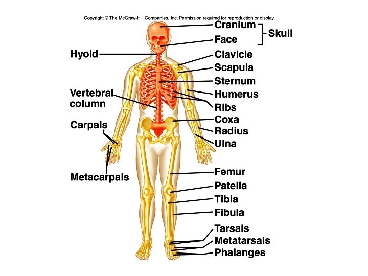
Axial skeleton supports and protects organs of head, neck and trunk Axial skeleton: skull (cranium and facial bones) hyoid bone (anchors tongue and muscles associated with swallowing) vertebral column (vertebrae and disks) bony thorax (ribs and sternum)
Appendicular skeleton includes bones of limbs and bones that anchor them to the axial skeleton Appendicular skeleton: pectoral girdle (clavicle, scapula) upper limbs (arms) pelvic girdle (sacrum, coccyx) lower limbs (legs) Articulation- where joints meet, connect, and are formed.
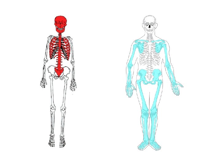
The Axial Skeleton · Forms the longitudinal part of the body · Divided into three parts · Skull · Vertebral Column · Rib Cage Copyright © 2003 Pearson Education, Inc. publishing as Benjamin Cummings Slide
The Axial Skeleton Figure 5. 6 Copyright © 2003 Pearson Education, Inc. publishing as Benjamin Cummings Slide
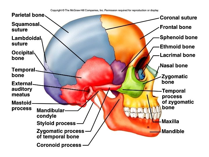
The Skull • 8 sutured bones in cranium • Facial bones: 13 sutured bones 1 mandible Cranium encases brain attachments for muscles sinuses
Bones of the Skull Figure 5. 11 Copyright © 2003 Pearson Education, Inc. publishing as Benjamin Cummings
Allows for growth
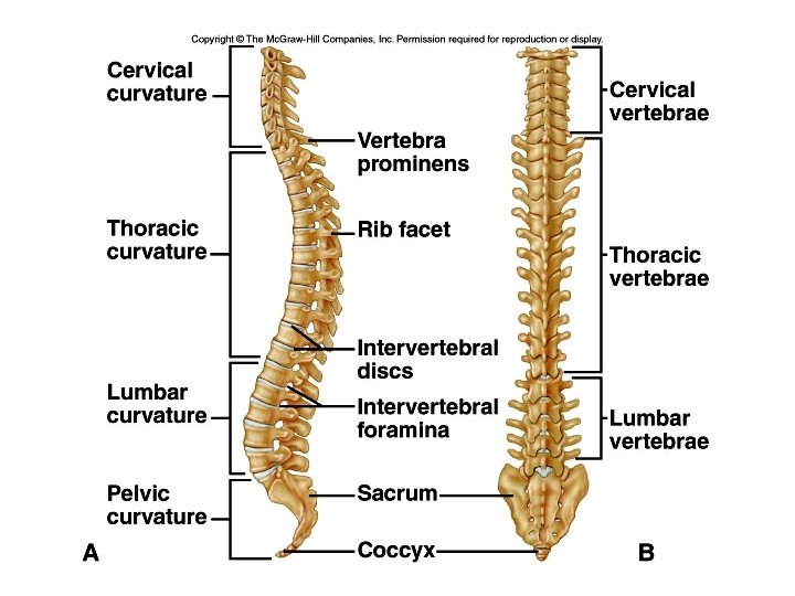
The Vertebral Column · Vertebrae separated by intervertebral discs made of cartilage · The spine has a normal S curvature · Each vertebrae is given a name according to its location Copyright © 2003 Pearson Education, Inc. publishing as Benjamin Cummings Figure 5. 14 Slide 5. 28
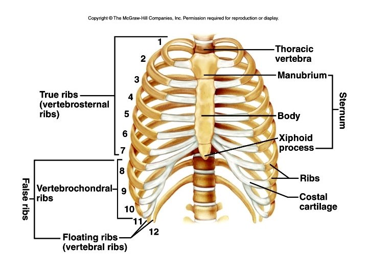
Thoracic cage ribs thoracic Vertebrae sternum costal cartilages • True ribs are directly attached to the sternum (first seven pairs) • Three false ribs are joined to the 7 th rib • Two pairs of floating ribs
Joints A joint, or articulation, is the place where two bones come together. • Fibrous- Immovable: connect bones, no movement. (skull and pelvis). • Cartilaginous- slightly movable, bones are attached by cartilage, a little movement (spine or ribs). • Synovial- freely movable, much more movement than cartilaginous joints. Cavities between bones are filled with synovial fluid. This fluid helps lubricate and protect the bones.
The Synovial Joint Figure 5. 28 Copyright © 2003 Pearson Education, Inc. publishing as Benjamin Cummings Slide 5. 51
Types of Synovial Joints Based on Shape Figure 5. 29 a–c Copyright © 2003 Pearson Education, Inc. publishing as Benjamin Cummings Slide
Types of Synovial Joints Based on Shape Figure 5. 29 d–f Copyright © 2003 Pearson Education, Inc. publishing as Benjamin Cummings Slide
Types of Joints Hinge- A hinge joint allows extension and retraction of an appendage. (Elbow, Knee)
Ball and Socket- A ball and socket joint allows for radial movement in almost any direction. They are found in the hips and shoulders. (Hip, Shoulder)
Gliding- In a gliding or plane joint bones slide past each other. Mid-carpal and midtarsal joints are gliding joints. (Hands, Feet)
Saddle- This type of joint occurs when the touching surfaces of two bones have both concave and convex regions with the shapes of the two bones complementing one other and allowing a wide range of movement. (Thumb)