The Skeletal System Structure Function and Diseases of


 Outline the gross and cellular anatomy and physiology of the musculoskeletal, neurological,")

 · Joints")


")
 • Here the broken bone has broken through tissues below the")


 •")


- Slides: 56
The Skeletal System: Structure, Function, and Diseases of the bones and joints
New Seating Chart Adyson: 12 Ashlyn: 2 Emily: 4 Elsayed: 1 Alex: 5 Parker: 9 Rachel: 13 Kayla: 3 Ben: 11 Lakyn: 6 Mason: 18 Kirsten: 10 Carson: 17 Hailey: 19 Courtney: 8 Wiley: 7
Bell Work 3 -1 -18 1. How many bones are in the body? 2. Have you ever broken a bone? If so, elaborate.
Standard 8) Outline the gross and cellular anatomy and physiology of the musculoskeletal, neurological, and cardiovascular systems. Review the gross anatomy of the other systems studied in previous courses.
Objectives • Learn the major bones • Understand different types of fractures.
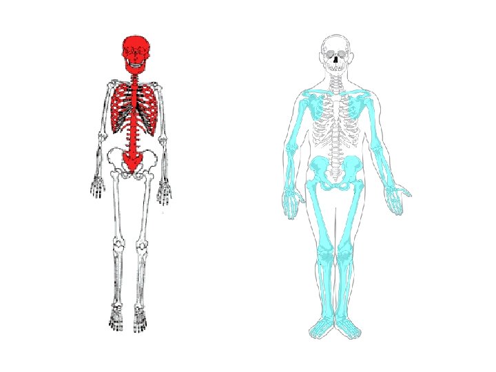
The Skeletal System · Parts of the skeletal system · Bones (skeleton) · Joints · Cartilages · Ligaments (bone to bone)(tendon=bone to muscle) · Divided into two divisions · Axial skeleton- skull, spinal column · Appendicular skeleton – limbs and girdle Copyright © 2003 Pearson Education, Inc. publishing as Benjamin Cummings
The Axial Skeleton · Forms the longitudinal part of the body · Divided into three parts · Skull · Vertebral Column · Rib Cage Copyright © 2003 Pearson Education, Inc. publishing as Benjamin Cummings Slide
Axial skeleton supports and protects organs of head, neck and trunk Axial skeleton: • skull (cranium and facial bones) • hyoid bone (anchors tongue and muscles associated with swallowing) • vertebral column (vertebrae and disks) • bony thorax (ribs and sternum)
Appendicular skeleton includes bones of limbs and bones that anchor them to the axial skeleton Appendicular skeleton: • pectoral girdle (clavicle, scapula) • upper limbs (arms) • pelvic girdle (sacrum, coccyx) • lower limbs (legs) Articulation- where joints meet, connect, and are formed.
Functions of Bones · Support of the body · Protection of soft organs · Movement due to attached skeletal muscles · Storage of minerals and fats · Blood cell formation Copyright © 2003 Pearson Education, Inc. publishing as Benjamin Cummings
Bones of the Human Body · The skeleton has 206 bones · Two basic types of bone tissue · Compact bone · Dense · Spongy bone · Small needle-like pieces of bone · Many open spaces Copyright © 2003 Pearson Education, Inc. publishing as Benjamin Cummings Figure 5. 2 b
Bones are classified by their shape: 1. Long- bones are longer than they are wide (arms, legs) 2. Short- usually square in shape, cube like (wrist, ankle) 3. Flat- flat , curved (skull, Sternum) 4. Irregular- odd shapes (vertebrae, pelvis)
Classification of Bones on the Basis of Shape Figure 5. 1 Copyright © 2003 Pearson Education, Inc. publishing as Benjamin Cummings
Cranium Clavicle Mandible Scapula Sternum Ribs Humerus Ilium Sacrum Radius Pubis Carpals Ulna Metacarpals Phalanges Femur Patella Tibia Fibula Tarsals Metatarsals Phalanges
Simon Says TOUCH THAT BONE!
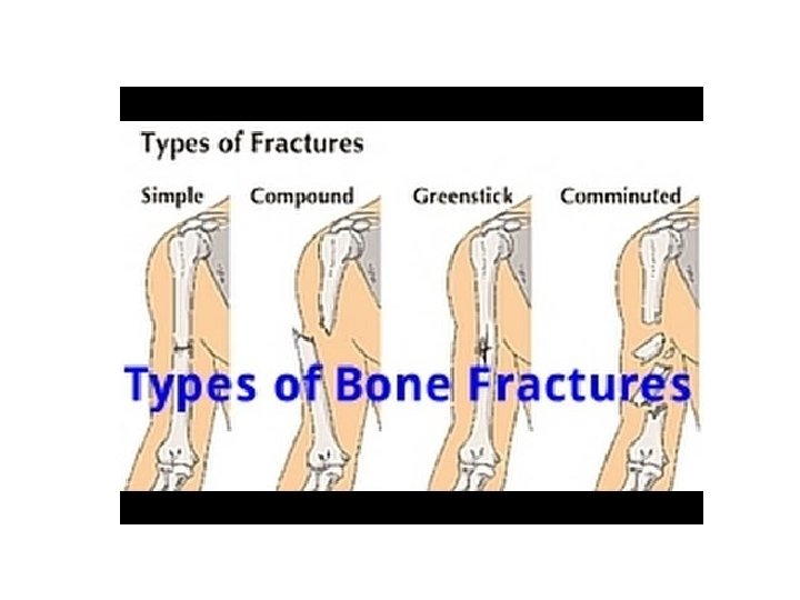
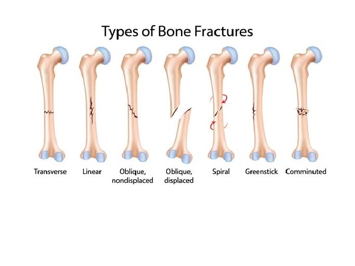
Bone Fractures A break or crack in a bone Types of bone fractures · Closed (simple) fracture – break that does not penetrate the skin. Can be transverse, oblique or spiral. · Open (compound) fracture – broken bone penetrates through the tissue or skin · Comminuted – Broken in three or more · Greenstick - frays, hard to repair, breaks like a green twig Copyright © 2003 Pearson Education, Inc. publishing as Benjamin Cummings
Descriptors • Complete: 2 separate pieces • Incomplete: Not in pieces • Non-displaced: bone cracks either part or all of the way through, but does move and maintains its proper alignment • Displaced: bone has not only cracked, but the pieces have separated.
Simple Closed Break
Simple Closed - Spiral
Open Compound (Skin)
Open Compound (tissues) • Here the broken bone has broken through tissues below the surface of the skin, but has not broken through the skin itself.
Open fractures
Comminuted • Bone is broken in 3 or more fragments • fractures of this degree occur after high-impact trauma such as in vehicular accidents.
Hairline Fracture A fine crack that appears in the bone due to overuse. Commonly seen in athletes, particularly in the foot and shin bones (where it is also known as a stress fracture).
Greenstick Fracture A greenstick fracture occurs when a bone bends and cracks, instead of breaking completely into separate pieces. The fracture looks similar to what happens when you try to break a small, "green" branch on a tree.
Greenstick • Most greenstick fractures occur in children younger than 10 years of age. • This type of broken bone most commonly occurs in children because their bones are softer and more flexible than are the bones of adults.
Causes of Fractures The most common causes of fractures are: • Trauma. A fall, a motor vehicle accident, or a tackle during a football game can all result in fractures. • Disease (Osteoporosis) This disorder weakens bones and makes them more likely to break. • Overuse. Repetitive motion can tire muscles and place more force on bone. This can result in stress fractures. Stress fractures are more common in athletes.
Symptoms Many fractures are very painful and may prevent you from moving the injured area. Other common symptoms include: • Swelling and tenderness around the injury • Bruising • Deformity — a limb may look "out of place" or a part of the bone may puncture through the skin
Healing Time • 3 -12 Weeks • Less for children due to Periosteum (stronger, thicker and flexible) • Periosteum is a dense layer of vascular connective tissue enveloping the bones except at the surfaces of the joints.
Create a Skeleton • Work in groups of two to create a skeleton. Color Axial and Appendicular and label • Work in pairs
BELL WORK 3 -2 -18 1. What are the four main complications of fractures? 2. What type of fracture is mostly found in children? 3. What type of fracture is broken in 3 or more fragments?
Complications • • Infection Compartment Syndrome Fat embolism Nerve vessel damage
Infection = OSTEOMYELITIS • Osteomyelitis is an infection of the bone, a rare but serious condition. Bones can become infected in a number of ways: Infection in one part of the body may spread through the bloodstream into the bone, or an open fracture or surgery may expose the bone to infection.
Compartment Syndrome • Compartment syndrome occurs when elevated pressure within a compartment of the body results in an insufficient amount of blood to supply the muscles and nerves with oxygen
FAT EMBOLISM • • Usually in long bone fractures Change of mental status Restless, Change in breathing status Increase in Respiratory Rate
Is it Broken? Bruising with pain and swelling Reduced movement Odd appearance Krackling sound (bone fragment moving) Edema (swelling)or Erythema (redness) Neurovascular impairment (6 P’s)
6 P’s • Pain – compartment syndrome if unrelieved by pain meds • Pallor – dusky, pale, compare unaffected extremity • Paralysis - movement • Paresthesia – tingling numb • Pulselessness – late sign in effective extremity. Put Sharpie over pulse • Poikilothermia – compare temperature. Cool may mean compartment syndrome
Compartment syndrome • Individual compartment in the fascia • Fascia is connective membrane keeps muscle, blood, nerve supply all together. It doesn’t expand to alleviate stress. • Not irreversible after 6 hours – lose muscle function.
If suspect Compartment Syndrome • Keep extremity at heart level (maintain arterial pressure) • Loosen and remove restrictive clothing • Needle biopsies to monitor pressure • If severe, do a fasciotomy- slice compartment open to relieve pressure
Is it Broken? Bruising with pain and swelling Reduced movement Odd appearance Krackling sound (bone fragment moving) Edema (swelling)or Erythema (redness) Neurovascular impairment (6 P’s)
Complications • • Infection Compartment Syndrome Fat embolism Nerve vessel damage
Initial Injury? • Immobilize - the process of holding a joint or bone in place with a splint, cast, or brace. This is done to prevent an injured area from moving while it heals. • Splint - above and below facture • STOP Bleeding – apply pressure, cover open fracture with sterile dressing or clean
Initial injury • Elevate extremity to reduce swelling – If CS keep at heart level to maintain arterial pressure • Apply Ice wrapped in towel to reduce swelling – prevents compartment syndrome. • NPO (might need surgery) • Pain Management
Treatment options Cast Immobilization A plaster or fiberglass cast is the most common type of fracture treatment, because most broken bones can heal successfully once they have been repositioned and a cast has been applied to keep the broken ends in proper position while they heal. Functional Cast or Brace The cast or brace allows limited or "controlled" movement of nearby joints. This treatment is desirable for some, but not all, fractures.
Treatment Options Traction is usually used to align a bone or bones by a gentle, steady pulling action. The use of a system of weights and pulleys to gradually change the position of a bone.
Traction
External Fixation • In this type of operation, metal pins or screws are placed into the broken bone above and below the fracture site. The pins or screws are connected to a metal bar outside the skin. This device is a stabilizing frame that holds the bones in the proper position while they heal. • In cases where the skin and other soft tissues around the fracture are badly damaged, an external fixator may be applied until surgery can be tolerated.
External Fixation
ORIF or Open Reduction and Internal Fixation During this operation, the bone fragments are first repositioned (reduced) in their normal alignment, and then held together with special screws or by attaching metal plates to the outer surface of the bone. The fragments may also be held together by inserting rods down through the marrow space in the center of the bone.
ORIF
Closed Reduction • Use anesthesia • Manually back in place and put in cast
Recovery • 3 -12 weeks, depending on the extent of the injury and how well you follow your doctor's advice. • Pain usually stops long before the fracture is solid enough to handle the stresses of normal activity. • Even after your cast or brace is removed, you may need to continue limiting your movement until the bone is solid enough for normal activity. • During your recovery you will likely lose muscle strength in the injured area. Specific exercises will help you restore normal muscle strength, joint motion, and flexibility.
Assignment Finish Skeleton Draw the bone fractures on a sheet of copy paper. Paste in notebook. Label and Define.