The Skeletal System Part 4 Joints Honors Anatomy


 �autoimmune disease: immune system attacks joint linings �characterized by:")

- Slides: 43
The Skeletal System Part 4 Joints Honors Anatomy & Physiology
Joints & Homeostasis �Joints contribute to homeostasis by holding bones together in ways that allow movement & flexibility
Joints �aka: “articulation” or “arthrosis” �a point of contact between 2 bones, a bone & cartilage, or between a bone & tooth
Joint Classification � 1. structural classification • based on anatomical characteristics �Fibrous joints �Cartilagenous joints �Synovial joints � 2. functional classification • based on type of movement they permit �Synarthrosis �Amphiarthrosis �Diarthrosis
Fibrous Joints �Articulating bones held very closely together by fibrous CT � 3 types: 1. Sutures • skull bones Syndesmoses 2. • interosseous membrane Gomphoses 3. • dentoalveolar joint
Gomphoses
Cartilagenous Joints �allows little or no movement �bones are tightly connected by either hyaline cartilage or fibrocartilage � 2 types: 1. Synchondoses • • hyaline cartilage connects bones epiphyseal plate Symphyses 2. • • fibrocartilage connects bone pubic symphysis
Symphyses
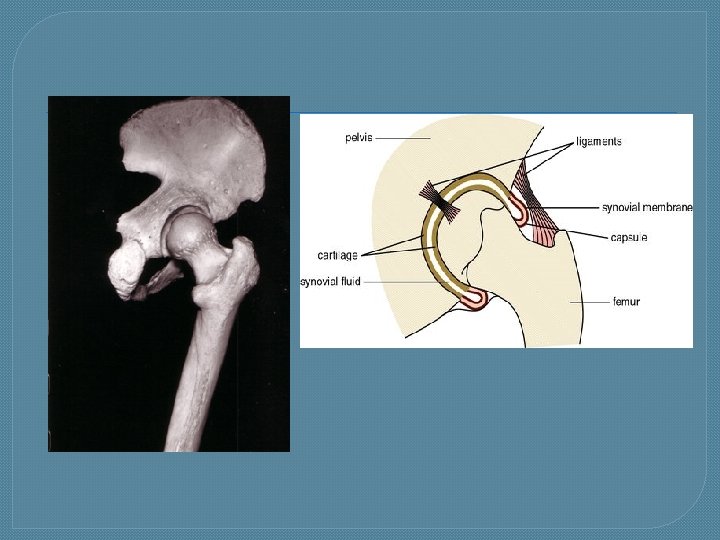
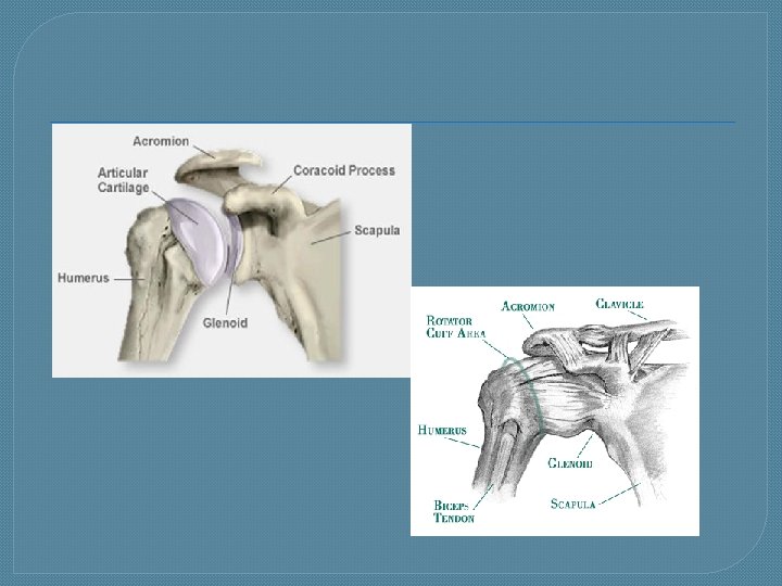
Synovial Joints �distinguishing characteristics: • synovial joint cavity �filled with synovial fluid • bones covered by articular cartilage �reduces friction
Parts of a Synovial Joint
Synovial Fluid �secreted by synovial membrane �viscous, clear, pale yellow fluid �forms film over surfaces w/in articular capsule �functions: 1. reduce friction 2. absorbing shocks 3. supporting chondrocytes in w/in articular cartilage
By the way…. �cracking sounds heard as joints move or popping sounds people make when the “crack” their knuckles explanation: • When synovial cavity expands creates partial vacuum suction from that draws CO 2 & O 2 out of blood vessels in synovial membrane form bubbles in synovial fluid bubbles pop
Sprains �a forcible wrenching or twisting of a joint that stretches or tears ligaments but does not dislocate the bones �occurs when ligaments are stressed beyond their capacity �may have associated damage to surrounding blood vessels, muscles, tendons, or nerves
Strains �stretched or partially torn muscle �often due to muscle contracting suddenly & powerfully
Bursae �saclike structures situated to alleviate friction in some joints �filled with fluid similar to synovial fluid �located between: • • skin & bones tendons & bones muscles & bones ligaments & bones
Bursae
Bursitis �inflammation of a bursa �usually caused by irritation from repeated, excessive exertion of a joint �or by: trauma, infection (syphilis or TB), RA �symptoms: pain, swelling, tenderness, limited movement
Types of Movements @ Synovial Joints � 1. Gliding �relatively flat surfaces move back-and-forth & side-to-side
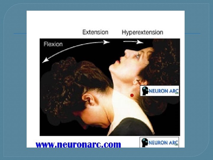
Types of Movements @ Synovial Joints � 2. flexion/extension/ hyperextension: �opposite movements • flexion: decrease in angle between articulating bones • extension: increase in angle between articulating bones • hyperextension: continuation of extension beyond the anatomical position
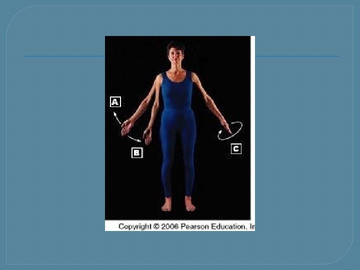
Types of Movements @ Synovial Joints � 3. abduction/adduction/ circumduction • abduction: movement of bone away from midline • adduction: movement of bone toward midline • circumduction: movement of distal end of a body part in a circle
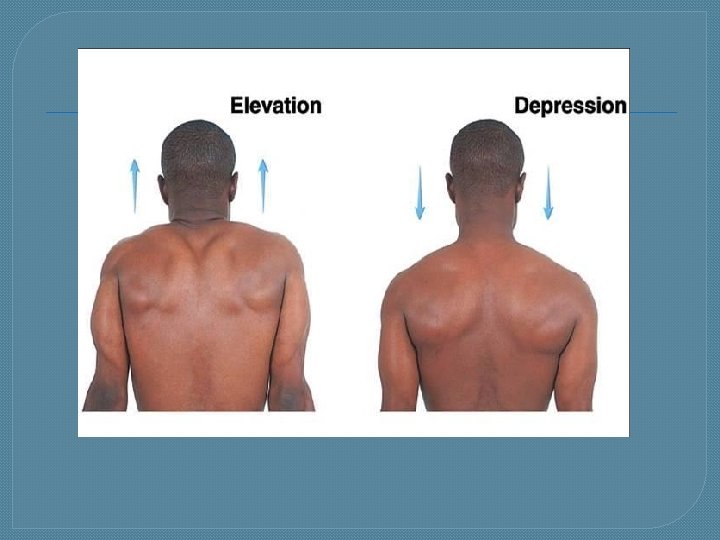
Types of Movements @ Synovial Joints � 4. elevation/depression: �elevation: upward movement of part of body (closing mouth, shrugging shoulders) �depression: downward movement of part of body (opening mouth, returning elevated shrugged shoulders to anatomical position)
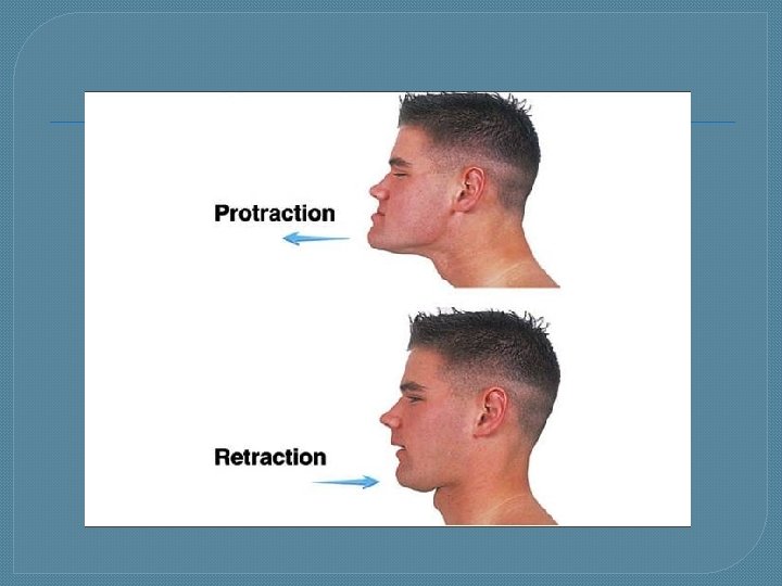
Types of Movements @ Synovial Joints � 5. protraction/retraction �protraction: movement of part of body anteriorly in transverse plane �retraction: returning a protracted part of body to anatomical position
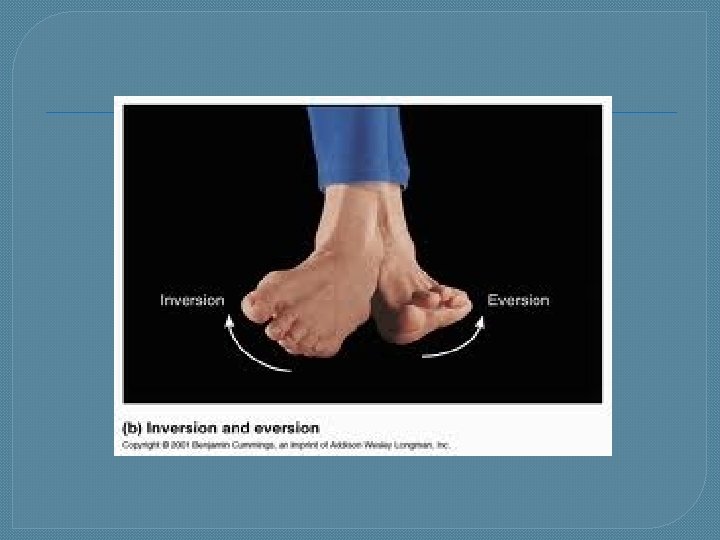
Types of Movements @ Synovial Joints � 6. inversion/ eversion �inversion: movement of soles medially @ intertarsal joints (soles face each other) �eversion: movement of soles laterally @ intertarsal joints
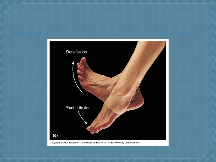
Types of Movements @ Synovial Joints � 7. dorsiflexion/ plantar flexion �dorsiflexion: bending foot @ ankle in direction of dorsum (superior surface) �plantar flexion: bending foot @ ankle in direction of plantar surface
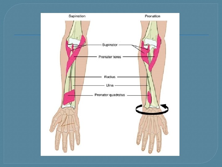
Types of Movements @ Synovial Joints � 8. supination/pronation �supination: movement of forearm in which palm is turned anteriorly �pronation: movement of forearm in which distal end of radius crosses over distal end ulna & palm is turned posteriorly
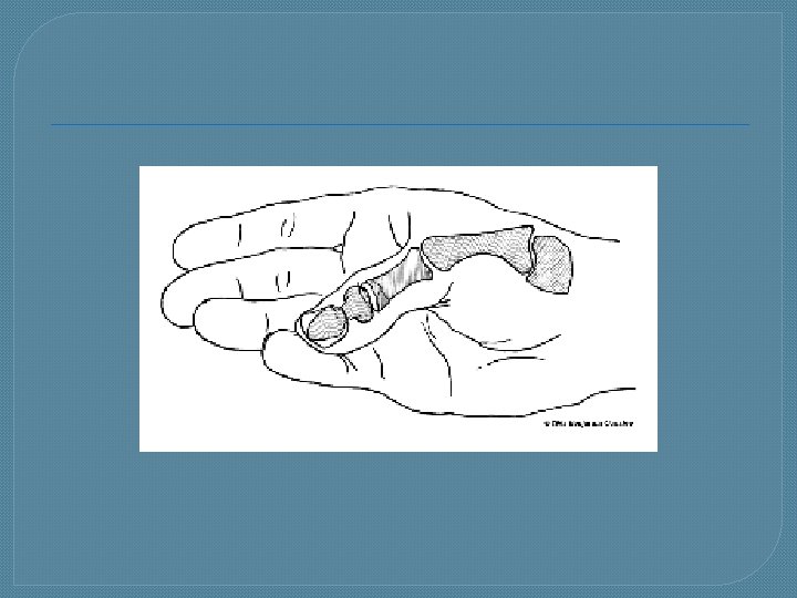
Types of Movements @ Synovial Joints � 9. opposition �movement of thumb in which thumb moves across palm to touch tips of the fingers on same hand
Identify Movements
Ball - & - Socket Joint �ball-like surface of one bone fits into a cuplike depression of another bone �permits movement around 3 axis + all directions in between • • • flexion extension abduction adduction circumduction rotation
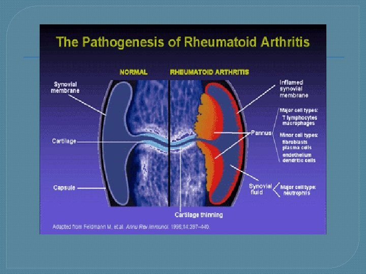
Homeostatic Imbalances �Rheumatoid Arthritis (RA) �autoimmune disease: immune system attacks joint linings �characterized by: • inflammation of joint swelling, pain, loss of function • usually bilateral joints involved but may not be to same degree
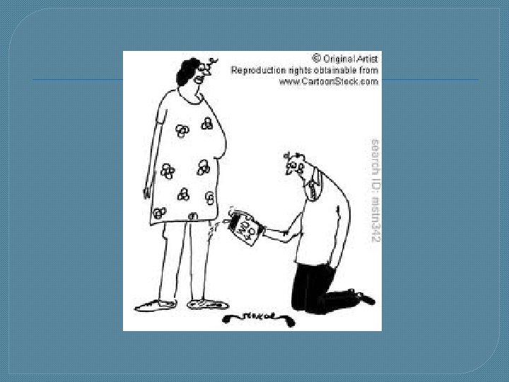
Medical Terminology �arthralgia: pain in a joint �subluxation: partial or incomplete dislocation of a joint