The Skeletal System Mrs Higgins LVT Locust Trace


")


 or vertebrae (plural) •")


 but are heavily muscled • Consist")


- Slides: 32
The Skeletal System Mrs. Higgins, LVT Locust Trace Agriscience Center Veterinary Assistant Program
Functions • External structure and appearance for most vertebrate animals Vital organs • Provide protection of _______ (take a guess) • Give rigidity and form to the body • Act as levers Calcium • Store minerals ________ and Phosphorus ________ • Form the cellular elements of blood
What am I made of? • The skeletal system is made of various forms of connective tissue o They all work together to provide structure and movement • Consists of: o o Bone Joints Cartilage Ligaments/Tendons
Terminology Osteoblasts Osteoclasts Bone • Oste/o ______ • Oste/o _____ Immature • -blasts ______ Break • -clasts ______ • Immature bone cells that produce bony tissue, become osteocytes • Eat away bony tissue from medullary cavity
Terminology • Parts of the bone o Diaphysis • Shaft of the long bone o Epiphysis • Either end of the long bone o Epiphyseal Cartilage • Layer of cartilage within the metaphysis of an immature bone that separates the diaphysis from the epiphysis. Location of growth o Metaphysis • In a mature bone; flared area by the epiphysis o Periosteum • Fibrous membrane that covers the surface of the bone o Medullary Cavity • “marrow cavity”; In young animals, filled with red marrow (hematopoietic tissue)
Bone Marrow • Hematopoietic o Hemat/o- blood o -poietic: pertaining to formations • Forms blood cells (red cells and white cells) • In adult animals, red marrow is replaced by yellow marrow. This is mostly fat cells, serves as fat storage area.
Cartilage • A form of connective tissue • More elastic and flexible than bone • Articular cartilage o Covers the joint surfaces of the bone • Meniscus o Curved fibrous cartilage found in some joints o Acts as cushion from force o Example…. . Stifle/knee
Cartilage
Joints • Also called articulations • Form the connection between bones • Different types depending on degree of movement 1. Fibrous 2. Cartilaginous 3. Synovial • There are many within each category above, but we will cover a few from each
Fibrous Joints • No joint cavity, no movement • Example o Suture joint: between bones of the skull. Suture joints often completely ossify in maturity
Cartilaginous Joints • Bones are united by cartilage with no joint cavity • Limited movement • Example o Symphyses joints: joined by flattened disks of fibrocartilage as found between the pelvic bones (birth canal) or between vertebrae
Synovial Joints • Moveable joints • Examples o Ellipsoid Joint: when a row of small bones fit against a long bone (carpus or tarsus) o Spheroid Joint: “ball and socket”; movement in nearly any direction. Spherical head of one bone fits into the depression of another bone. (Example is…. . ___________) Hip joint o Hinge joints: allows for movement in one direction. Example Knee or stifle _________ o Pivot joint: movement occurs around one axis. Example Atlas/axis joint of neck ______________
Synovial Joints Ellipsoid
Synovial Joint Spheroid (ball and socket)
Synovial Joins Hinge Joint
Synovial Joint Pivot Joint
Terms of Movement • Adduction o Movement towards midline • Abduction o Movement away from midline • Flexion o Closure of a joint angle o Reducing angle • Extension o Straightening of joint o Increasing the angle • Hyperflexion o When a joint is flexed or extended too far
Movement Terms
Tendons • Connective tissue that attaches muscle to bone.
Ligament • Connective tissue that attaches bone to bone
The Skeleton
The Skeleton • Broken into two parts • Axial Skeleton: the central skeleton consisting of the skull, vertebral column, and ribs • Appendicular skeleton: Limbs/Appendages (thoracic limbs and pelvic limbs) **Most of the skeletal system will be the same for different species of animals…. However, there will be differences in the feet/legs and the vertebral column of each animal. Why? ?
Vertebral Column • Made up of many individual vertebra (singular) or vertebrae (plural) • Numbered from head to tail and grouped into sections o o o Cervical Thoracic Lumbar Sacral Coccygeal • Two of the vertebra have names o C 1: atlas o C 2: axis
Thoracic Limbs • The forequarters carry up to 70% of the body weight of animals • Consists of o o o o Scapula Humerus Radius Ulna Carpal bones Metacarpal bones Phalanges
Pelvic Limbs • Carry less weight (about 30%) but are heavily muscled • Consist of o o o o Pelvis Femur Patella Tibia Fibula Tarsal Bones Metatarsal bones Phalanges *Sesmoid bone: small bone held in place by tendon (patella)
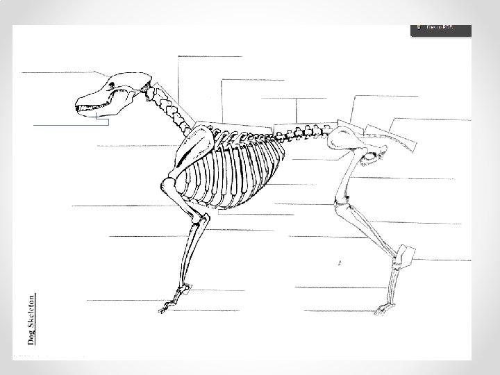
Let’s Label!!
Do you see the differences?
Dog Swine Cattle Horse Different numbers of metacarpal bones and phalanges present
Horse Lower Leg • P 1 or long pastern or proximal phalanx • P 2 or short pastern or middle phalanx • P 3 or coffin bone or distal phalanx **The horse industry uses long and short pastern and coffin bone
Great Interactive Websites • http: //www 2. ca. uky. edu/agripedia/agmania/interactive/ • http: //www. real 3 danatomy. com/bones/dog-skeleton 3 d. html • http: //www. vet. osu. edu/assets/flash/education/outreach/ games/skeleton. html