The Skeletal Muscular and Integumentary Systems Chapter 36


 •")


")


 in axons of motor neuron")


 • Two types of glands • Sebaceous")


- Slides: 50
The Skeletal, Muscular and Integumentary Systems Chapter 36
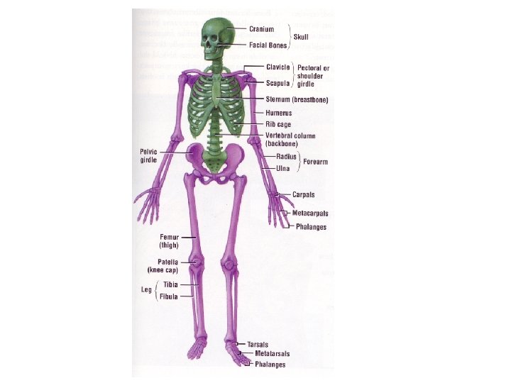
36 -1 The Skeleton • • • Functions: Supports body Protects organs Provides movement Stores minerals Provides site for blood cell formation
36 -1 The Skeleton • Structure • 206 bones in adult • Axial-supports center of body, includes skull, vertebral column and rib cage • Appendicular-includes arms, legs, pelvis and shoulders
36 -1 Structure of Bones • Most of the mass is minerals (salts) • A solid network of living cells and protein fibers surrounded by calcium and phosphorus
36 -1 Structure of Bones • Outer layer • Periosteum-tough layer of connective tissue • Blood vessels inside to carry oxygen and nutrients to bones
36 -1 Structure of Bones • Compact bone • Thick, dense, not solid • Haversian canals run through that have blood vessels and nerves • Spongy layer on inside at end of long bones and center of short flat bones, provides latticework/structure without a lot of mass
36 -1 Structure of Bones • Bone marrow • Yellow-mainly fat cells • Red-produce red blood cells, some white blood cells and platelets
36 -1 Structure of Bones
36 -1 Structure of Bones
36 -1 Development of Bones • As newborns, comprised of cartilage • Cartilage cells in a network of protein fiberscollagen, elastin. Dense, fibrous supportive and flexible • As we age, ossification (bone formation) occurs-cartilage is replaced by bones
36 -1 Development of Bones • • • Osteoblasts create bone Osteo-bone Blast-cells that form new cells Osteoclasts break down bone Osteo-bone Clast-cells that break cells
36 -1 Development of Bones • Ossification-begins 7 months before birth • Osteoblasts secrete mineral deposits • Osteoblasts become surrounded by bone and become osteocytes (-cyte=cell) • Occurs at end of bones at growth plates • Stops when you stop growing
36 -1 Development of Bones • Also occurs when bones are broken – Osteoclasts remove damaged bone – Osteoblasts provide new bone • Cartilage remains in places where flexibility is needed – Tip of nose, external part of ears, where rib bones are attached to sternum (breast bone at center of rib cage)
36 -1 Development of Bones
36 -1 Development of Bones
36 -1 Types of Joints • Immovable-bones in skull • Slightly movable-joints between vertebrae • Freely moving joints-ball and socket, pivot, hinge, saddle joints
36 -1 Types of Joints
36 -1 Structure of Joints • In freely movable joints, ends are covered by cartilage to protect bones from rubbing against each other • Surrounded by a fibrous joint capsule – Ligaments – Layer of cells that produce synovial fluid – Bursa are sacs of synovial fluid (knees, some other joints)
36 -1 Structure of Joints • When tissue is damaged, inflammation is the body’s response – Swelling, redness, heat, pain – Bursitis-occurs in bursa – Arthritis-100 types, affects 10% world’s population
36 -1 Structure of Joints
36 -2 The Muscular System • 40% of mass of your body is muscle • Function: • Moves bones, maintains blood pressure, moves food through digestive system, all movement
36 -2 The Muscular System • • Three types of muscle Skeletal Smooth Cardiac
36 -2 The Muscular System Skeletal muscle Attached to bone Voluntary movement, most controlled by CNS Under microscope, alternating light and dark bands-striations • Large (1 mm-30 cm) and multinucleated • Made of muscle fibers, connective tissue, blood vessels and nerves • •
36 -2 Skeletal Muscle
36 -2 Skeletal Muscle • Muscle – Bundle muscle fibers • Muscle fiber (cell) – Myofibril » Z disc and sarcomere • Myosin, actin
36 -2 Smooth Muscle Usually not under voluntary control Spindle shaped Not striated Not multinucleated Found in hollow structures-intestines, blood vessels • Most can function without nervous stimulation, are connected to each other trough gap junctions so nerve impulses can pass from one cell to the next • • •
36 -2 Smooth Muscle
36 -2 Cardiac muscle • • • Found only in heart Striated Cells smaller than skeletal muscle One or two nuclei Can function without nervous stimulation, are connected to each other trough gap junctions so nerve impulses can pass from one cell to the next
36 -2 Cardiac muscle
36 -2 Muscle Contraction • Myofibrils – Myofilaments • Thick and thin • Myosin and actin
36 -2 Muscle Contraction • Contraction occurs when thick and thin filaments (myosin and actin proteins) move past each other • Cross bridges form between the two protein molecules • Cross bridge changes shape, pulls on actin filament which slides toward center of sarcomere (smallest unit of muscle proteins that can contract) and distance between the Z discs (ends of sarcomeres) decreases • Cross bridge released • Process repeated
36 -2 Muscle Contraction
36 -2 Control of muscle contraction • Skeletal muscle must contract in a controlled fashion • Controlled by CNS and PNS, brain to motor neurons to muscle
36 -2 Control of muscle contraction • Neuromuscular junction-where motor neuron meets muscle
36 -2 Control of muscle contraction • Pockets (vesicles) in axons of motor neuron release acetycholine • Acetycholine molecules diffuse across synapse • Causes an “impulse” that causes the release of Ca ++ (Ca 2+, calcium ions) • Ca 2+ causes proteins to make actin and myosin form cross bridges to cause contraction
36 -2 Muscle Contraction • https: //www. youtube. com/watch? v=BMT 4 Pt XRCVA
36 -2 How muscles and bones interact • Skeletal muscles generate force by contracting and pulling on bones • Muscles are attached to bone by connective tissue called tendons • Tendons pull on bones and make them act as levers • Joint is the fulcrum • Usually several muscle pull the lever (bone) in different directions • Most skeletal muscles work as opposing pairs, when one contracts the other relaxes
36 -2 How muscles and bones interact
36 -2 How muscles and bones interact • Skeletal muscle mainly partially contracted, some of the cells are contracted, some are not -resting muscle tone • Keeps back and legs straight • Regular exercise increases muscle tone • Muscles grow by making new material within the muscle cells • Muscles not used get smaller and weaker
36 -3 The Integumentary System • • • The Skin-Functions Barrier against infection and injury Regulates body temperature Removes waste products Provides protection against UV radiation
36 -3 The Skin • 2 main layers-epidermis, dermis • Underneath subcutaneous fat, connective tissue
36 -3 The Skin • • • Epidermis Outer layer of skin No blood vessels Outside made of dead cells Inside made of living cells – Cells reproduce rapidly – Cells form keratin – Melanocyte-pigment cells-melanin
36 -3 The Skin • Dermis • Inner layer of skin • Contains collagen, blood vessels, nerve endings, glands, sense organs, smooth muscles and hair follicles • Blood vessels narrow when cold, widen when hot to regulate body temperature (change surface area of heat exchange between cells and blood)
36 -3 The Skin • Dermis (continued) • Two types of glands • Sebaceous glands-oil-secrets sebum which makes keratin waterproof • Sweat glands-perspiration (water, salts, minerals) evaporates to regulate body temperature
36 -3 UV Index
36 -3 Monthly UV Index in San Jose 2014 • • • • Month January February March April May June July August September October November December Average 2. 0 2. 79 4. 74 7. 3 8. 42 9. 61 10. 03 8. 55 7 4. 57 2. 63 1. 69
36 -3 Hair and Nails • Basic structure is keratin (same as bird feathers, reptile scales) • Hair-protects from UV, provides insulation, prevent dirt and particles from entering eyes, nose, ears – Grow from hair follicles that are in close contact with sebaceous glands – Cells fill in with keratin as they grow, then die
36 -3 Hair and Nails • • Nails-protect tips of fingers and toes Grow from nail root Cells fill in with keratin as they grow, then die Fingernails-3 mm per month, toenails 4 X as fast
36 -2 Uses and Making of artificial skin • https: //www. youtube. com/watch? v=5 A 3 Vlw. N HGII