The sixth aortic arch the pulmonary arch it
 it forms the pulmonary arteries.")


- Slides: 10
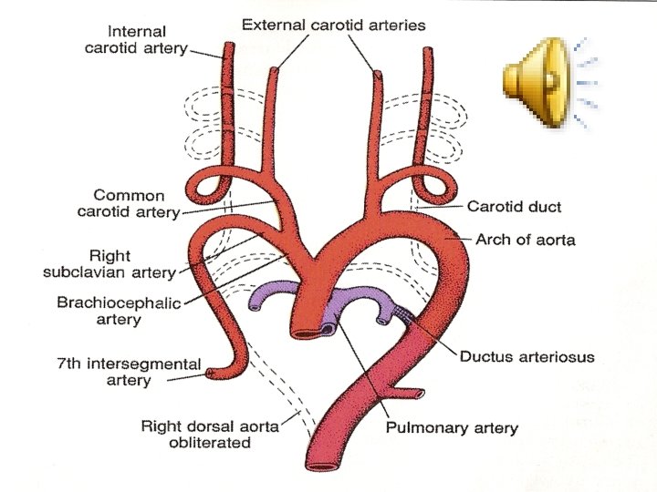
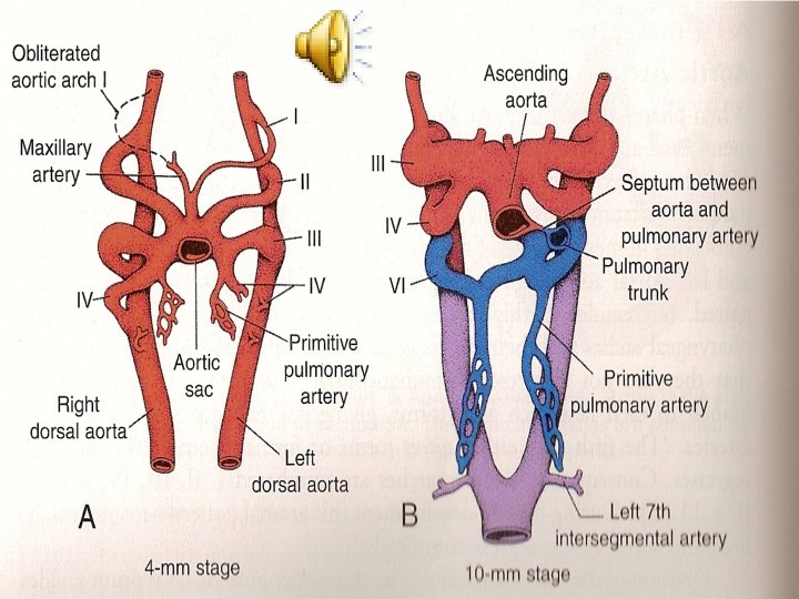
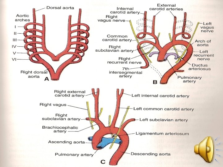
• The sixth aortic arch: (the pulmonary arch) it forms the pulmonary arteries. The connection of the Rt. sixth arch and the Rt dorsal aorta will disappears, while the connection of the 6 th arch with the dorsal aorta forms the ductus arteriosus on the left side.
• During the development of the aortic arches, there will be disappearance of the following: • 1. the bilateral dorsal aortae between the 3 rd and the 4 th arches (called the carotid arch). • 2. the Rt dorsal aorta between the origin of the Rt 7 th intersegmental artery and the junction with the left dorsal aorta.
• Also during this developemtal period, the heart descends from the neck to the thoracic cavity, as a result of that, the following two changes occur: • 1. the Lt subclavian artery (originating from the 7 th intersegmental artery) become nearer to the left common carotid artery.
• 2. recurrent laryngeal nerve (which is the nerve of the 6 th pharyngeal arch) hooks around the distal part of the left 6 th arch (forming the ductus arteriosus) to reach the larynx. • On the right side, the nerve hook ascends superiorly because of the disappearance of the distal part of the 6 th aortic arch, thus hooking around the Rt subclavian artery.
• The vitelline arteries: • These are a number of paired vessels, supplying the yolk sac. Later on these arteries will supply the derivatives of the gut tube, by forming the celiac trunk, the superior & inferior mesenteric arteries.
• The umbilical arteries: • These are the two branches of the dorsal aorta, connect during the 4 th wk with the common iliac arteries & loose its initial connection with the dorsal aorta. • After birth the umbilical artery form the internal iliac artery & the sup. vesical arteries. The distal parts of the umbilical a. , is obliterated forming the medial umbilical ligament.
• Abnormalities of the arterial system: • Patent ductus arteriosus: • • It occurs either alone or with: tetralogy of Fallot, transposition of great blood vessels, pulmonary valve atrasia, aortic valve atresia, pre-ductal coarctation of aorta, or interrupted aortic arch.