The silhouette sign Felson And its derivatives Etienne
 And its derivatives Etienne Leroy Terquem – Pierre L’Her SPI")


 Right posterior alveolar opacity. Notice the positive silhouette")


")


")


")


 Normal left hilus")
")


")
")


- Slides: 79
The silhouette sign (Felson) And its derivatives Etienne Leroy Terquem – Pierre L’Her SPI / ISP Soutien Pneumologique International / International Support for Pulmonology
The silhouette sign • When 2 opacities of the same density are in contact with each other, their contours disappear. • When they are separated by any tissue of a different density (air), their respective contours are visible.
Normal chest radiography
Anterior or posterior opacity?
Anteriorororposterioropacity? Anterior opacity: medium lobe, in contact with heart (and small pleural effusion in posterior cul-de-sac )
Anterior or posterior opacity?
Anterior or posterior opacity? Anterior opacity: middle lobe. The right heart contour disappears
Anterior or posterior opacity?
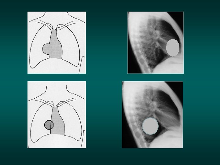
Anterior or posterior opacity? Posterior opacity: the right contour of the heart is visible. On the lateral view the posterior part of the diaphragm in contact with the opacity has disapeared.
Anterior or posterior opacity?
Anterior or posterior opacity? Posterior opacity: the left contour of the heart is visible
Anterior or posterior opacity?
Anterior or posterior opacity? The left contour of the heart is visible: posterior opacity
Anterior or posterior opacity Superior or inferior lobe?
Left upper lobe Positive silhouette sign with cardiac edge Left upper lobe Remember in left lung : Forward the fissure = upper lobe Behind the fissure = lower lobe
Anterior or posterior opacity?
Right posterior opacity (right lower lobe) Right posterior alveolar opacity. Notice the positive silhouette sign on the lateral view With the posterior part of the right diaphragm…
Application of the silhouette sign to the diaphragm Right hemi diaphragm: no silhouette sign with the heart Left hemi diaphragm Positive silhouette sign with the heart
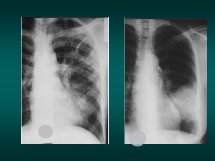
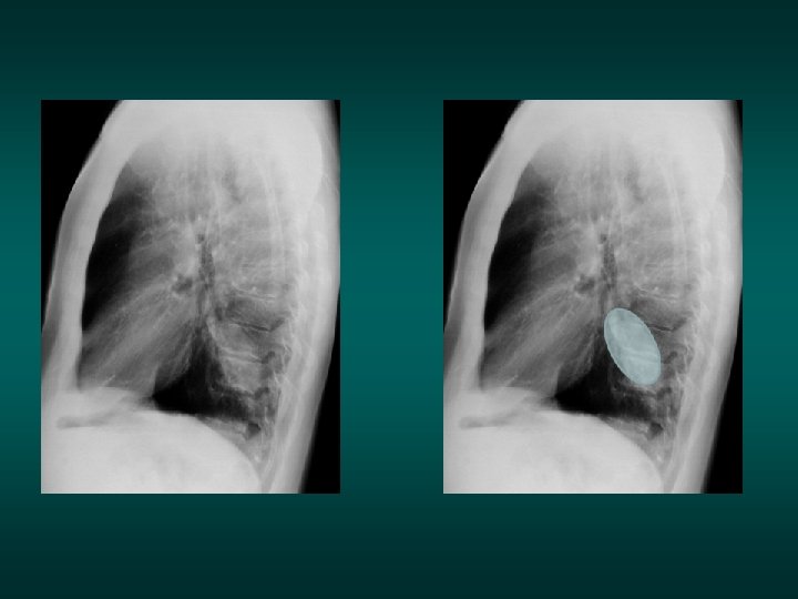
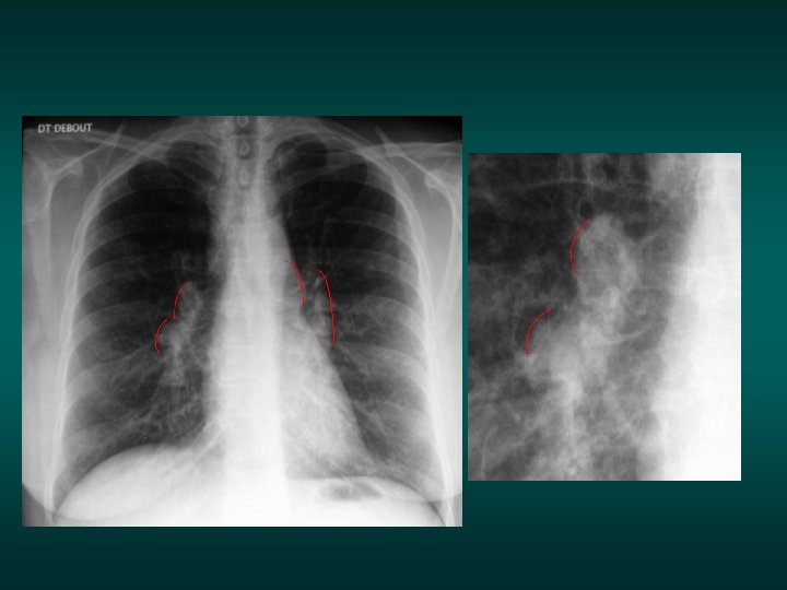
Male, heavy smoker, weight loss, hemoptisis AFB sputum negative
Male, heavy smoker, weight loss, hemoptisis AFB sputum negative Round left opacity. Negative silhouette sign. The opacity is visible behind The heart, in the posterior cul de sac (bronchial cancer)
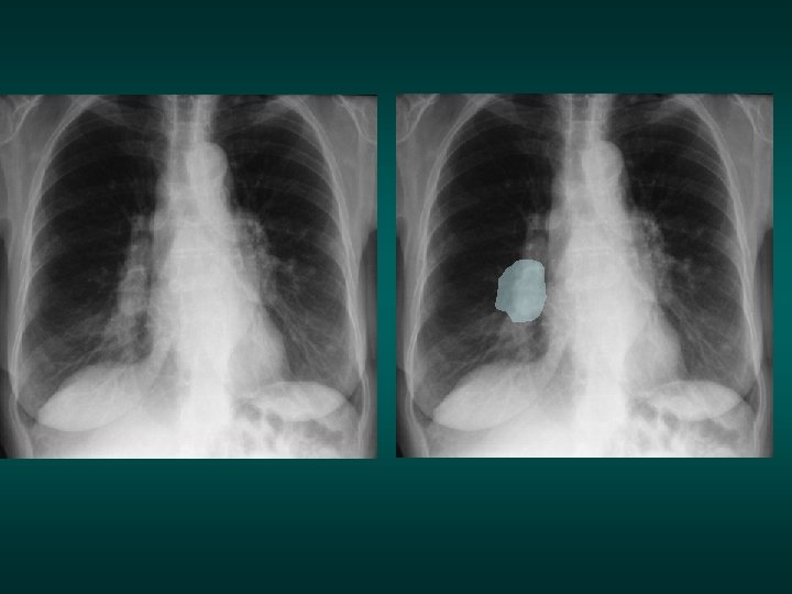
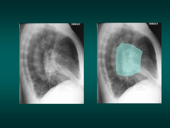
Male, one episode of hemoptisis, AFB sputum negative
Male, one episode of hemoptisis, AFB sputum negative Alveolar opacity visible behind heart silhouette: posterior opacity (possible cancer)
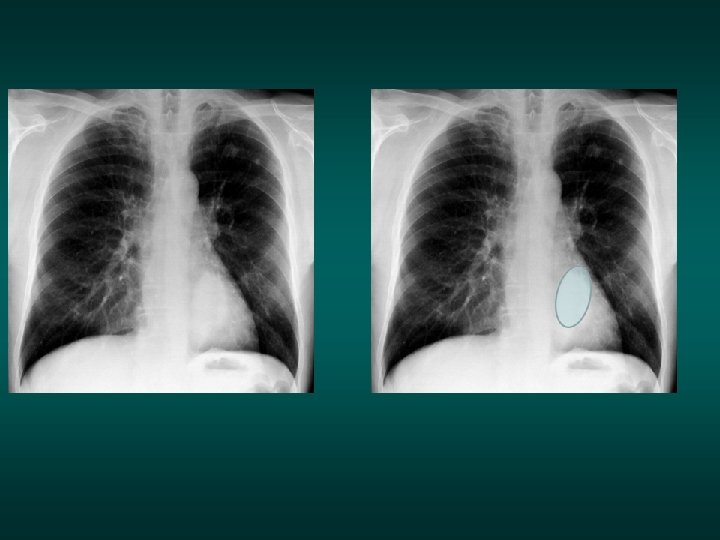
Application of the silhouette sign: Iceberg sign The opacity is above the diaphragm: the inferior edge is well visible because air density surrounding The opacity is above and under the diaphragm: the inferior limit is lost in the abdominal opacities
What is abnormal on this CXR
The opacity is completely intra-thoracic, behind the right diaphragm Caution to the hidden zones
Male, 38 years old, increasing paint in the dorsal and lumbar area fot 3 monthes 1998 1999
Male, 38 years old, increasing paint in the dorsal and lumbar area fot 3 monthes 1998 1999 Pott’s disease: the opacity is above and under the diaphragm
Pott’s disease
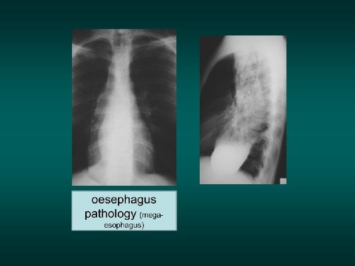
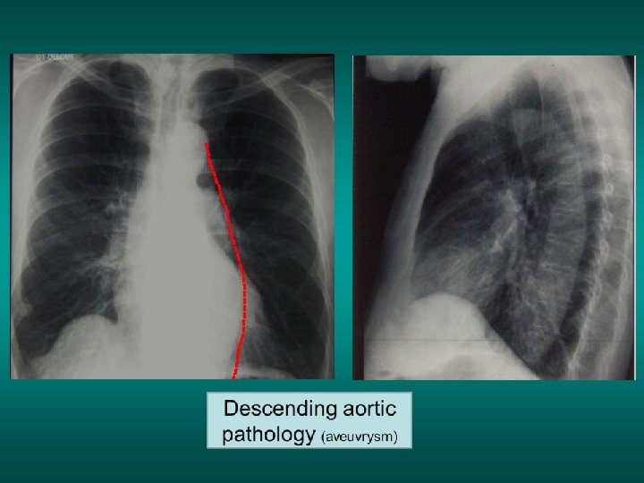
Iceberg sign Descending aortic pathology Oesophagus pathology Spinal column pathology
Spinal column pathology (Pott disease)
Female, no symptoms except sometimes regurgitations
Female, no symptoms except sometimes regurgitations The opacity is posterior
Hiatal hernia
Application of the silhouette sign: The cervico-thoracic pass sign 2 1 clavicle 2 1 1: The external and superior edges of the mediastinal opacity disappear above the clavicles. This sign means that the opacity is anterior in the superior mediastinum 2 2 The superior edge of the opacity is visible in the pulmonary air: the opacity is posterior
Anterior intrathoracic goitre
Anteror intrathoracic goitre. This goitre is compressive: notice the compression of the trachea
Posterior goitre (courtesy of Dr Bellamy)
Posterior or anterior opacity ?
Posterior or anterior opacity ? Posterior: bronchial cancer of the left lung apex +++ notice the destruction of the posterior arch of the third rib
Posterior (bronchial cancer of the left lung apex)
Young woman. Asthenia, weight loss and nocturnal sweet.
Silhouette sign applied to the mediastinum: disappearance of the aorta arch: contact with a tissular mass (Hodgkin’s adenopathy)
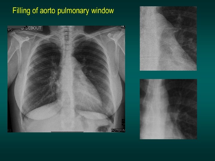
Application of the sihouette sign: Filling of the aorto-pulmonary space aorta Pulmonary artery Adenopathy in aorto-pulmonary space
Filling of aorto pulmonary window Normal hilus
Filling of the aorto-pulmonary space(adenopathy) Normal left hilus
How to make diagnosis of a « big hilus » (silhouette sign application)
Hilus enlargment Vascular enlargment? Adenopathies? Overlap by anterior or posterior opacity?
Convergence sign of the hilus Aorto pulmonary space Pulmonary artery The ramifications of the pulmonary artery loose their silhouette on the edge of the opacity: this opacity is the pulmonary artery
Convergence sign of the hilus (vascular opacity, pulmonary hypertension)
Convergence sign of the hilus (vascular opacity, pulmonary hypertension)
Adenopathies? Hilar adenopathies: q Opacities with convexe external edge q Opacities overlapping normal vascular opacities
Adenopathies? Hilar adenopathies: q Opacities with convexe external edge q Opacities overlapping normal vascular opacities
Bilateral TB adenopathies
Bilateral TB adenopathies
Bilateral adenopathies
Hilar adenopathy Normal hilus
Lateral view is usefull to analyse the involvment of the hilar and mediastinum lymph node
Lateral view is usefull to analyse the involvment of the hilar and mediastinum lymph node Especially in young children for TB adenopathies diagnosis From Dr Pavvas Andronikou. MBBCh, FCRad, FRCR, Ph. D
TB adenopathies Normal lateral view
Normal lateral view
Opacitty overlaping the hilus Normal vessels are visible through The hilus is overlapped by • a posterior mass • or an anterior lmass (posterior or anterior overlap) The opacity Lateral view is helpfull for diagnosis.
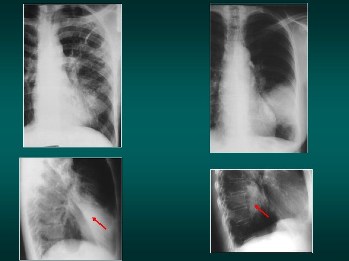
Right hilar adenopathy ?
Posterior overlap sign: right hilar opacity. The pulmonary artery is visible through the opacity, which does not erase the heart contour: This opacity is posterior. On front view this opacity could also suggest adenopathy. Lateral view make correct diagnosis: posterior mass (cancer) When you doubt about hilar opacity Ask for the lateral view
Left hilar opacity. Adenopathy or not?
Posterior overlap sign: left hilar opacity. The pulmonary artery is visible through the opacity, which does not erase heart contour: This opacity is posterior.
Left hilar opacity, Adenopathy? Anterior mass? Posterior mass?
Anterior overlap: Pulmonary artery is visible through the opacity. Cardiac edge is erased : anterior opacity, with filling of the retro sternal space on lateral view
Posterior overlap Anterior overlap