The Sensory Receptors The sensory receptors are cells


- Slides: 85

The Sensory Receptors

§The sensory receptors are cells that capture all information about the environment that is processed by the brain. §They are an integral part of the sensory organs which are. Eye Ear Skin sight Hearing touch Tongue taste Nose smell

§ The path followed by a sensory signal always follows the same path. § Sensory receptor captures information (stimulus) § A “transformer “ changes info into a nervous impulse § Sensory neurons send info to the brain for analysis

The Eye (sight) §The eye is a complex organ that is sensitive to light.

Parts of the eye § The eyeball is covered by 3 membranes: § The sclera § The choroid § The retina

The sclera • The sclera- thick opaque (white) outside layer that protects the eye and give it its shape • The thick layer becomes thinner and transparent forming the cornea at the front of the eye.

sclera

Choroid layer • a blood vessel rich layer that turns into the iris • The iris is the coloured part the eye which is composed groups of muscles that the amount of light the eye of of two control entering • The pupil is the opening formed by the iris which widens in the dark and contracts in bright light

sclera choroid

The Retina § The Retina – is a pink-beige coloured membrane that contains specialized nerve cells called photoreceptors sensitive to light § The retina covers 2/3 of the inside of the eyeball ends in a jagged edge. § this makes it susceptible to detaching

sclera choroid retina

The Retina § The photoreceptors cover the retina in an irregular manner

§ There are 2 types of photoreceptors: § Cones § Rods Cones § There about 20 x less cones than rods § They are concentrated in the center of the retina in a spot (a cone filled pit) approx 2 mm in diameter called the macula.

The retina The macula

• Cones are responsible for color and very detailed vision but need lots of light for this. (you see less color detail in when it gets dark) • There are 3 types of cones: § Red – detect red § Green – detect green § Blue – detect blue § Red and some green = orange § All three stimulated equally results in white color

Rods § Rods are detect contrast (not color) therefore are important for night vision § The farther away from the macula you go the more dense the rods become. § Rods cover the retina except in the macula and the “blind spot”. § The blind spot is where the optic nerve leaves the eye and there are NO photoreceptors.

The “blind spot” where optic nerve leaves the eye

sclera choroid retina Optic nerve

The transparent parts of the eye • The transparent parts of the eye allow light to pass through the eye and converge on the retina. • These are in order: – – The cornea The aqueous humor The lens The vitreous humor

lens Vitreous humor cornea a Aqueous humor

The cornea • The cornea is the transparent part of the sclera which covers the front of the eyeball. • Its slightly domed shape allows the convergence of the light rays. • Malformations of the cornea can lead to vision problems. ( myopia and astigmatism)

sclera choroid retina Optic nerve iris

Aqueous Humor • The aqueous humor is a transparent liquid between the cornea and the iris. • It is water based and contains some glucose • It brings nutrients and oxygen to the lens and bordering retinal cells. • It is constantly being renewed.

sclera choroid retina Optic nerve Aqueous humor iris

Lens • The lens is located behind the iris. • It is flexible and convex on both sides • Approx 9 mm dia. and changes its shape from 4 -5 mm thick depending on whether you are looking at a far object or a close one.

Lens • Lens hardens with age and is no longer flexible (requiring corrective lenses-glasses) • A cataract is the turning opaque (white) of the lens

sclera choroid retina Optic nerve lens Aqueous humor iris

Vitreous Humor • The vitreous humor is a transparent jelly-like substance that fills the eye between the lens and the retina. • Because of its consistency, the vitreous humor exerts a constant pressure on the membranes of the eyeball keeping its shape. • It is also constantly renewed and supplies retina and lens with oxygen and glucose, and removes waste.

sclera iris choroid retina Optic nerve Vitreous humor lens Aqueous humor cornea

Auxiliary structures § The eye is surrounded by various structures that aid in its protection § The eyebrow § Keeps sweat from going into the eyes § The eyelashes § Keep dust out

§ The eyelids § wipe the eye and spread the tears over the eye § The tear(lacrimal) glands § produce tears that clean and lubricate the eye § Tears are antibacterial § Tears contain water and mineral salts § Tears wash across the eye and excess liquid drains into the lachrymal duct (tear duct) leading to the nose

How the eye sees • When light travels through a dense transparent substance with different curvature, it is deviated • A concave lens – light diverges

• Light through a convex lens is convergent (coming together)

Lens accommodation • When looking at a faraway object the incoming light is parallel • so the lens does not have to change its shape to focus image on the retina *

• When looking at close objects the light from the object is diverging • so the lens must become “fatter” in order to focus the light on the retina


Vision anomalies and their correction • Myopia – near sightedness occurs when the cornea is too curved or the eye is too long. This causes light to focus in front of the retina, resulting in blurry distance vision.


Concave lens needed

Hyperopia • farsightedness • occurs when the cornea is too flat in relation to the length of the eye. • This causes light to focus at a point beyond the retina, resulting in blurry close vision and occasionally blurry distance vision as well. *


Hyperopia Convex lens for correction

Presbyopia § is a vision condition in which the lens loses its flexibility, making it difficult to focus on close objects.

• in later years of life, the crystalline lens of the eye looses the ability to focus both near and distant images

Presbyopia (similar to hyperopia) Convex lens for correction

Astigmatism • occurs when the cornea is shaped like a football (more curved in one direction than the other)


Astigmatism • often occurs in combination with myopia (near sightedness) and hyperopia (farsightedness). • This causes light to focus in more than one point on the retina, resulting in blurry and distorted vision. *


Web Links • Optical Illusions: http: //www. youtube. com/watch? v=y 2 EB 4 PGUs 8 • Colour Blind “Test”: http: //www. youtube. com/watch? v=CWyrp 3 hu 4 KE • Mind Tricks: http: //www. youtube. com/watch? v=u 1 SX 2 -e. Qe 4&feature=related

The Ear (hearing) The ear is a complex organ that has 3 functions: • To Receive sound • To Help us maintain our balance • To Detect the body’s spatial movement *

• The ear is divided into 3 sections: – The external ear – The middle ear – Inner ear

The outer ear • The outer ear is composed of 2 parts: • The pinna (outer ear) • The auditory canal

The pinna • acts as a funnel that captures the sound The auditory canal • the auditory canal brings the sound to the middle ear • It protects the ear drum and internal ear from insects, bacteria and dust • The canal has hairs and secretes a wax that capture the foreign particles *

The Middle Ear Composed of : • The ear drum • The ossicles • The Eustachian tube

The Ear Drum • The ear drum is a very sensitive membrane that vibrates when sound hits it • It seals the middle ear from the outside world • The ear drum is transmits the vibrations to three small bones *

The Ossicles (smallest bones in the body) • The first bone is called the “hammer” • The second is the “anvil” • The third is the “stirrup” • The bones transmit the sound vibrations of the ear drum to the inner ear *

The Eustachian tube • This tube is about 4 cm long that leads from the middle ear to the pharynx • The tube is normally closed when one swallows or yawns • Its purpose is to equalize the pressure on each side of the ear drum * Eustachian tube

The Inner Ear • The inner has three parts – The vestibule – The semicircular canals – The cochlea Semi-circular canals vestibule stirrup anvil hammer cochlea

The Vestibule • The vestibule is located at the entrance to the cochlea • It contains nerve cells that are sensitive to body movements and the earth’s gravitational force • Info from the vestibule is processed in the cerebellum (balance and posture) *

Semi circular canals • 3 rings filled with fluid oriented in different planes that allow us to orient ourselves in space *

• When the head moves the liquid in the canals moves and this is sensed by the sensor cells and info is sent to and computed by the brain *

Cochlea • The main hearing organ (shaped like a snail shell) • The cochlea is filled with liquid and lined with cilia that transform sound vibrations into nerve impulses • Each cilia reacts to its own frequency (high near the vestibule low at the extremity) • The sound info is sent to the brain via the auditory nerve *

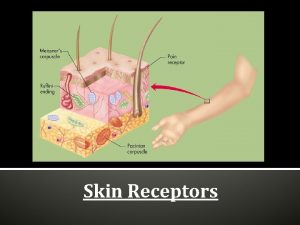
The Skin (touch, heat, pain sensors) • The skin, the largest organ, is about 7% of our body mass. • It is made of 2 layers: – Epidermis – Dermis*

The Epidermis • Is the waterproof outer skin layer • made of dead cells, varying in thickness depending where they are found • Are continuously replaced by cells from underneath • At the bottom of the epidermal layer is a layer of basal cells • Among these basal cells are melanocytes containing melanin *

• Melanin is the brown pigment that protects us from the sun’s harmful radiation • The more sun exposure the more melanin is produced, “tanning” • Over exposure to these harmful rays can cause mutations in the melanocytes causing melanoma (a deadly form of skin cancer) or basal cell carcinoma • The epidermis is the main line of defence against the invasion by bacteria and other environmental dangers. *

The Dermis • Constitutes the second layer of the skin

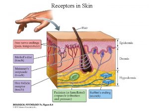
The Dermis • This layer contains many structures like: • • blood vessels - to supply nutrients and O 2 sweat glands - ending in pores on skin surface Hair follicles - where the hair starts to grow Sebaceous glands - oil glands to protect hair and skin • Nerve endings - pressure, pain and temp • Muscles – connected to hair follicles to erect hairs *

The Subcutaneous Tissue • • Found under the dermis Made of adipose tissue (fat) Not uniformly spread in the body Thicker in the hips and abdomen than in the eyelids *

Skin Physiology Tactile function of the skin • Different structures in the skin layers permit humans to have the sense of touch. • In the dermis there are sensors that detect light pressure and change it into a nerve impulse

Skin Physiology • Free nerve endings detect temp and pain • The touch receptors are not spread out evenly over the body *

• There are more touch receptors in the fingers and face, especially the lips, than in any of the other areas or the body • If the human body were drawn proportionally according to the number of touch receptors we would look like this *

The Non Tactile function of the skin Besides being a tactile receptor organ the skin has other functions: 1) First line of defence against bacteria ( see circulatory system notes) 2) Protect the body against ultraviolet radiation 3) Thermal regulation of the body 4) Excretion of certain substances 5) Production of vitamin D *

Thermal regulation (see excretion notes) • When the body temp reaches a certain level the blood vessels under the skin dilate (open up) allowing more blood to flow and give off body heat. • Sweat glands produce sweat that evaporates from the skin producing a cooling effect. • When the body temp drops below a certain level the blood vessels under the skin constrict slowing down the blood flow and conserving the body heat. *

Excretion of certain substances (see excretion notes) • The sweat glands are involved with maintaining the mineral balance in the blood by excreting sweat, which contains urea, water and minerals. (similar to dilute urine) Production of vitamin D • The skin contains cholesterol which when exposed to the sun’s radiation is converted to Vitamin D. • *

• Vitamin D is needed for the absorption of calcium and phosphorus essential for proper bone development • in the winter when we are not exposed to the sun’s radiation, it is important to drink milk enriched with Vitamin D or take cod liver oil which is also rich in vitamin D. *

The Nose (smell) • Besides filtering, warming and humidifying air the nose’s other job is as an olfactory (smell) organ. • The top of the nose is formed by 2 bones – nasal bones • The end of the nose is made of cartilage *

• The olfactory(smell) area is located in the top of the nasal cavity • this area has specialized nerve cells that detect odours called the olfactory bulb. • The nose can detect about 10, 000 different odours • The information is sent directly to the olfactory area of the brain *

For a substance to be perceived by the nose 4 conditions must be met: 1. It must be odoriferous, of a chemical nature that stimulates the smell receptor cells 2. It must be volatile (gaseous), transported by the air 3. It must be in a sufficient concentration to stimulate receptor cells 4. It must come into contact with the receptor cells(not blocked by mucus)

The Tongue (taste) • The tongue is the organ that has receptors to capture flavour molecules • The tongue is a muscle that is covered with a layer of epithelial cells (moist) that form rough bumps called papillae

Types of taste buds

Flavour detection • The tongue can only detect flavourful molecules of the following 4 basic flavours: • Bitter • Sour • Salty • Sweet

For a substance to be tasted, 4 conditions must be met: 1. It must have a taste 2. It must be able to dissolve in saliva 3. It must be in a sufficient concentration to stimulate receptor cells 4. It must come into contact with the taste receptor cells

Non tasting function of the tongue • The tongue is used to move food around for chewing • It is composed of muscle fibers that are oriented in different directions making it very versatile (flexible)

The tongue a complex muscle Transverse muscle fibers Longitudinal muscle fibers Vertical muscle fibers
 Antigentest åre
 Proprioceptors
 Types of sensory receptors
 Classification of sensory receptors
 Fluid that bathes the sensory receptors of the inner ear
 Classification of sensory receptors
 Somatic nervous system
 Eye sensory receptors
 Sensory receptors
 Classification of somatic senses
 Classification of sensory receptors
 Masses of cells form and steal nutrients from healthy cells
 Thyroid parafollicular cells
 Chapter 8 cellular reproduction cells from cells
 Similarities between plant and animal cells venn diagram
 Label
Label Why dna is more stable than rna?
 How are somatic cells different from gametes
 Cells and life lesson 1 answer key
 Prokaryotic cells vs eukaryotic cells
 Olfactory groove keros classification
 What animals have prokaryotic cells
 Chlorocruorin
 Organelle trail
 Dr saja
 Which organisms are prokaryotes
 Prokaryotic v. eukaryotic cells
 Nicotinic receptors in the brain
 Iontropic
 Cholinergic agents
 Olfactory receptor
 Tonic receptors
 Micturition
 Pain receptors
 Adrenergic receptors
 Somatic and autonomic nervous system
 4 types of image receptors
 Graded dose response curve
 Ionotropic vs metabotropic receptors
 J receptors
 Somatic motor pathway
 Parasimpaticolitice
 Alpha adrenergic agonist
 Hearing and equilibrium
 T3 receptor
 Benowitz
 3 types of lung receptors
 Malleus incus stapes
 Difference between alpha and beta receptors
 Oldcart pain
 Mesatonum drug
 Medulla respiration
 Hormone receptors
 Jan j shim md
 Macula ear
 Difference between alpha and beta receptors
 Olfactory epithelium
 Viral receptors
 Nicotine acetylcholine receptors
 Tobacco treatment specialist certification
 Muscarinic vs nicotinic
 Alpha 1 vs alpha 2 receptors
 Cutaneous receptors
 Head paradoxical reflex
 Glut receptors
 Opioid receptors location
 Nicotinic vs muscarinic receptors
 Nicotinic vs muscarinic receptors
 Chemoreceptor example
 Spare receptors
 Hapsis receptors
 Mechanism of drug action
 Respiratory receptors
 Tuberous receptors
 Umami taste receptors on tongue
 Pain receptors in brain
 Peptide hormone receptors
 Chemoreceptor trigger zone
 Intracellular receptors
 Difference between ionotropic and metabotropic receptors
 Definition of respiration
 Derine
 Effexor side effects
 Nociceptive sensation
 Alpha 2 receptors location
 Terminal ganglia

