The Senses Part 3 Eye The Eye The


")


 …vision correction laser surgery… Video Clip")

- Slides: 25
The Senses Part 3 Eye
The Eye The eye is in the skull for protection. Eye movement is controlled by 6 eye muscles
Visual Accessory Organs do not contribute to your sense of sight but play a role in thr function of the eye. Can you guess the celebrity Eyes?
Eyelid Covers and protect the eye Thin layer of skin Skin will not protct you from intense radiation that‘s why we use special goggles in tanning beds.
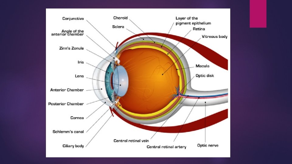
Conjunctivia • Is a covering around the eye and under the eyelids. Conjunctivitis • Aka Pink Eye • Infection of the Conjunctivia • Bacterial or viral infection • Very contagious
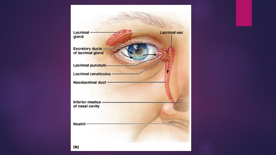
Glands of the Eye Lacrimal Glands are tear glands. Producing tears, which drain into the nasal cavity. Function: • to moisten eye • Contains enzymes to kill bacteria
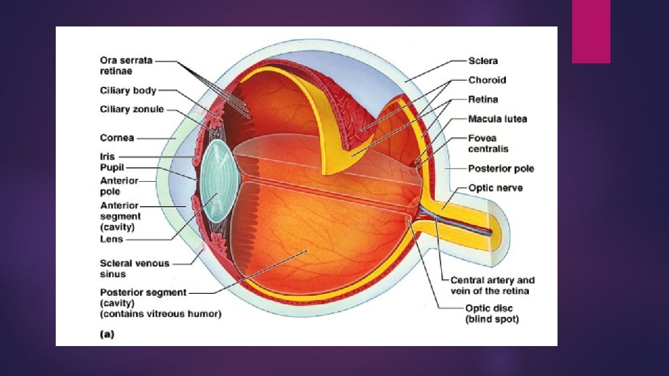
Outer Tunic Cornea • Transparent • Focuses light Sclera • Continuation of cornea • Going toward the back of the eye (white of eye) Optic Nerve • Transmits visual information from eye to the brain (Occipital lobe)
Middle Tunic Choroid • Ciliary Body • Colored portion of eye Aqueous humor • Focus vision Iris • Holds the lens in place Lens • Contains blood vessels Liquid surrounding lens Pupil • Opening for light to enter eye
Pupils – Fun Fact When you are looking at someone you love, your pupils dilate, and they do the same when you are looking at someone you hate.
Inner Tunic Retina • Contains visual receptor cells • photoreceptor cells (Rods & Cones) The retina is made up of PHOTORECEPTORS, which are sensors for light. Rods are for light /dark vision Cones are for color vision
Work on the Eye Diagram Worksheet before moving on… link Label and color the Eye diagram Mini Research on Eye Diseases Ideas: • Cataracts • Retinal Detachment • Glaucoma • Hyperopia • Myopia • Astigmatism § Etc….
Colorblindness A genetic trait that affects boys more than girls. The location of the gene is on the x chromsome.
Inner Tunic Fovea • Centralis Region of the sharpest vision in the back of the eye Optic Disc • Where nerve fibers leave the eye to send message to brain • Blind spot Vitreous Humor • Supports internal parts • Fluid filled
How does light pass through the eye? Cornea -> Aquaeus Body -> Lens -> Vitreous Body -> Retina -> Rods & Cones are stimulated.
How does vision work? 1. Light penetrats the Retina. 2. Inside the Retina Rods and Cones are stimulated. 3. Rods and Cones send a nerve impulse down the Optic Nerve to the Brain. 4. Brain interprets the stimulus in the visual cortex of the Occipital lobe.
Accomodation Lens changes shape to facilitate focused vision Shape of lens depends on movement of cilary muscle
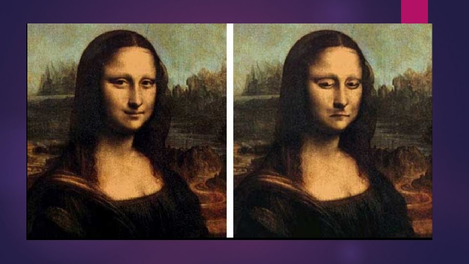
We have difficulty interpreting images that are upside down…. Which one is the real Mona Lisa?
Why are babies born with blue eyes? Melanin is a brownish pigment that adds color to your hair, eyes, and skin. At the time babies are born, melanin hasn't yet been "deposited" in the eyes' iris. Hence, they appear blue. After about six months, eyes change color depending on the amount of melanin. If you have a lot of it, your eyes will turn dark brown. If you have little, they'll stay blue. And if you have no melanin, your eyes may appear pink (albino). .
Lasik Surgery… (not for the squeamish) …vision correction laser surgery… Video Clip
Cow Eye Dissection Day!!! Video of Cow Dissection – watch the day prior to dissection day! Have Fun. . !! Every group of 2 needs the following 2 Lab sheets: 1. Lab Instructions 2. Sheet for Drawings