The sectional anatomy of limbs and MRI The

The sectional anatomy of limbs and MRI The medical college of shandong university

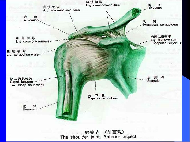
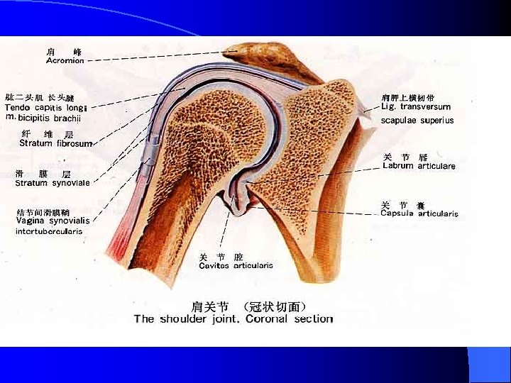
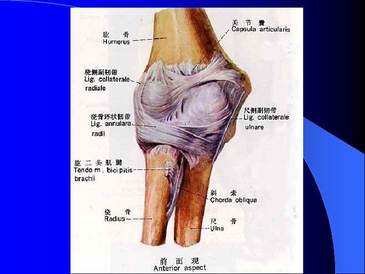
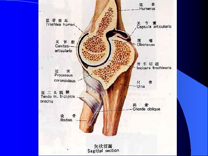
The sectional anatomy of upper limb and MRI

Acromial end deltoid Head of humerus Glenoid cavity Articular capsule Coronal scan of R. shouder by MRI Greater tubercle

Greater tubercle deltoid Long head of biceps brachii Head of Glenoid cavity humerus Transverse section of R. shouder (specimen)

deltoid Pectoralis major Pectoralis minor Head of humerus Glenoid cavity Transverse scan of R. shouder by MRI

Cephalic V. Brachial A. /V. brachioradialis brachialis Pronator teres Medial epicondyle Lateral epicondyle olecranon Transverse section of R. elbow joint (specimen)

brachioradialis brachialis Lateral epicondyle Basilic V. Trochlea of humerus olecranon Medial epicondyle Transverse scan of R. elbow joint by MRI
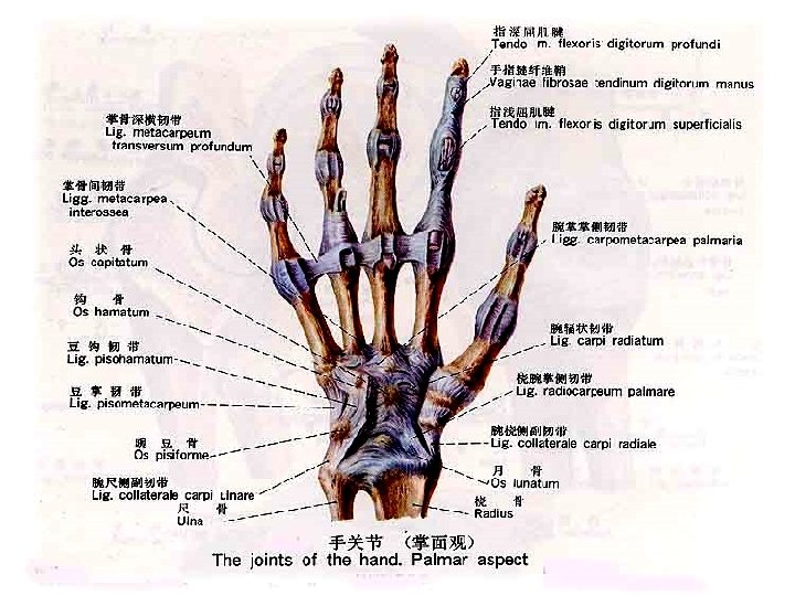

1 st. metacarpal B. Trapezium B. Scaphoid B. Trapezoid B. Capitate B. Hamate B. Pisiform B. radius ulna

1 st. metacarpal B. Trapezium B. Trapezoid B. Scaphoid B. Capitate Hamate B. Triquetral B. Lunate radius ulna
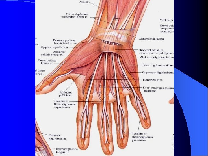

Capitate Hamate 2 nd. Radial A. V. hypothenar 5 th. Tendons of flexor digitorum Ulnar A. V. Palmar aponeurosis Median N. 1 st. metacarpal B. Carpal canal Transverse section of hand joint (specimen)

Capitate 2 nd. Hamate 1 st. metacarpal thenar hypothenar Palmar aponeurosis Median N. Transverse scan of R. hand joint by MRI

The sectional anatomy of lower limb and MRI

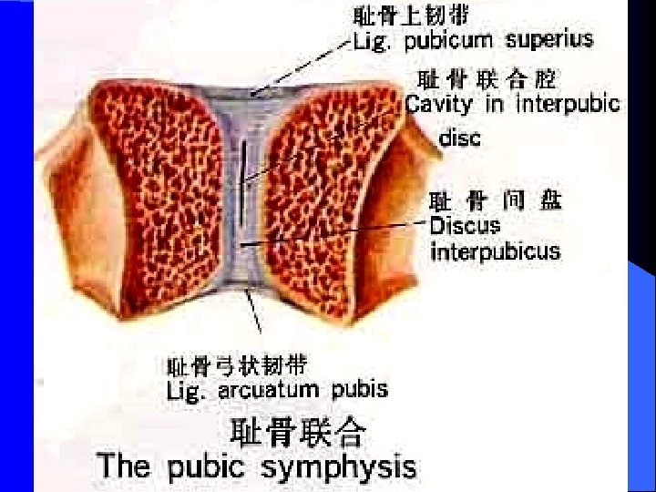
Greater sciatic foramen Sacrospinous lig. Lesser sciatic foramen Sacrotuberous lig. Pubic symphsis

Ant. sup. iliac spine Iliofemoral lig. Greater trochanter Obturator membrane Lesser trochanter Hip joint

Iliofemoral lig. Greater trochanter Neck of femur Ischial tuberosity Lesser trochanter

acetabulum Head of femur Greater trochanter Lig. of Head of femur Ischial tuberosity Neck of femur

acetabulum Head of femur Greater trochanter Neck of femur Lig. of head of femur

acetabulum Head of femur Lig. of head of femur Greater trochanter

Neck of femur Head of femur acetabulum Greater trochanter Transverse section of hip joint (specimen)

Pubic symphsis Head of femur Urinary bladder acetabulum rectum Transverse scan of R. hip joint by MRI

rectum Head of femur acetabulum Urinary bladder Greater trochanter penis Lesser trochanter Coronal scan of R. hip joint by MRI

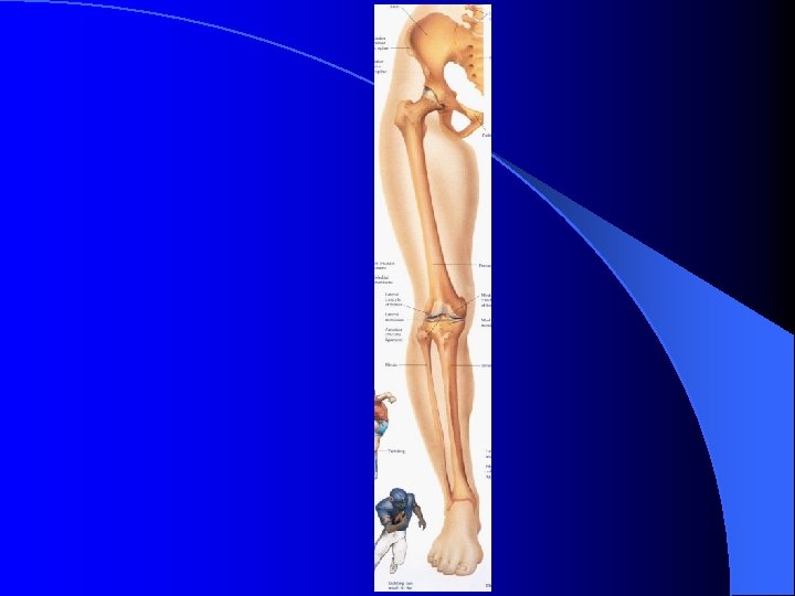
femur fibula patella

femur tibia

femur Med. Lateral condyle Ant. cruciate lig. Lat. meniscus med. meniscus tibia Patellar lig. fibula patella

Lateral condyle femur Med. med. condyle meniscus Lat. meniscus post. cruciate lig. tibia fibula

tibia Ant. cruciate lig. Lat. meniscus med. meniscus

patella femur Patellar lig. tibia

patella Lateral femur condyle Transverse section of knee joint

patella femur Lat. Med. condyle Transverse scan of knee joint by MRI

patella Patellar lig. femur Ant. cruciate lig. tibia Sagittal section of knee joint

patella femur tibia Sagittal section of knee joint fibula

patella femur tibia fibula Sagittal section of knee joint

femur Patellar lig. tibia Sagittal scan of knee joint by MRI

patella femur Patellar lig. tibia Sagittal scan of knee joint by MRI T 2

Med. condyle femur Lateral condyle Lat. meniscus tibia Coronal section of knee joint ( anterior part )

femur Ant. cruciate lig. Med. Lateral condyle post. cruciate lig. tibia fibula Coronal section of knee joint (middle

Med. Lat. condyle Lat. meniscus med. meniscus tibia fibula Coronal section of knee joint ( posterior part )

Med. Lat. condyle Lat. meniscus tibia fibula Med. Lat. condyle Coronal scan of knee joint by MRI tibia

talus tibia calcaneus

tibia fibula talus navicular Cuboid B. Cuneiform B. 5 4 3 2 1 metatarsal
")
tibia fibula talus calcaneus Coronal section of foot joint ( posterior part )

tibia Tendo calcaneus talus navicular calcaneus Sagittal scan of foot joint by MRI

Thanks!!
- Slides: 53