The Role of Cell Organelles in Chlamydias Life


 – ~3. 5")
 Signs & Symptoms • 85 -90% do not show symptoms –")
 Possible Complications 1. Pelvic inflammatory disease 1. Infertility (10 K women/yr")
 Pelvic Inflammatory Disease • Chlamydia over stimulates the body’s immune system")

")
")
 1. What happens when Chlamydia enters")


 1. Most Chlamydia infect columnar epithelial cells 2. 3.")


 of DNA and RNA As")

- Slides: 38
The Role of Cell Organelles in Chlamydia’s Life Cycle Goals • • Study Chlamydia as a vehicle to understand the interrelationships and functioning of various cell organelles. Identify potential future strategies for treating Chlamydia infections
Chlamydia Resources Optional Reading • “Can Chlamydia Be Stopped? ” In the May 2005 edition of Scientific American • Good overview of Chlamydia : http: //www. cdc. gov/std/Chlamydia/STDFact-Chlamydia. htm http: //www. niaid. nih. gov/factsheets/stdclam. htm http: //pathmicro. med. sc. edu/mayer/chlamyd. htm
Chlamydia—a bacterial infection Chlamydia trachomatis 1. Common sexually transmitted disease (STD) – ~3. 5 million Americans are infected with Chlamydia yearly 2. World's leading cause of preventable blindness – – – Flies transmit the bacterium to the eye Causes painful eye condition known as conjunctivitis Conjunctivitis may lead to Trachoma and then blindness 3. ~600 million infected world-wide with one or more Chlamydia strains
Chlamydia (Chlamydia trachomatis) Signs & Symptoms • 85 -90% do not show symptoms – Leads to irreversible damage before detected • • • Testicular or abdominal pain Painful urination in men Burning and/or or itching of genitals Discharge from genitals Fever (late in disease)
Chlamydia (Chlamydia trachomatis) Possible Complications 1. Pelvic inflammatory disease 1. Infertility (10 K women/yr in USA!) 2. Ectopic or tubal pregnancy 3. Death of fetus 2. 3. 4. 5. 6. Eye infections Blindness Liver problems Heart problems Infant pneumonia 8. What “normally” happens when bacteria enter a cell?
Chlamydia (Chlamydia trachomatis) Pelvic Inflammatory Disease • Chlamydia over stimulates the body’s immune system • Leads to inflammation of the fallopian tubes • Blocks passage of eggs to uterus • Possible Ectopic and tubal pregnancy • Back to previous slide Source: http: //adam. about. com/reports/000046. htm
Macrophages: “Big Eaters” – – Eat dead, injured, and foreign cells Engulfed cells transported to lysosome for digestion ID each of the following 1= 2= 3= 4= 5= Phagocytosis—a macrophage snacking on bacteria
The formation and functions of lysosomes (Layer 1)
The formation and functions of lysosomes (Layer 2)
The formation and functions of lysosomes (Layer 3) 1. What happens when Chlamydia enters a cell?
Life Cycle of Chlamydia trachomatis • Most Chlamydia infect columnar epithelial cells • Why not all cells? • Some may infect macrophages —the very cell that is supposed to kill bacteria! Source: http: //pathmicro. med. sc. edu/mayer/chlamyd. htm
Chlamydia Attachment Click here for detailed diagram of membrane structure Source: http: //www. chlamydiae. com/images/gifs 6 dec 00/ctattach 2. gif
The detailed structure of an animal cell’s plasma membrane
How does Chlamydia hide itself within its host cell? Chlamydia… 1. May use a tube (type III secretion apparatus) to secrete proteins that block protein receptors on vesicle surface • Adaptive value? 2. Divert glycolipids from golgi apparatus • Glycolipids used to “remodel” the surface of the vesicle—adaptive value? Source: “Can Chlamydia Be Stopped? ” Scientific American. May 2005
Future strategies for treating Chlamydia • • • New strategies are required since vaccines are ineffective—why don’t they work? Knowledge of Chlamydia’s life cycle allows for the development of future drugs How might a new drug work that would… 1. Interfere with Chlamydia entering its host cell 2. Allow a lysosome to attach to a phagocytotic vesicle that contains Chlamydia? 3. Inhibit Chlamydia’s reproduction and growth within infected cells? 4. Halt Chlamydia’s ability to spread from cell to cell Source: “Can Chlamydia Be Stopped? ” Scientific American. May 2005
Life Cycle of Chlamydia pneumoniae • Colds • Bronchitis • !0% of all pneumonias acquired outside of hospitals (300 K in US/yr) • Possibly linked to Arteriosclerosis leads to strokes & heart attacks Source: http: //www. chlamydiae. com/docs/biology/biol_devcycle. asp
Chlamydia’s Life Cycle (cont. ) 1. Most Chlamydia infect columnar epithelial cells 2. 3. Some may infect macrophages. Elementary Bodies (EB) • • 4. Rigid outer membrane that is extensively cross-linked by disulfide bonds. Makes resistant to harsh environmental conditions both inside and outside of the cell Reticular Bodies (RB) • • • Non-infectious intracellular form of the Chlamydia. Metabolically active replicating form of the Chlamydia. Possess a fragile membrane lacking the extensive disulfide bonds characteristic of the EB. Source: http: //pathmicro. med. sc. edu/mayer/chlamyd. htm
Summary of Chlamydia’s Life Cycle 1. 2. 3. 4. 5. 6. 7. The EBs bind to receptors on susceptible cells Enter cell by phagocytosis EBs reorganize and become RBs while inside vesicle The chlamydia inhibit the fusion of the vesicle with the lysosomes and thus resist intracellular killing. RBs replicate by binary fission and reorganize into EBs. Each vesicle may contain 100 - 500 progeny Eventually the RB and EB within the vesicle leave the cell by exocytosis Source: http: //pathmicro. med. sc. edu/mayer/chlamyd. htm
Eukaryotic Cell Structure 1. Nucleus: » » Site of DNA, the genetic material Controls cellular activities 2. Smooth Endoplasmic Reticulum: » Makes lipids and lipids used in plasma membranes 3. Ribosomes: » Site of protein synthesis
Eukaryotic Cell Structure 4. Rough Endoplasmic Reticulum: » » Membrane bound channel studded with Ribosomes Makes proteins found in other organelles and proteins exported from the cell 5. Golgi Apparatus: » Modifies newly made proteins, lipids, and carbohydrates 6. Vesicles: » Membrane-bound “balloons” that transport or store substances in cells
Eukaryotic Cell Structure 7. Lysosomes: » » Sacs containing enzymes that digests worn out cell parts Digests food ingested by phagocytosis 8. Cytoskeleton: » » Protein fibers that help a cell maintain its shape Some fibers involved with transport of vesicles 9. Mitochondria: » Harvests energy from organic molecules (e. g. sugars and fats) to produce ATP—the energy “currency” of all cells!
Trace the pathway of a digestive enzyme from the protein’s gene to the lysosome
Pathway of a digestive enzyme from the protein’s gene to the lysosome
Pathway of a digestive enzyme from the protein’s gene to the lysosome 1. 2. 3. 4. 5. 6. 7. 8. Gene read to make m. RNA goes to Rough E. R Ribosomes read RNA Inactive protein produced Protein packaged into vesicles Vesicles transported to Golgi Protein modified in Golgi Protein packaged into vesicles Vesicles fuse with lysosomes
Ribosome on Rough ER Producing a Protein such as GCase A ribosome reads m. RNA to produce a protein molecule ID of structures. . . 1. __________ 2. __________ 3. __________ 4. __________ –
Rough E. R. to Golgi Apparatus
Transport from Golgi Apparatus - Proteins modified by Golgi Apparatus are either. . . Used inside cell e. g. ___________ Or Exported from cell e. g. ___________
Membrane Bound Glycolipids – – Glycolipids are normally found on membrane surfaces. Involved with cell – cell recognition
Synthesis of Glycolipids in Cells 1. Which organelle synthesizes lipids? 2. Where are sugars added to newly made biochemicals? i. e. where do chemical modifications occur? (note: sugars are synthesized in the cytoplasm) 3. Trace the biosynthetic pathway of a glycolipid through the cell from where the lipid is produced to the glycolipids home in the plasma membrane
Glycolipid Synthesis and Transport
Glycolipid Synthesis and Transport
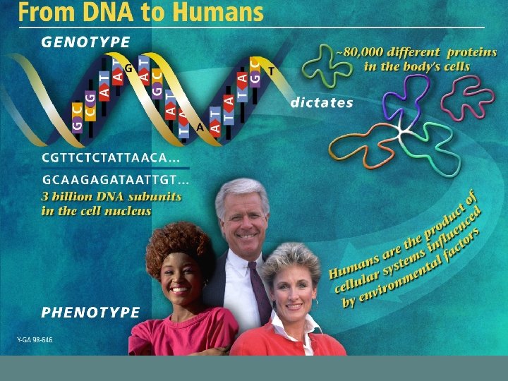
Nucleic Acids: DNA and RNA Nucleic acids 1. » » are long chains of nucleotides the “genetic molecules” Nucleotides 2. » » the building blocks (monomers) of DNA and RNA As monomers they power almost all processes in all cells – e. g. ATP
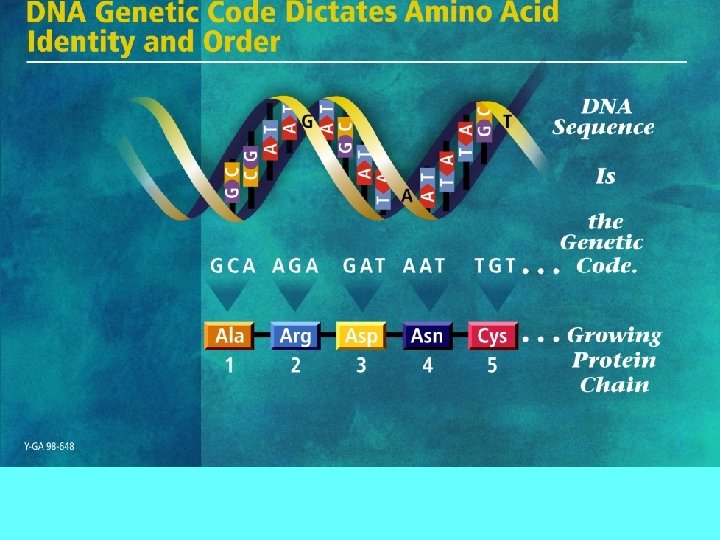
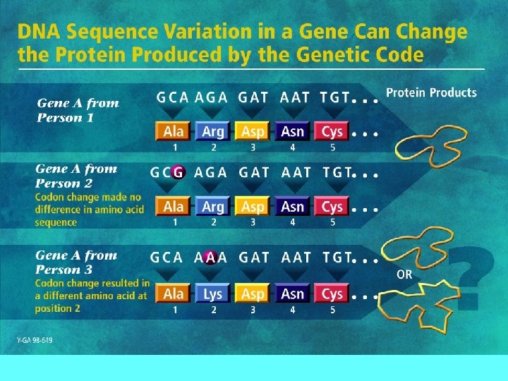
DNA Nucleotides 1. 2. Four Kinds of nucleotides in DNA A = Adenine T = Thymine G = Guanine C = Cytosine Central dogma of Biology a. The order of nucleotides in a gene determines the order of ________________ in a protein – The order of ___________ in a protein determines ____________ of a protein which in turn determines the ___________ of the protein.
Nucleotide Structure Nucleotides are. . the building blocks (monomers) of DNA and RNA As monomers they transfer energy to power almost all processes in all cells –e. g. ATP
~26, 000 genes code for proteins that perform all life functions