
The Rhesus Rh Blood Group system Mohammed Laqqan
The Rhesus (Rh) Blood Group system Mohammed Laqqan
The Rh(D) Antigen l l l Rh is the most complex system, with over 45 antigens. The complexity of the Rh blood group Ags is due to the highly polymorphic genes that encode them. Discovered in 1940 after work on Rhesus monkeys. The 2 nd most important after ABO in the crossmatch test. Only the most clinically significant Ags will be discussed. Mohammed Laqqan
Rh Genetics l The genes that control the system are autosomal codominant located on the short arm of chromosome 1. Mohammed Laqqan
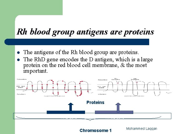
Rh blood group antigens are proteins l l The antigens of the Rh blood group are proteins. The Rh. D gene encodes the D antigen, which is a large protein on the red blood cell membrane, & the most important. Proteins RHD gene RHCE gene Chromosome 1 Mohammed Laqqan
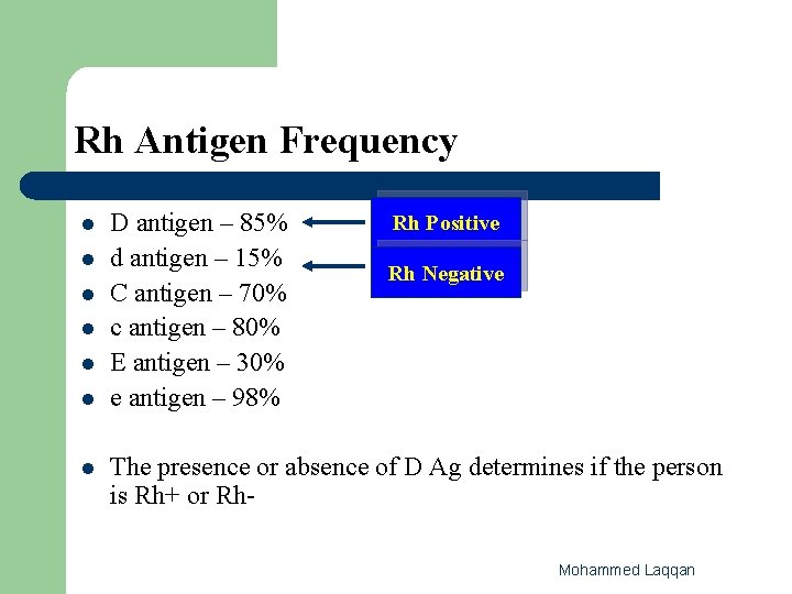
Rh Antigen Frequency l l l l D antigen – 85% d antigen – 15% C antigen – 70% c antigen – 80% E antigen – 30% e antigen – 98% Rh Positive Rh Negative The presence or absence of D Ag determines if the person is Rh+ or Rh. Mohammed Laqqan
Nomenclature of the RH system 3 Different nomenclatures: 1 - Fisher-Race 2 - Weiner 3 - Rosenfield Nomenclature Mohammed Laqqan
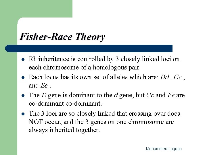
Fisher-Race Theory l l Rh inheritance is controlled by 3 closely linked loci on each chromosome of a homologous pair Each locus has its own set of alleles which are: Dd , Cc , and Ee. The D gene is dominant to the d gene, but Cc and Ee are co-dominant. The 3 loci are so closely linked that crossing over does NOT occur, and the 3 genes on one chromosome are always inherited together. Mohammed Laqqan
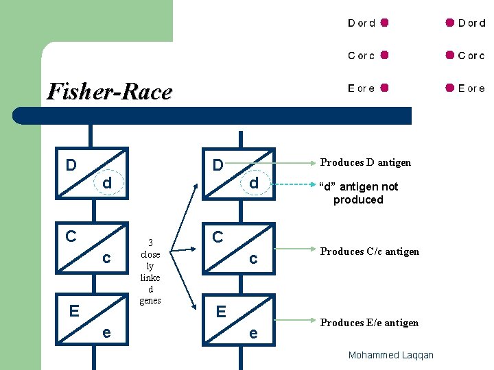
Fisher-Race D D d C c E e 3 close ly linke d genes Produces D antigen d C c E e “d” antigen not produced Produces C/c antigen Produces E/e antigen Mohammed Laqqan
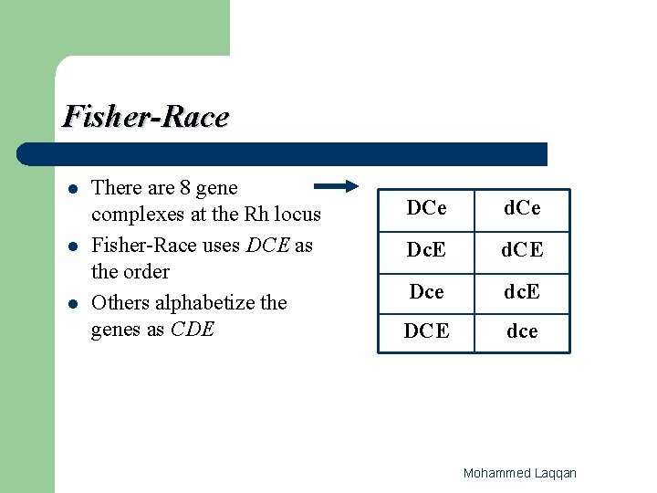
Fisher-Race l l l There are 8 gene complexes at the Rh locus Fisher-Race uses DCE as the order Others alphabetize the genes as CDE DCe d. Ce Dc. E d. CE Dce dc. E DCE dce Mohammed Laqqan
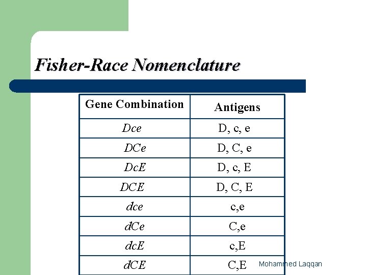
Fisher-Race Nomenclature Gene Combination Antigens Dce D, c, e DCe D, C, e Dc. E D, c, E DCE D, C, E dce c, e d. Ce C, e dc. E c, E d. CE C, E Mohammed Laqqan
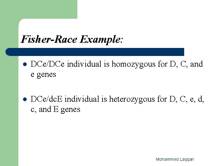
Fisher-Race Example: l DCe/DCe individual is homozygous for D, C, and e genes l DCe/dc. E individual is heterozygous for D, C, e, d, c, and E genes Mohammed Laqqan
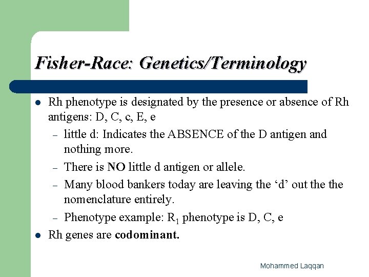
Fisher-Race: Genetics/Terminology l l Rh phenotype is designated by the presence or absence of Rh antigens: D, C, c, E, e – little d: Indicates the ABSENCE of the D antigen and nothing more. – There is NO little d antigen or allele. – Many blood bankers today are leaving the ‘d’ out the nomenclature entirely. – Phenotype example: R 1 phenotype is D, C, e Rh genes are codominant. Mohammed Laqqan
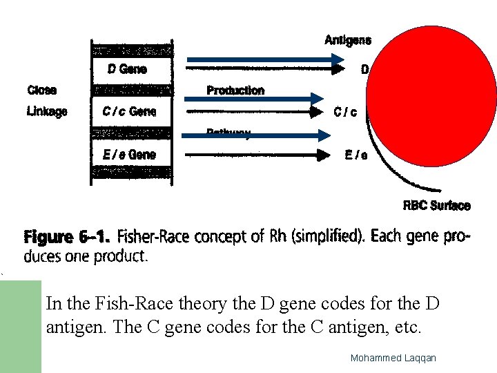
In the Fish-Race theory the D gene codes for the D antigen. The C gene codes for the C antigen, etc. Mohammed Laqqan
Wiener Theory l l l Good for describing phenotype There is one Rh locus at which occurs one Rh gene, but this gene has multiple alleles. For example, one gene R 1 produces one agglutinogen (antigen) Rh 1 which is composed of three "factors" The three factors are analogous to C, D, and e respectively The main difference between the Fisher-Race and Wiener theories is that the: – – Fisher-Race theory has three closely linked loci, the Wiener theory has only one gene locus at which multiple alleles occur. Mohammed Laqqan
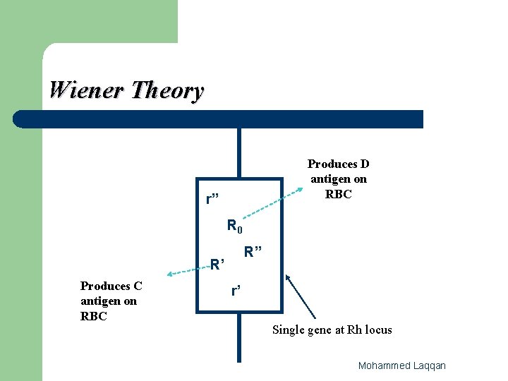
Wiener Theory Produces D antigen on RBC r” R 0 R” R’ Produces C antigen on RBC r’ Single gene at Rh locus Mohammed Laqqan

Wiener l Wiener further theorized that 8 major genes led to different combinations of antigens (D, C, E, c, e): – – R 0, R 1, R 2, Rz r, r′, r″, ry Mohammed Laqqan

2 - Weiner Nomenclature expressed by the use of a single letter. R D present r D absent Prime ′ or 1 C Double ″ or 2 E Mohammed Laqqan
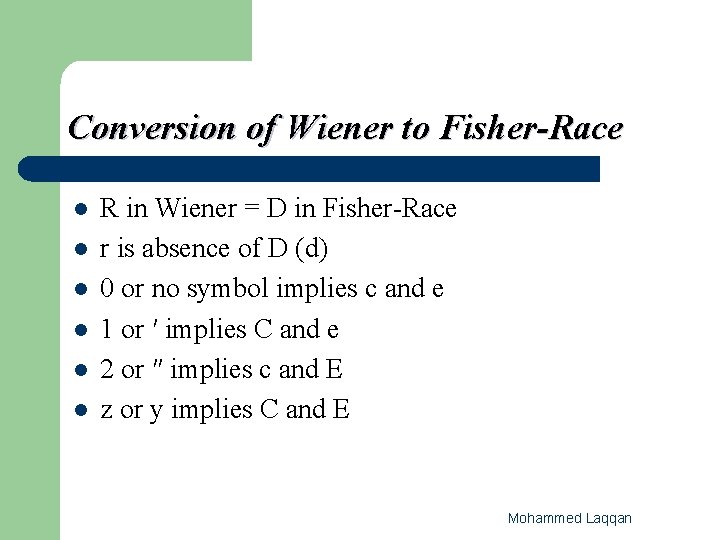
Conversion of Wiener to Fisher-Race l l l R in Wiener = D in Fisher-Race r is absence of D (d) 0 or no symbol implies c and e 1 or ′ implies C and e 2 or ″ implies c and E z or y implies C and E Mohammed Laqqan
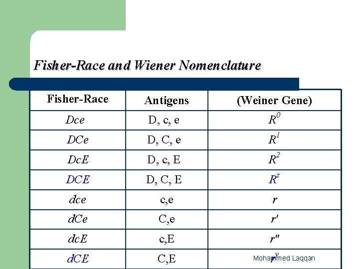
Fisher-Race and Wiener Nomenclature Fisher-Race Antigens (Weiner Gene) Dce D, c, e R 0 DCe D, C, e R 1 Dc. E D, c, E R 2 DCE D, C, E Rz dce c, e r d. Ce C, e r′ dc. E c, E r″ d. CE C, E ry Mohammed Laqqan
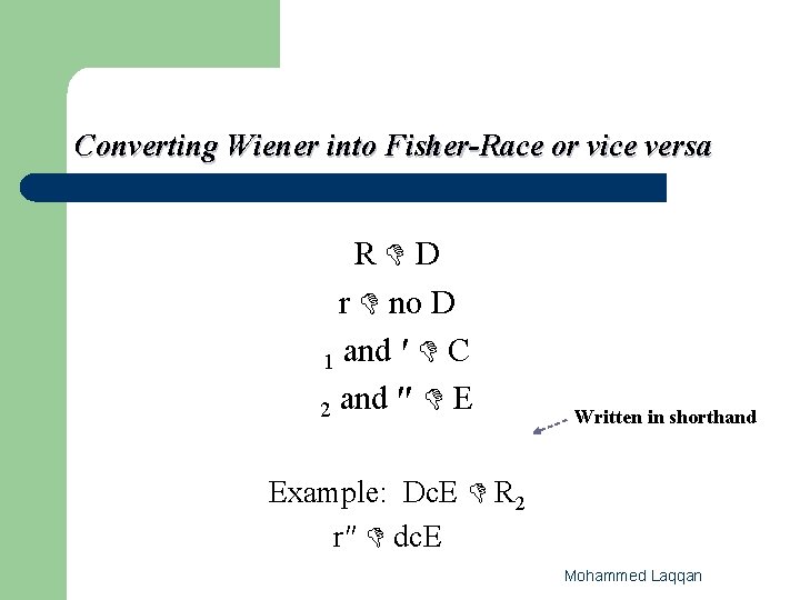
Converting Wiener into Fisher-Race or vice versa R D r no D 1 and ′ C 2 and ″ E Written in shorthand Example: Dc. E R 2 r″ dc. E Mohammed Laqqan
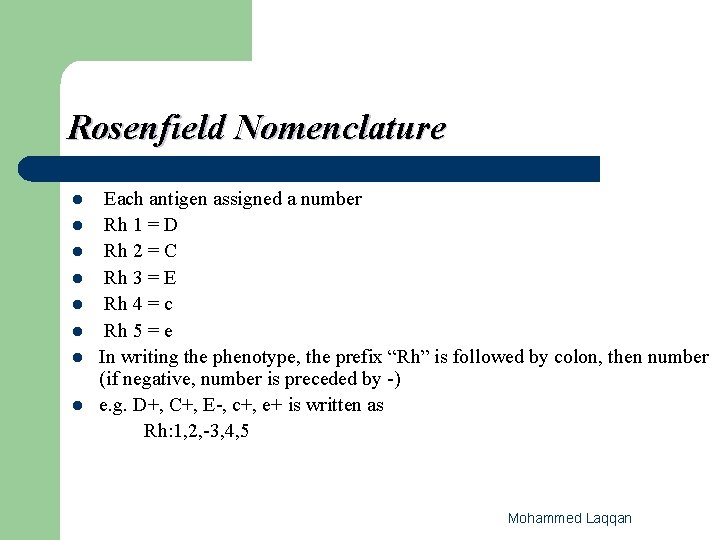
Rosenfield Nomenclature l l l l Each antigen assigned a number Rh 1 = D Rh 2 = C Rh 3 = E Rh 4 = c Rh 5 = e In writing the phenotype, the prefix “Rh” is followed by colon, then number (if negative, number is preceded by -) e. g. D+, C+, E-, c+, e+ is written as Rh: 1, 2, -3, 4, 5 Mohammed Laqqan
Significance l l l After ABO, the Rh system is the second most important system. This is because: The D antigen is extremely immunogenic. It causes the production of anti-D in 50 - 70% of Rh(D) negative people who are exposed to the D antigen. Moreover, anti-D is the most common cause of severe HDN and can cause in Utero death. Because of this, in blood transfusion, the patient and donor are matched for Rh(D) type as well as ABO groups. The C and E Ags are not as immunogenic as D, routine typing for these Ags is not performed Mohammed Laqqan
Rh Deleted l l l Red cells that express no Ags at the C & E loci ( D ) Number of D Ags greatly increase Anti-D Ig. G Abs can agglutinate these cells Mohammed Laqqan
Rh null l RH null: individual that appears to have no Rh antigens ( �, �, �) RBC has fragile membrane- short lived Must use autologous blood products – l Demonstrate mild hemolytic anemia (Rh antigens are integral part of RBC membrane and absence results in loss of membrane integrity) – l No D, C, c, E, e antigens present on the RBC membrane Stomatocytosis. When transfusion is necessary ONLY Rh Null blood can be used to transfuse. Mohammed Laqqan
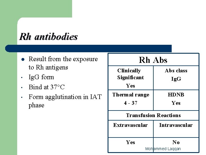
Rh antibodies l • • • Result from the exposure to Rh antigens Ig. G form Bind at 37°C Form agglutination in IAT phase Rh Abs Clinically Significant Yes Abs class Ig. G Thermal range 4 - 37 HDNB Yes Transfusion Reactions Extravascular Intravascular Yes No Mohammed Laqqan
Clinical Significance of Rh antibodies • • • Related to Hemolytic transfusion reactions Re-exposure to antigen cause rapid secondary response Always check patients history for previous transfusion or pregnancy to avoid re-exposure. Mohammed Laqqan
Hemolytic disease of the Newborn (HDN) l l Usually related to D antigen exposure and the formation of anti-D Usually results from D negative female and D positive male producing and offspring. – l 1 st l The baby will probably be D positive. pregnancy not effected, the 2 nd pregnancy and on will be effected-results in still birth, severe jaundice, anemia related to HDN. To prevent this occurrence the female is administered RHIG. Mohammed Laqqan
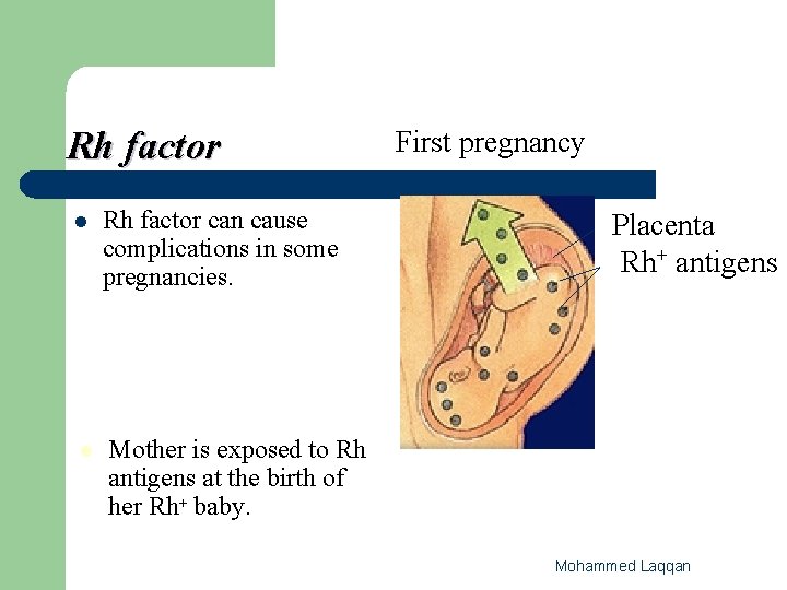
Rh factor l l Rh factor can cause complications in some pregnancies. First pregnancy Placenta Rh+ antigens Mother is exposed to Rh antigens at the birth of her Rh+ baby. Mohammed Laqqan
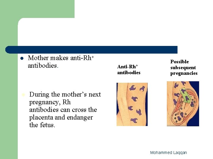
l Mother makes anti-Rh+ antibodies. Anti-Rh+ antibodies l Possible subsequent pregnancies During the mother’s next pregnancy, Rh antibodies can cross the placenta and endanger the fetus. Mohammed Laqqan
Weak D Phenotype l Most D positive rbc’s react macroscopically with Reagent anti-D at immediate spin – – l These patients are referred to as Rh positive Reacting from 1+ to 3+ or greater HOWEVER, some D-positive rbc’s DO NOT react (do NOT agglutinate) at Immediate Spin using Reagent Anti-D. These require further testing (37 o. C and/or AHG) to determine the D status of the patient. Mohammed Laqqan
Variants of D • • Weak expression of the Rh system on the RBC, (Du) Du red cells can be classified into three categories according to the mechanism that account for the Weak D antigen Mohammed Laqqan
Categories of Du red cells 1 - Acquired Du (Position Effect) 2 - Du Variant (Partial D) 3 - Hereditary Du (Genetically Transmissible) Mohammed Laqqan
1 - Acquired Du (Position Effect) l C allele in trans position to D allele – Example: Dce/d. Ce, Dc. E/d. CE In both of these cases the C allele is in the trans position in relation to the D allele. l D antigen is normal, C antigen appears to be crowding the D antigen. (Steric hindrance) l Does NOT happen when C is in cis position l Example: DCe/dce Can safely transfuse D positive blood components. – Mohammed Laqqan

2 - Du Variant (Partial D) l l The D- Ag consists of at least 4 parts Missing one or more PARTS (epitopes) of the D antigen remaining Ag is weakly expressed Alloantibodies are produced to the missing parts Du variants should receive Rh –ve blood when transfused Mohammed Laqqan
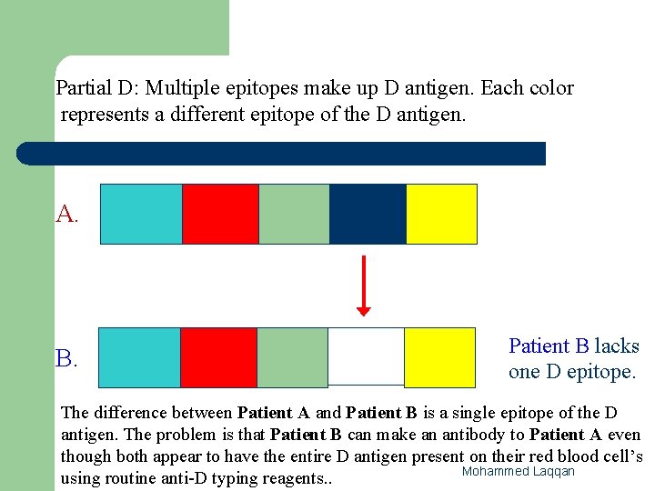
Partial D: Multiple epitopes make up D antigen. Each color represents a different epitope of the D antigen. A. B. Patient B lacks one D epitope. The difference between Patient A and Patient B is a single epitope of the D antigen. The problem is that Patient B can make an antibody to Patient A even though both appear to have the entire D antigen present on their red blood cell’s Mohammed Laqqan using routine anti-D typing reagents. .
3 - Hereditary Du (Genetically Transmissible) l The RHD gene codes for weakened expression of D antigen in this mechanism. – – l l l D antigen is complete, there are just fewer D Ag sites on the rbc. Quantitative! Common in Black population (usually Dce haplotype). Very rare in White population. Agglutinate weakly or not at all at immediate spin phase. Agglutinate strongly at AHG phase. Can safely transfuse D positive blood components. Mohammed Laqqan
- Slides: 36