
The Respiratory System Use this ppt to complete


- Slides: 43
The Respiratory System Use this ppt to complete notes pgs 1 -4. Study the information as you go & discuss each slide with each other. Ask me if you need any explanation or have a question about the info.
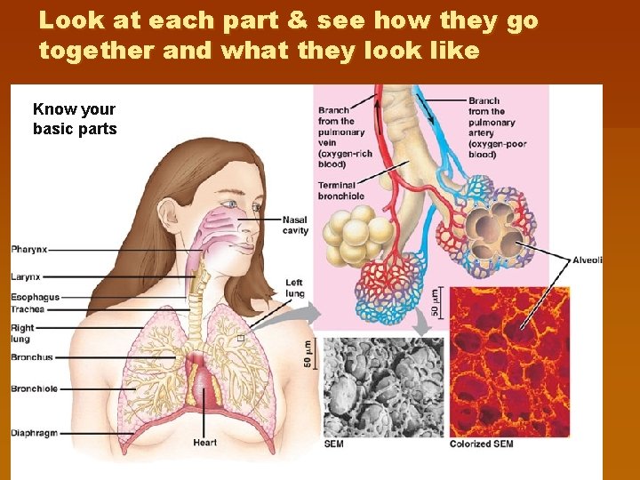
Look at each part & see how they go together and what they look like Know your basic parts
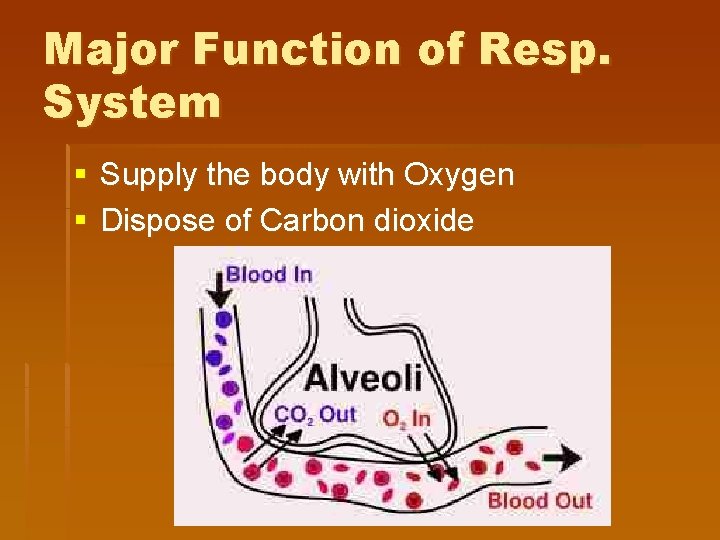
Major Function of Resp. System § Supply the body with Oxygen § Dispose of Carbon dioxide
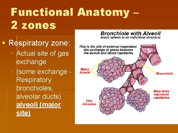
Functional Anatomy – 2 zones § Respiratory zone: § Actual site of gas exchange § (some exchange Respiratory bronchioles, alveolar ducts) alveoli (major site)
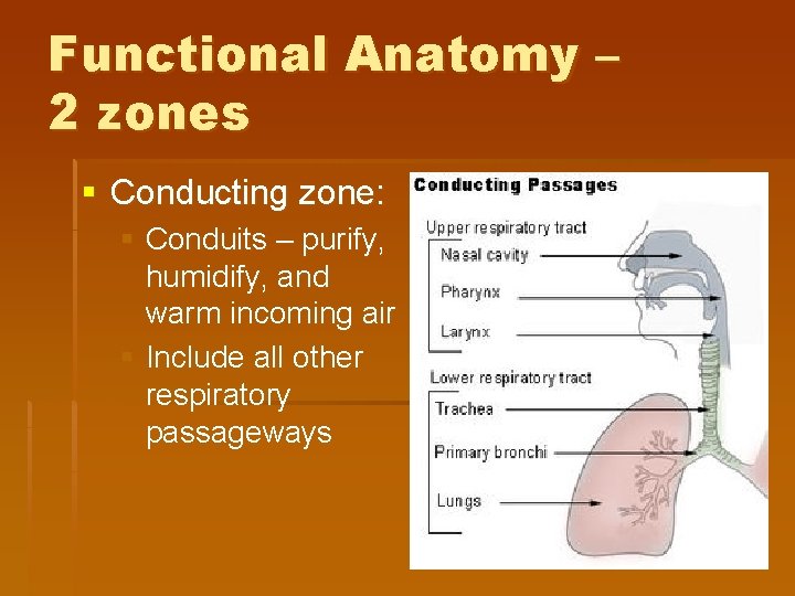
Functional Anatomy – 2 zones § Conducting zone: § Conduits – purify, humidify, and warm incoming air § Include all other respiratory passageways
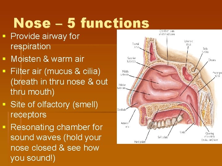
Nose – 5 functions § Provide airway for respiration § Moisten & warm air § Filter air (mucus & cilia) (breath in thru nose & out thru mouth) § Site of olfactory (smell) receptors § Resonating chamber for sound waves (hold your nose closed & see how you sound!)
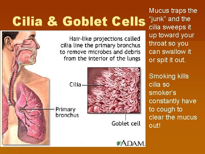
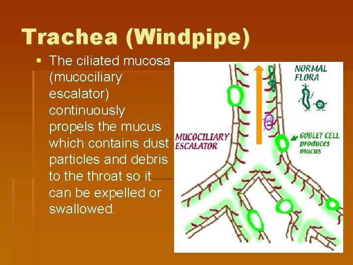
Cilia & Goblet Cells Mucus traps the “junk” and the cilia sweeps it up toward your throat so you can swallow it or spit it out. Smoking kills cilia so smoker’s constantly have to cough to clear the mucus out!
Cold day = Runny nose § The cilia in your nose become sluggish & slow when they are cold & do not move the mucus down into your throat § Mucus in the nasal cavity accumulates & dribbles out
Nasal Conchae § Nasal Conchae aka. Nasal. Turbinates= increase SA of mucosa exposed to air to help warm & filter it – also increase turbulence (mini tornado effect) of air – more inhaled particles swirled onto mucus and trapped
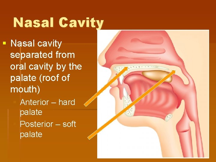
Nasal Cavity § Nasal cavity separated from oral cavity by the palate (roof of mouth) § Anterior – hard palate § Posterior – soft palate
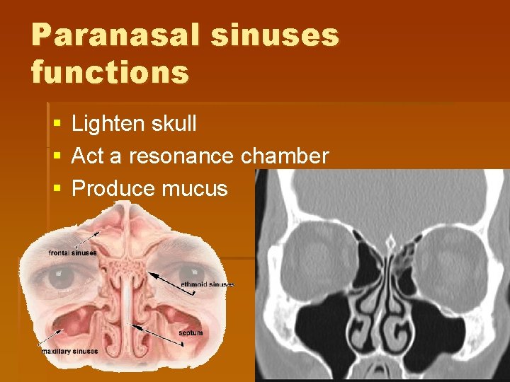
Paranasal sinuses functions § § § Lighten skull Act a resonance chamber Produce mucus
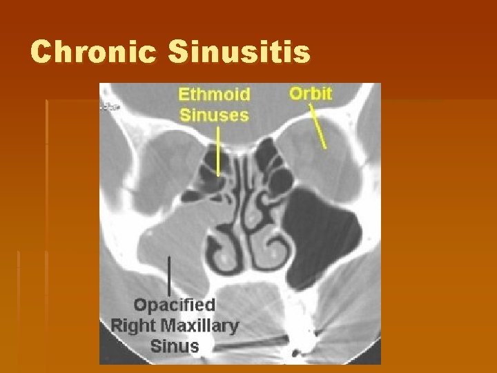
Chronic Sinusitis
Stop and be sure you completely understand this page before you move on
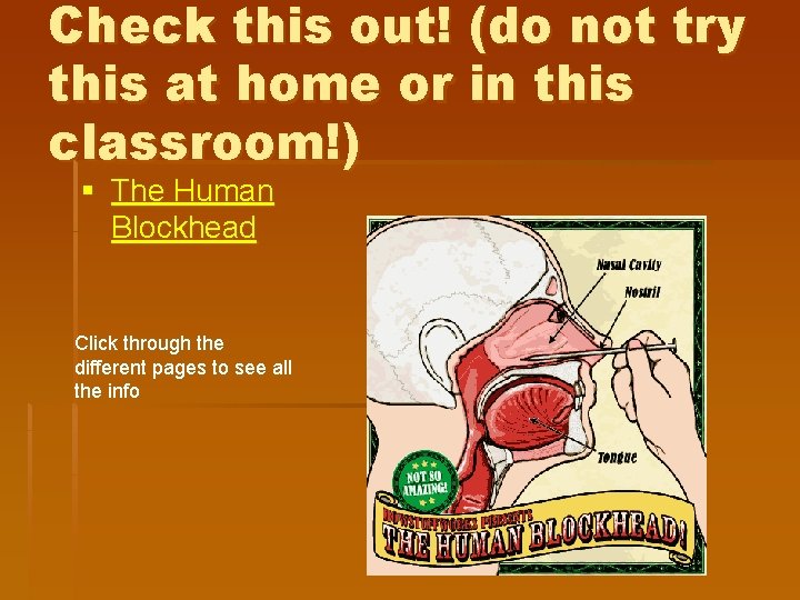
Check this out! (do not try this at home or in this classroom!) § The Human Blockhead Click through the different pages to see all the info
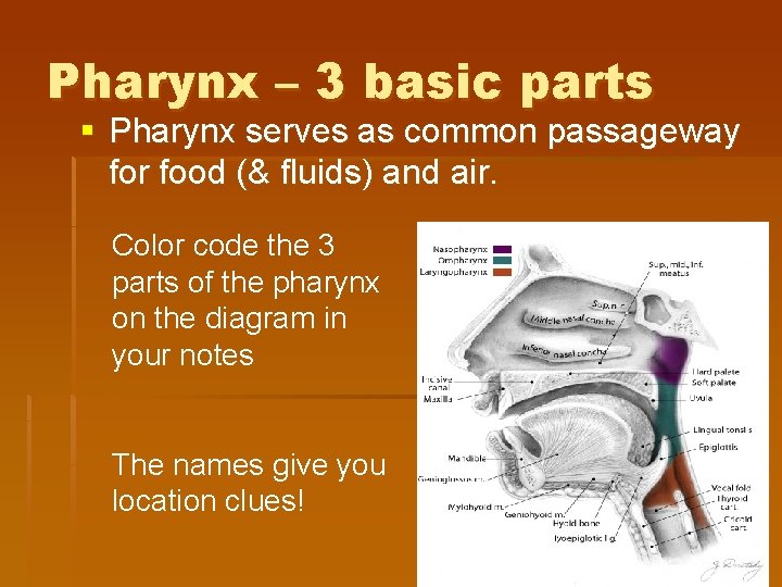
Pharynx – 3 basic parts § Pharynx serves as common passageway for food (& fluids) and air. Color code the 3 parts of the pharynx on the diagram in your notes The names give you location clues!
Pharynx – 3 basic parts § Nasopharynx – air only § During swallowing, Soft palate & uvula rise upward to close off nasopharynx which prevents food & fluids from entering it § Oropharynx & Laryngopharynx – food, liquids & air § Food will be directed posteriorly to the esophagus § Air will go anteriorly into the larynx
Tonsils (think about the name – it tells you the location) § Pharyngeal tonsils: aka. Adenoids – located in nasopharynx § Palatine tonsils: located in oropharynx § Lingual tonsils: located at base of tongue § All tonsils are lymph nodes & work with immune system § You will be labeling these on the back page diagram
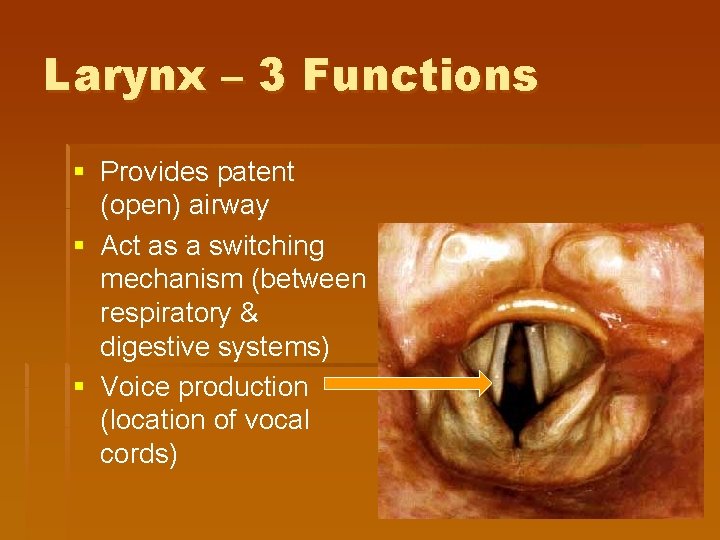
Larynx – 3 Functions § Provides patent (open) airway § Act as a switching mechanism (between respiratory & digestive systems) § Voice production (location of vocal cords)
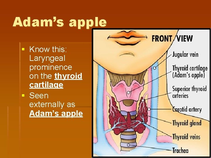
Adam’s apple § Know this: Laryngeal prominence on the thyroid cartilage § Seen externally as Adam’s apple
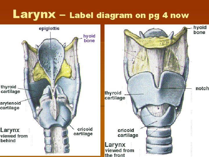
Larynx – Label diagram on pg 4 now
Epiglottis § 9 th cartilage § When air is flowing into the larynx – free edge projects upward § During swallowing: § Larynx is pulled upward § Epiglottis is tipped back and down to cover laryngeal inlet into trachea § Routes food/fluid into esophagus
Cough Reflex § Initiated if anything other than air enters the larynx § Pressure from air moves object upward out of the larynx § Reflex does not work when unconscious so not a good idea: § To give fluids to an unconscious person § Also a reason why people in an alcoholic coma often die from aspirating their own vomit.
Trachea (Windpipe) § The ciliated mucosa (mucociliary escalator) continuously propels the mucus which contains dust particles and debris to the throat so it can be expelled or swallowed.
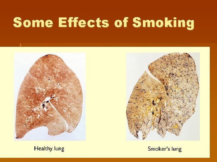
Smoking § Diminishes ciliary activity § Coughing is ONLY method of preventing mucus accumulation in the lungs § Smokers should never be given medications that INHIBIT the cough reflex.
Some Effects of Smoking
Stop and be sure you completely understand this page before you move on
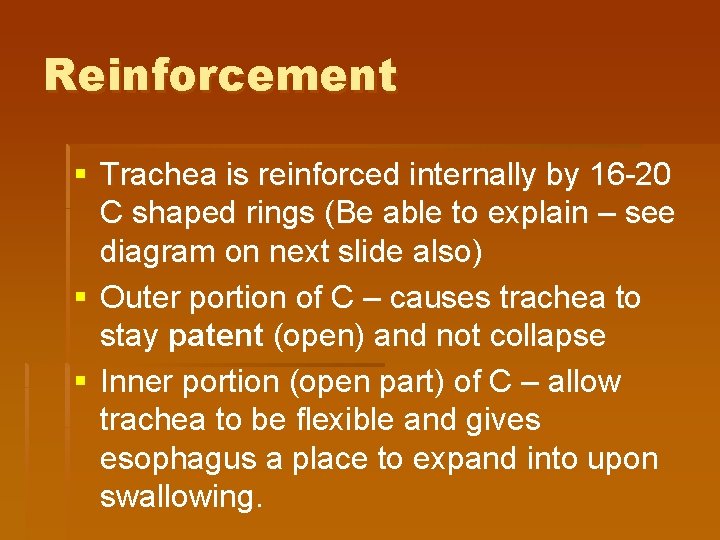
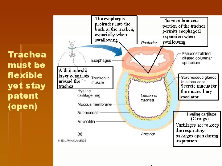
Reinforcement § Trachea is reinforced internally by 16 -20 C shaped rings (Be able to explain – see diagram on next slide also) § Outer portion of C – causes trachea to stay patent (open) and not collapse § Inner portion (open part) of C – allow trachea to be flexible and gives esophagus a place to expand into upon swallowing.
Trachea must be flexible yet stay patent (open)
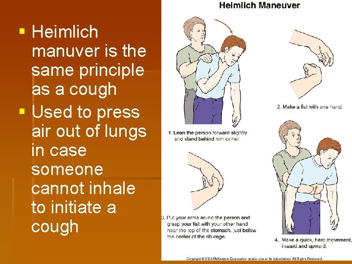
§ Heimlich manuver is the same principle as a cough § Used to press air out of lungs in case someone cannot inhale to initiate a cough
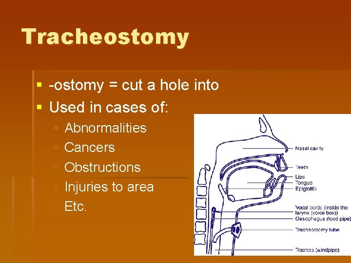
Tracheostomy § -ostomy = cut a hole into § Used in cases of: § Abnormalities § Cancers § Obstructions § Injuries to area § Etc.
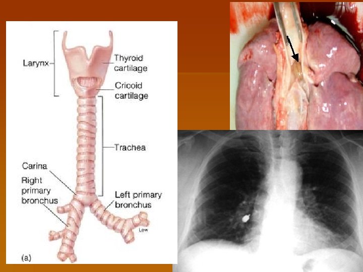
Bronchial Tree § Trachea divides into right and left primary bronchi at the level of the sternal angle (where manubrium and body of sternum meet). § Inhaled objects usually lodge in the right primary bronchus since it is wider, shorter, and at a more vertical angle
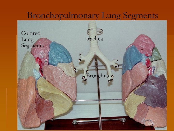
Lungs § Left lung is smaller, consisting of 2 lobes and contains a cardiac notch § Right lung has 3 lobes § FYI: Bronchopulmonary segments § Served by own artery, vein, and individual segmental bronchus § Left lung has 8 segments while right lung has 10.
FYI: Important Info § Respiratory therapists and surgeons use this info about the different bronchopulmonary segments so they can treat the patient as needed § Even to the point of removing the diseased segment and leaving the good tissue § The lungs weigh approximately 2. 5 pounds
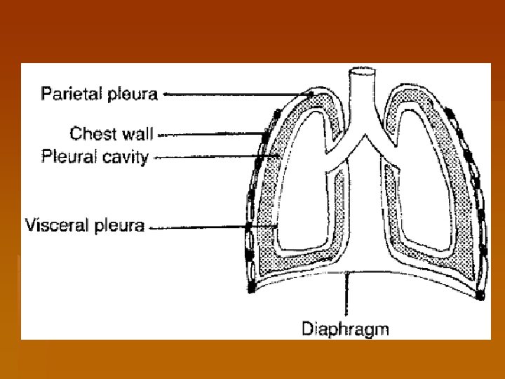
Pleurae: Review § Parietal vs. visceral § Function of pleural fluid § Lubricate layers so they can slide across each other § Cause them to cling tightly to each other through surface tension (helps maintain pressure differences necessary for inhaling/exhaling)
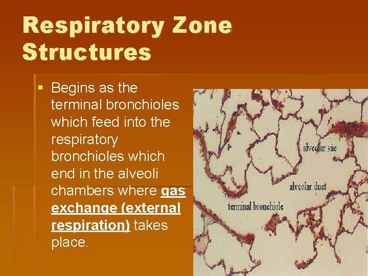
Respiratory Zone Structures § Begins as the terminal bronchioles which feed into the respiratory bronchioles which end in the alveoli chambers where gas exchange (external respiration) takes place.
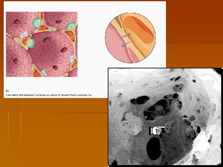
Alveoli § Composed of simple squamous – much thinner than a sheet of paper § Membrane has gas on one side and blood on the other. § Account for the largest portion of lung volume and provide a tremendous surface area for gas exchange
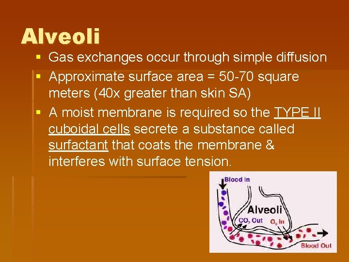
Alveoli § Gas exchanges occur through simple diffusion § Approximate surface area = 50 -70 square meters (40 x greater than skin SA) § A moist membrane is required so the TYPE II cuboidal cells secrete a substance called surfactant that coats the membrane & interferes with surface tension.
Page 4 diagram § Use the lab book or the text book or the internet to label the head diagram § Label only the ones that have a dot on the end. § Be very specific about the structures. § May check with me when done.
Study for Quiz #1 Remember – it includes the diagrams!!