THE RESPIRATORY SYSTEM MARAM ABDALJALEEL MD FACULITY OF

THE RESPIRATORY SYSTEM MARAM ABDALJALEEL, MD FACULITY OF MEDICINE UNIVESITY OF JORDAN Maram Abdaljaleel, MD Dermatopathologist. AND and Neuropathologist DERMATOPATHOLOGIST NEUROPATHOLOGIST https: //www. 123 rf. com/photo_38644498_stock-illustration-mascot-illustration-of-the-lungs-coughing-violently. html

OBJECTIVES: • Function and anatomy • Microscopic structure of the alveolar wall • Atelectasis (Collapse) • Acute respiratory distress syndrome (ARDS) • Restrictive vs. Obstructive lung diseases

FUNCTION AND ANATOMY: The major function of the lung is to replenish oxygen and remove carbon dioxide from blood.
https: //teachmeanatomy. info/thorax/organs/tracheobronchial-tree/")
By Teach. Me. Series Ltd (2019)https: //teachmeanatomy. info/thorax/organs/tracheobronchial-tree/

http: //www. medicinehack. com/2011/05/microscopic-structure-of-alveolar-wall. html

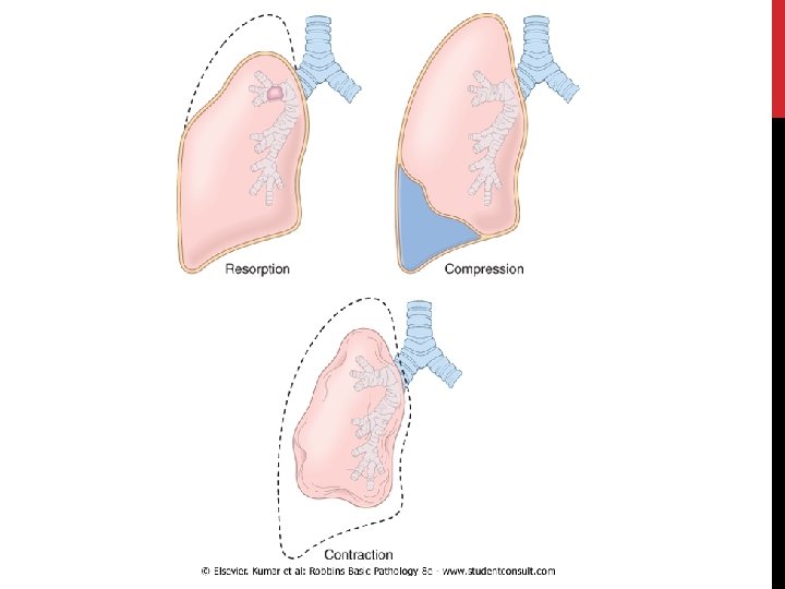
ATELECTASIS Robbins and Cotran Atlas of Pathology - 3 rd Edition

ATELECTASIS • is loss of lung volume caused by inadequate expansion of air spaces. • It results in shunting of inadequately oxygenated blood from pulmonary arteries into veins, thus giving rise to a ventilation perfusion imbalance and hypoxia.
")
THREE TYPES: • Resorption atelectasis • Compression atelectasis • Contraction atelectasis (cicatrization atelectasis)
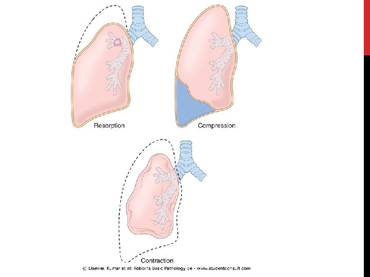

1. RESORPTION ATELECTASIS • Due to total obstruction of a bronchus preventing air from reaching distal airways. • The air already present gradually becomes absorbed, and alveolar collapse follows.

RESORPTION ATELECTASIS, CAUSED BY: • The most common cause is Obstruction of a bronchus by: ü Intrabronchial mucous or mucopurelant plugs in post operative patients. ü Foreign body aspiration, especially in children ü Obstructive lung disease: bronchial asthma, bronchiectasis, chronic bronchitis ü Intrabronchial tumors.
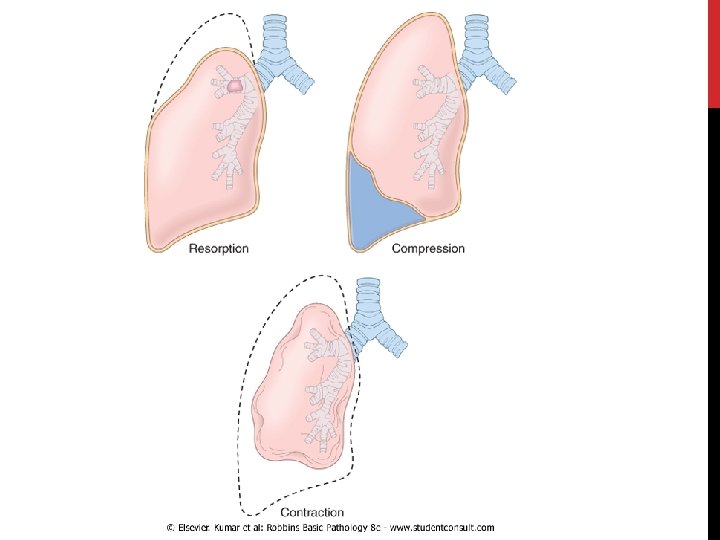

2. COMPRESSION ATELECTASIS • caused by accumulation of fluid, blood, or air within pleural cavity, which mechanically collapse adjacent lung. a. Pleural effusion like in Congestive Heart Failure b. Pneumothorax: air in the pleural cavity
 • Occurs due to local or generalized fibrosis of")
3. CONTRACTION ATELECTASIS (CICATRIZATION ATELECTASIS) • Occurs due to local or generalized fibrosis of the lung or pleura that prevents full expansion of the lung. Atelectasis (except when caused by contraction) is potentially reversible and should be treated promptly to prevent hypoxemia and superimposed infection of the collapsed lung.
 • The epidemiology and definition are evolving. • Formerly")
ACUTE RESPIRATORY DISTRESS SYNDROME (ARDS) • The epidemiology and definition are evolving. • Formerly considered to be the severe end of a spectrum of acute lung injury • Defined as respiratory failure occurring within 1 week of a known clinical insult with bilateral opacities on chest imaging, NOT fully explained by effusions, atelectasis, cardiac failure, or fluid overload.
: • graded based on the severity of the changes")
ACUTE RESPIRATORY DISTRESS SYNDROME (ARDS): • graded based on the severity of the changes in arterial blood oxygenation. • extensive bilateral injury to alveoli known histologically as diffuse alveolar damage (DAD)

SEVERE ARDS: • characterized by rapid onset of life-threatening: a. respiratory insufficiency. b. Cyanosis c. Severe arterial hypoxemia that becomes refractory to oxygen therapy and may progress to multisystem organ failure.
 Ø sepsis (30%– 35) Ø Aspiration Ø trauma")
ARDS; TRIGGERS: Ø pneumonia (35%– 45%) Ø sepsis (30%– 35) Ø Aspiration Ø trauma (including brain injury, abdominal surgery, and multiple fractures) Ø pancreatitis Ø transfusion reactions.

ARDS should not be confused with respiratory distress syndrome of the newborn; the latter is caused by a deficiency of surfactant caused by prematurity.

Robbins and Cotran pathologic basis of disease, 9 th edition

PATHOGENESIS: • the integrity of the alveolar-capillary membrane is compromised by endothelial and epithelial injury. • As early as 30 minutes after an acute insult, there is increased synthesis and release of IL-8, IL-1 and TNF by pulmonary macrophages. • leading to endothelial activation and sequestration and activation & chemotaxsis of neutrophils in pulmonary capillaries.

PATHOGENESIS/CONT. • Activated neutrophils release reactive oxygen species & proteases that damage the alveolar epithelium and endothelium causing vascular leakiness and loss of surfactant that render the alveolar unit unable to expand. • the destructive forces are counteracted by endogenous anti-proteases and anti-oxidants

• In the end, it is the balance between the destructive and protective factors that determines the degree of tissue injury and clinical severity of the ARDS.

HISTOLOGY: • In the acute phase of ARDS : Ø The most characteristic finding is the presence of hyaline membranes Ø consists of fibrin-rich edema fluid admixed with remnants of necrotic epithelial cells (similar to respiratory distress syndrome of the newborn)

FIGURE 13. 3 A, ROBBINS BASIC PATHOLOGY, 10 TH EDITION

HISTOLOGY: In the organizing stage: Ø proliferation of type II pneumocytes Øintraalveolar fibrosis due to organization of the fibrin-rich exudates. ØMarked thickening of the alveolar septa due to proliferation of interstitial cells and collagen deposition.

FIGURE 13. 3 B, ROBBINS BASIC PATHOLOGY, 10 TH EDITION

CLINICAL FEATURES • Patients are hospitalized for one of the predisposing conditions • Profound dyspnea and tachypnea followed by increasing cyanosis and hypoxemia, respiratory failure, and the appearance of diffuse bilateral infiltrates on radiographic examination. • Hypoxemia may be refractory to oxygen therapy

OUTCOME: • The overall hospital mortality rate is 38. 5%. • Most patients who survive the acute insult recover normal respiratory function within 6 to 12 months, but the rest develop diffuse interstitial fibrosis leading to chronic respiratory insufficiency
 3. development of multiorgan")
PREDICTORS OF POOR PROGNOSIS 1. advanced age 2. bacteremia (sepsis) 3. development of multiorgan failure

OBSTRUCTIVE VS. RESTRICTIVE DIFFUSE PULMONARY DISEASES can be classified into two Categories: 1 - OBSTRUCTIVE AIRWAY DISEASES: characterized by an increase in resistance to airflow caused by partial or complete obstruction at any level 2 - RESTRICTIVE DISEASES: characterized by reduced expansion of lung parenchyma and decreased total lung capacity.

Restrictive defects occur in two general conditions: 1. chest wall disorders in the presence of normal lungs: • severe obesity, diseases of the pleura, and neuromuscular disorders that affect the respiratory muscles 2. acute or chronic interstitial lung diseases: Ø The classic acute restrictive disease is ARDS. Ø Chronic restrictive diseases include the pneumoconioses, interstitial fibrosis of unknown etiology, and sarcoidosis.

THANK YOU!
- Slides: 34