The Respiratory System for student copy Functions of


- Slides: 32
The Respiratory System for student copy
Functions of the Respiratory System • Gas Exchange
Organs of the Respiratory System • Upper Respiratory Tract – Nose – Pharynx – Larynx • Lower Respiratory Tract – Trachea – Bronchi – Lungs
Nose • framework composed of bone & cartilage • 2 nostrils called: external nares – where air enters the nasal cavity – rt & lt separated by nasal septum • site of nose bleeds @ internal edge: internal nares
Pharynx • throat • 3 divisions: 1. Nasopharynx – begins @ internal nares end of soft palate 2. Oropharynx – edge of soft palate hyoid bone 3. Laryngopharynx – hyoid bone upper edge of esophagus
3 Parts of the Pharynx
Larynx • “voice box” • Cartilage • Parts: – – Epiglottis Glottis Thyroid cartilage Cricoid cartilage
Larynx • moves upward when you swallow – tips epiglottis over the glottis (opening of trachea) – allows food esophagus (--/ down trachea to lungs) – if not swallowing: glottis is open allowing air lungs – http: //www. linkstudio. info/images/portfolio/medani/Swal low. swf
Trachea • rings of cartilage maintain its shape to prevent it from closing • forks into 2 bronchi
Bronchus • each enters a lung where it branches into smaller & smaller bronchioles resembling an inverted tree
Bronchioles • fine tubes that allow passage of air • smooth muscle surrounds them when contracts airways constrict • epithelium covered with cilia & mucus • mucus traps dust, particulates • cilia beat upward removing trapped particles from airways (moves particles ~1 -3 cm/hr)
Bronchioles
Gas Exchange in Lungs
Pulmonary Function Tests • “PFTs” • subject breathes into a closed system in which air is trapped w/in a bell floating in water • bell moves up when patient exhales / down when they inhale
Pulmonary Function Tests • Tidal Volume: – amt of air expired • Vital Capacity: – max amt of air that can forcefully exhaled after a max inhalation
Spirogram
Anatomical Dead Space • not all inspired air will get into the lungs • exhaling does not force all air out of the body
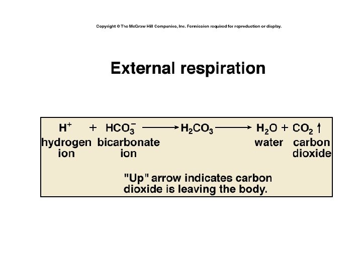
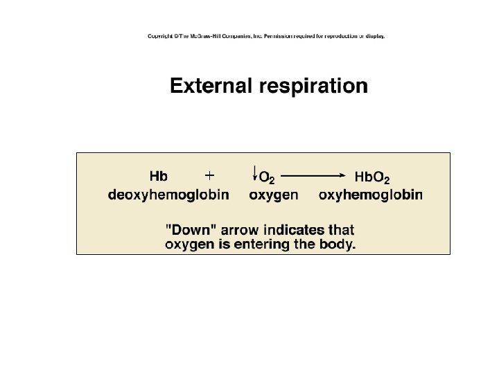
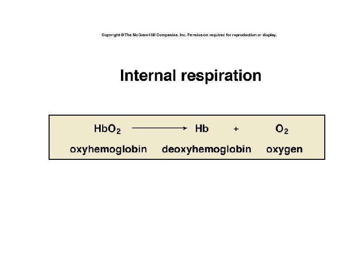
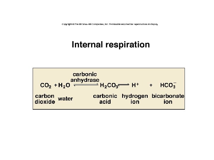
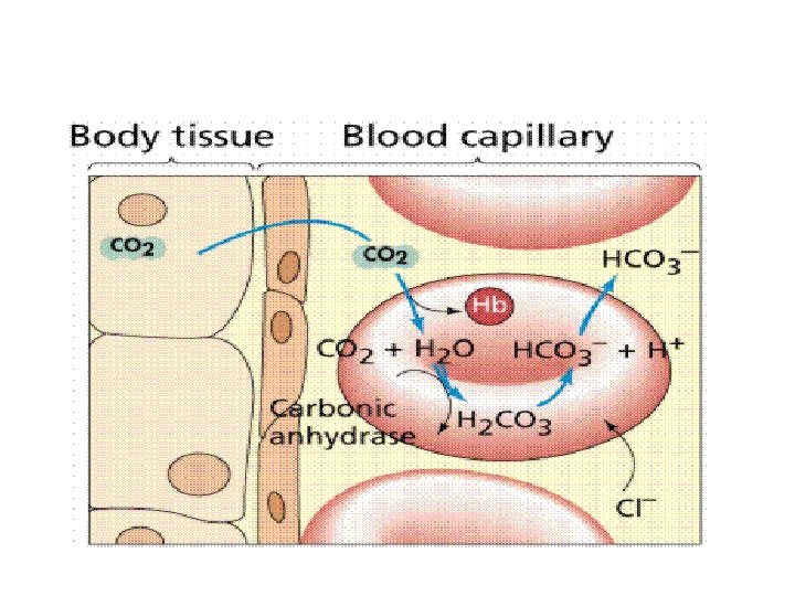
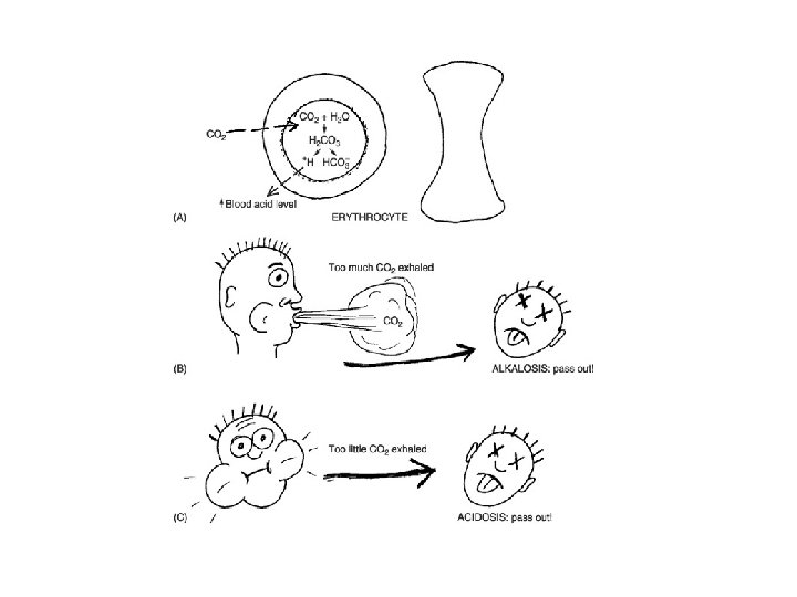
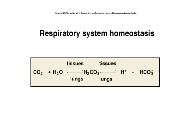
Hemoglobin • helps transport oxygen, carbon dioxide, & buffer blood • as carbon dioxide leaves cells & diffuses thru interstial fluid then into capillary it combines with water to form carbonic acid
Hgb Loading & Unloading Oxygen
Respiratory p. H Balance
Respiratory Acidosis • hypoventilation • accumulation of CO 2 in tissues • p. H decreases • plasma HCO 3 - increases
Respiratory Alkalosis • hyperventilation • excessive loss of CO 2 • p. H increases • plasma HCO 3 - decreases • CO 2 in blood increases