The Respiratory System AP Review Respiratory Overview Respiratory


 � 3” – 4” long in anterior neck �Superiorly attaches to")


 – cavity that accommodates the heart �Left lung – separated")


")


- Slides: 43
The Respiratory System AP Review
Respiratory Overview • Respiratory and circulatory systems are closely related structurally and functionally • External respiration – occurs at the alveoli of the lungs with the capillaries • This is where the O 2 and CO 2 exchange with the lung capillaries • Internal respiration – takes place between the blood capillaries and the tissue cells.
Major Functions of the Respiratory System q. To supply the body with oxygen and dispose of CO 2 q. Respiration – four distinct processes must happen ◦ Pulmonary ventilation – moving air into and out of the lungs ◦ External respiration – gas exchange between the lungs and the blood ◦ Transport – transport of oxygen and carbon dioxide between the lungs and tissues ◦ Internal respiration – gas exchange between systemic blood vessels and tissues
Respiratory System �Consists of the respiratory and conducting zones �Respiratory zone ◦ Site of gas exchange ◦ Consists of bronchioles, alveolar ducts, and alveoli
Respiratory System �Conducting zone ◦ Provides rigid conduits for air to reach the sites of gas exchange ◦ Includes all other respiratory structures (e. g. , nose, nasal cavity, pharynx, trachea) �Respiratory muscles – diaphragm and other muscles that promote ventilation
Functional Anatomy
Function of the Nose �The only externally visible part of the respiratory system that functions by: ◦ Providing an airway for respiration ◦ Moistening and warming the entering air ◦ Filtering inspired air and cleaning it of foreign matter ◦ Serving as a resonating chamber for speech ◦ Housing the olfactory receptors
Nasal Cavity �Olfactory mucosa ◦ Lines the superior nasal cavity ◦ Contains smell receptors �Respiratory mucosa ◦ Lines the balance of the nasal cavity ◦ Glands secrete mucus containing lysozyme and defensins to help destroy bacteria
Nasal Cavity
Nasal Cavity �Inspired air is: ◦ Humidified by the high water content in the nasal cavity ◦ Warmed by rich plexuses of capillaries �Ciliated mucosal cells remove contaminated mucus
Nasal Cavity �Superior, medial, and inferior conchae: ◦ Protrude medially from the lateral walls ◦ Increase mucosal area ◦ Enhance air turbulence and help filter air �Sensitive mucosa triggers sneezing when stimulated by irritating particles
Functions of the Nasal Mucosa and Conchae �During inhalation the conchae and nasal mucosa: ◦ Filter, heat, and moisten air �During exhalation these structures: ◦ Reclaim heat and moisture ◦ Minimize heat and moisture loss
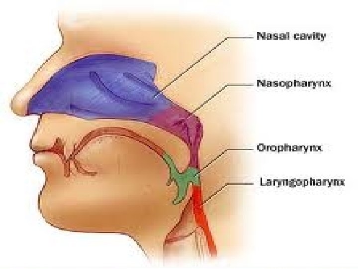
Pharynx �A fibromuscular tube from base of skull to C 6 �Funnel-shaped tube of skeletal muscle that connects to the: ◦ Nasal cavity and mouth superiorly ◦ Larynx and esophagus inferiorly �Extends from the base of the skull to the level of the sixth cervical vertebra
Larynx (Voice Box) � 3” – 4” long in anterior neck �Superiorly attaches to the hyoid bone �Inferiorly attaches to the trachea �The three functions of the larynx are: ◦ To provide a patent airway ◦ To act as a switching mechanism to route air and food into the proper channels ◦ To function in voice production
Framework of the Larynx Epiglottis – elastic cartilage that covers the laryngeal inlet during swallowing
Trachea • About 4”-5” long • Connects the larynx to the bronchi • Consists of a tube supported by 1620 “C” Shaped (hyaline) • Cartilage in anterior and smooth muscle in posterior to keep the lumen open
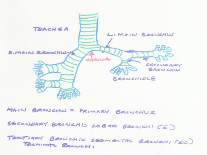
Conducting Zone: Bronchi �The carina of the last tracheal cartilage marks the end of the trachea and the beginning of the right and left bronchi �Air reaching the bronchi is: ◦ Warm and cleansed of impurities ◦ Saturated with water vapor �Bronchi subdivide into secondary bronchi, each supplying a lobe of the lungs �Air passages undergo 23 orders of branching in the lungs
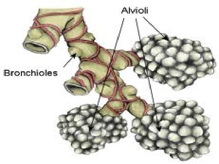
Respiratory Zone �Defined by the presence of alveoli; begins as terminal bronchioles feed into respiratory bronchioles �Respiratory bronchioles lead to alveolar ducts, then to terminal clusters of alveolar sacs composed of alveoli �Approximately 300 million alveoli: ◦ Account for most of the lungs’ volume ◦ Provide tremendous surface area for gas exchange
Respiratory Zone
Gross Anatomy of the Lungs �Lungs occupy all of the thoracic cavity except the mediastinum ◦ Root – site of vascular and bronchial attachments ◦ Costal surface – anterior, lateral, and posterior surfaces in contact with the ribs ◦ Apex – narrow superior tip ◦ Base – inferior surface that rests on the diaphragm ◦ Hilus – indentation that contains pulmonary and systemic blood vessels
Lungs �Cardiac notch (impression) – cavity that accommodates the heart �Left lung – separated into upper and lower lobes by the oblique fissure �Right lung – separated into three lobes by the oblique and horizontal fissures �There are 10 bronchopulmonary segments in each lung
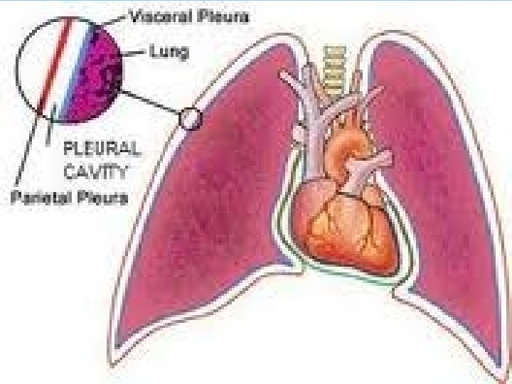
Pleurae �Thin, double-layered serosa �Parietal pleura ◦ Covers the thoracic wall and superior face of the diaphragm ◦ Continues around heart and between lungs
Pleurae �Visceral, or pulmonary, pleura ◦ Covers the external lung surface ◦ Divides the thoracic cavity into three chambers �The central mediastinum �Two lateral compartments, each containing a lung
Breathing �Breathing, or pulmonary ventilation, consists of two phases ◦ Inspiration – air flows into the lungs ◦ Expiration – gases exit the lungs
Pressure Relationships in the Thoracic Cavity �Respiratory pressure is always described relative to atmospheric pressure �Atmospheric pressure ◦ Pressure exerted by the air surrounding the body �Intrapulmonary pressure – pressure within the alveoli �Intrapleural pressure – pressure within the pleural cavity
Pressure Relationships �Two forces act to pull the lungs away from the thoracic wall, promoting lung collapse ◦ Elasticity of lungs causes them to assume smallest possible size ◦ Surface tension of alveolar fluid draws alveoli to their smallest possible size �Opposing force – elasticity of the chest wall pulls the thorax outward to enlarge the lungs
Airway Resistance �As airway resistance rises, breathing movements become more strenuous �Severely constricted or obstructed bronchioles: ◦ Can prevent life-sustaining ventilation ◦ Can occur during acute asthma attacks which stops ventilation �Epinephrine release via the sympathetic nervous system dilates bronchioles and reduces air resistance
Alveolar Surface Tension �Surface tension – the attraction of liquid molecules to one another at a liquid-gas interface �The liquid coating the alveolar surface is always acting to reduce the alveoli to the smallest possible size �Surfactant, a detergent-like complex, reduces surface tension and helps keep the alveoli from collapsing
Lung Compliance �The ease with which lungs can be expanded �Determined by two main factors ◦ Distensibility of the lung tissue and surrounding thoracic cage ◦ Surface tension of the alveoli
Factors That Diminish Lung Compliance �Scar tissue or fibrosis that reduces the natural resilience of the lungs �Blockage of the smaller respiratory passages with mucus or fluid �Reduced production of surfactant �Decreased flexibility of the thoracic cage or its decreased ability to expand �Examples include: ◦ Deformities of thorax ◦ Ossification of the costal cartilage ◦ Paralysis of intercostal muscles
Respiratory Volumes �Tidal volume ◦ Air that moves into and out of the lungs with each breath (approximately 500 ml) �Inspiratory reserve volume ◦ Air that can be inspired forcibly beyond the tidal volume (2100– 3200 ml) �Expiratory reserve volume ◦ Air that can be evacuated from the lungs after a tidal expiration (1000– 1200 ml) �Residual volume ◦ Air left in the lungs after strenuous expiration (1200 ml)
Respiratory Capacities �Inspiratory capacity ◦ Total amount of air that can be inspired after a tidal expiration �Functional residual capacity ◦ Amount of air remaining in the lungs after a tidal expiration �Vital capacity ◦ The total amount of exchangeable air ◦ TV + IRV + ERV �Total lung capacity ◦ sum of all lung volumes ◦
Oxygen Transport �Molecular oxygen is carried in the blood: ◦ Bound to hemoglobin (Hb) within red blood cells ◦ Dissolved in plasma
Carbon Dioxide Transport �CO 2 is transported in the blood in three forms ◦ Dissolved in plasma – 7 to 10% ◦ Chemically bound to hemoglobin – 20% is carried in RBCs ◦ Bicarbonate ion in plasma – 70% is transported as bicarbonate
Control of Respiration: Medullary Respiratory Centers �The dorsal respiratory group or inspiratory center ◦ Appears to be the pacesetting respiratory center ◦ Excites the inspiratory muscles and sets breath rates (12 -15 breaths/minute) ◦ Becomes dormant during expiration �The ventral respiratory group is involved in forced inspiration and expiration
Depth and Rate of Breathing: Higher Brain Centers �Hypothalamic controls act through the limbic system to modify rate and depth of respiration ◦ Example: breath holding that occurs in anger �A rise in body temperature acts to increase respiratory rate �Cortical controls are direct signals from the cerebral motor cortex that bypass medullary controls ◦ Examples: voluntary breath holding, taking a deep breath
Medullary Respiratory Centers
Lifespan Changes �By the 28 th week, a baby born prematurely can breathe on its own �During fetal life, the lungs are filled with fluid and blood bypasses the lungs �Gas exchange takes place via the placenta �At birth, respiratory centers are activated, alveoli inflate, and lungs begin to function �Respiratory rate is highest in newborns and slows until adulthood �Lungs continue to mature and more alveoli are formed until young adulthood �Respiratory efficiency decreases in old age
• Lifespan changes reflect an accumulation of environmental influences and the effects of aging in other organ systems, and may include: • The cilia become less active • Mucous thickening • Swallowing, gagging, and coughing reflexes slowing • Macrophages in the lungs lose efficiency • An increased susceptibility to respiratory infections • A “barrel chest” may develop • Bronchial walls thin and collapse • Dead space increasing