The Respiratory System A Functions 1 Smell receptor


 – covers the front")


 d. Lobules segments – lobules (has a lymphatic vessel,")


: This is the amount of air passing")


- Slides: 57
The Respiratory System
A. Functions 1. Smell receptor 2. Produces sound
https: //www. youtube. com/watch? v=g. Xk. GSc 6 d 0 x. Q
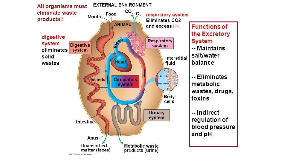
3. Gas exchange 4. Eliminates wastes
5. Respiration 6. Filters air
B. The exchange of gases 1. Respiration is the exchange of gases 2. Processes a. Pulmonary ventilation = breathing
b. External respiration - gas exchange between environment and lungs c. Internal respiration - gas exchange between blood and cells
C Portions 1. Conducting - moves air into lungs 2. Respiratory - where gas exchange occurs
https: //www. youtube. com/watch? v=La. OBc. F 6 N 7 e 4 Cow lungs 1: 37
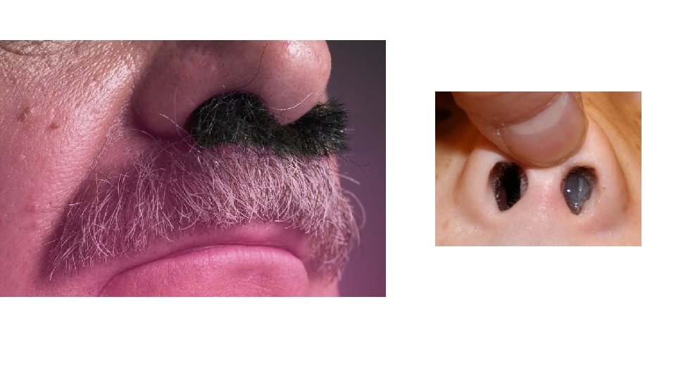
A. Nose 1. Structures a. External internal nares b. Nasal cavity c. Nasal septum d. Olfactory epithelium e. Nasal meatuses f. Nasal conchae
2. Functions a. Warms, moistens, filters incoming air b. Receives olfactory stimuli c. Resonates sound
B. Pharynx 1. Nasopharynx – equalizes pressure with the Eustachian tube/ sweeps dirty mucus down towards mouth 2. Oropharynx – common passageway for food and air 3. Common passageway for food and air
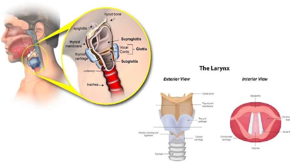
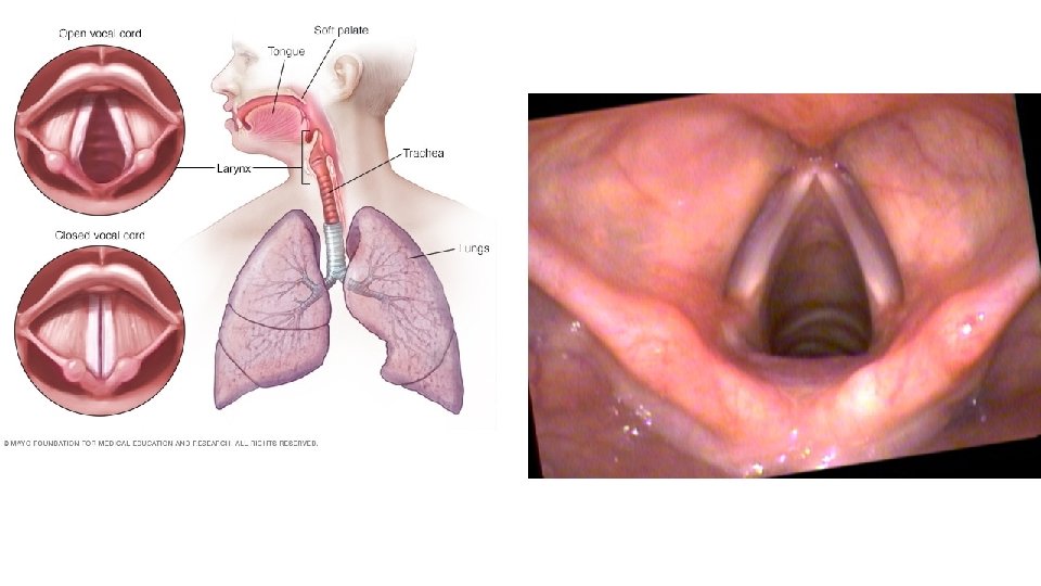
C. Larynx - voice box 1. Thyroid cartilage (Adam's apple) – covers the front and shapes it 2. Two mucus membrane folds - false vocal cords (pressure stabilizers and true vocal cords (ligaments that stretch to make sounds from vibrations)
https: //www. youtube. com/watch? v=v 9 Wdf-Rw. Lcs Vocal cors 2: 20
http: //medicalterms. info/anatomy/Trachea/ D. Trachea - windpipe 1. Passageway for air 2. Lined with mucus cilia and supported by cartilage
3. Cilia move mucus up it to be swallowed https: //www. youtube. com/watch? v=4 LRZv. VFBk 4 o
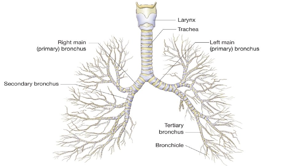
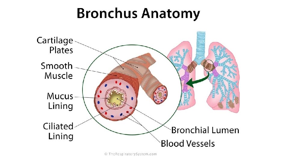
E. Bronchi 1. The 1 st 2 divisions of the trachea 2. Primary bronchus divide into secondary tertiary bronchioles terminal bronchioles – bronchial tree
3. Rings of cartilage in the bronchi get replaced by strips that disappear in the bronchioles. Smooth muscle increases as the cartilage decreases.
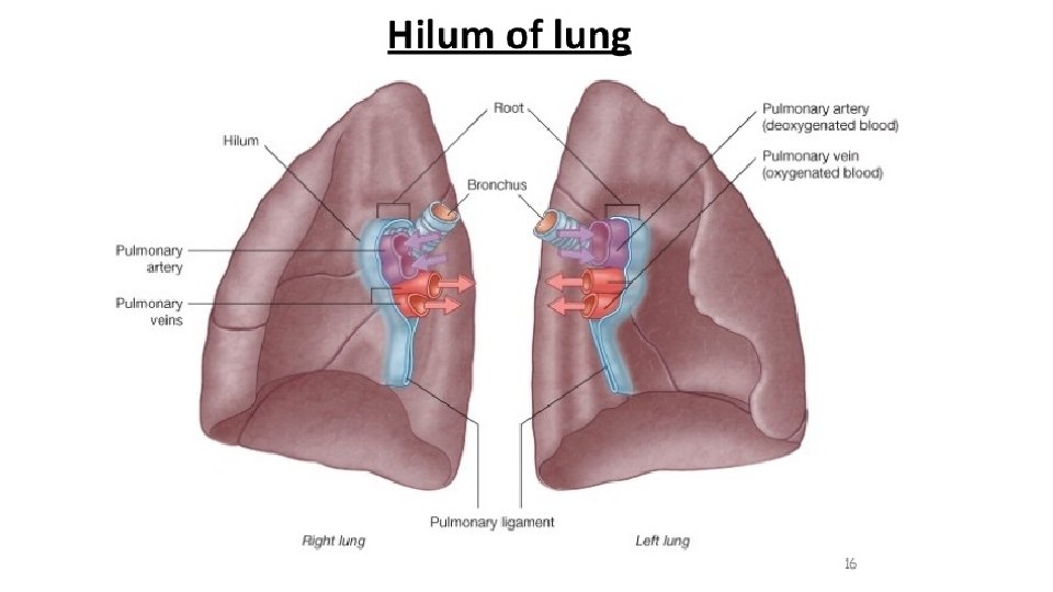
F. Lungs 1. Parts a. Pleural membrane encloses and protects the lungs, 2 layers inner visceral parietal with the pleural cavity in between b. Base, apex, hilus
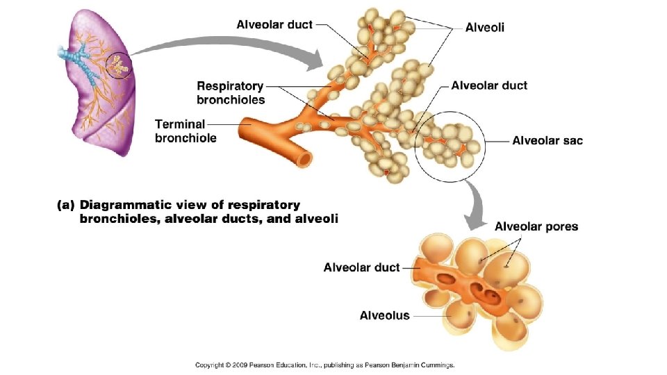
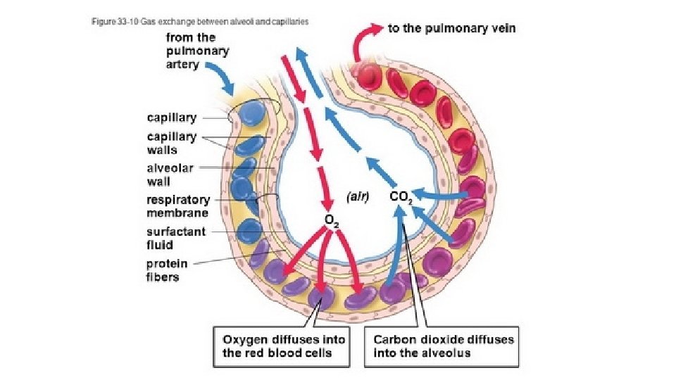
c. Lobes and fissures (grooves) d. Lobules segments – lobules (has a lymphatic vessel, arteriole, venule, a terminal bronchiole respiratory bronchiole alveolar ducts several alveolar sacs which have alveoli)
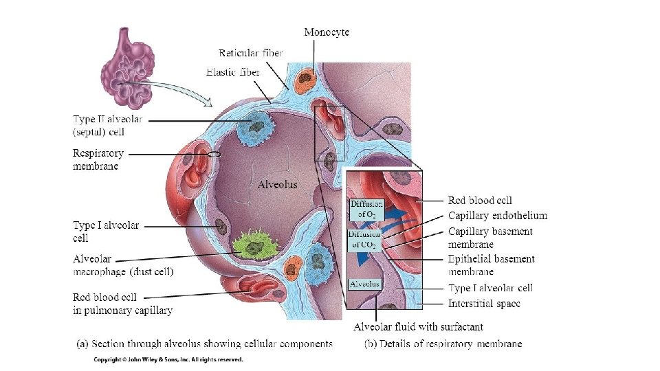
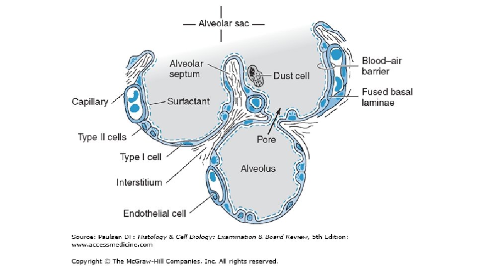
e. Alveoli are lined with squamous epithelium for gas exchange and septal cells to produce surfactant. Surfactant prevents the thin walled alveoli from sticking together.
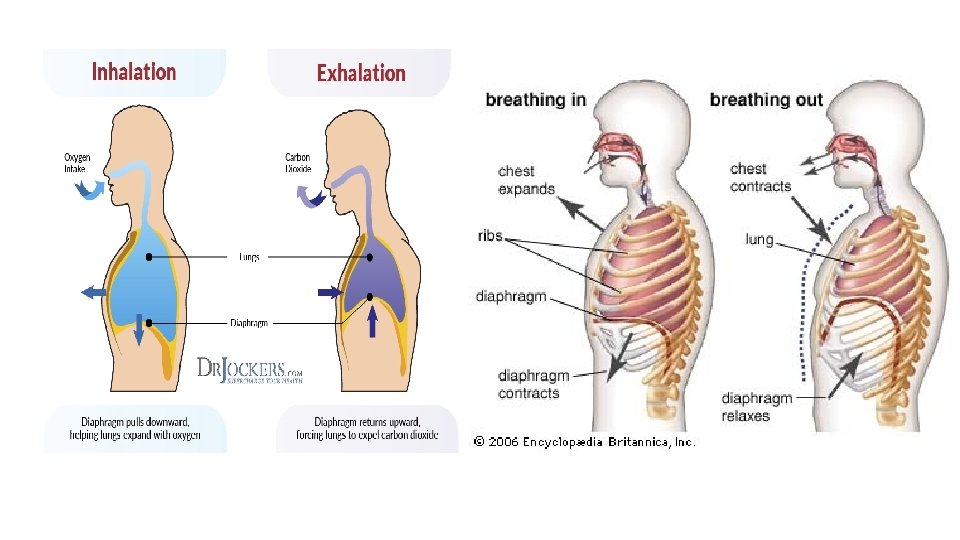
Respiration A. Purpose is the supply oxygen and remove CO 2 through 3 processes – pulmonary ventilation, external respiration and internal respiration B. Pulmonary ventilation - breathing https: //www. youtube. com/watch? v=Omo. U 3 Eex. FQQ
1. Inspiration diaphragm contracts, flattens itself, lowers itself and decreases pressure within the lung. External intercostal muscles contract and open up the thoracic cavity. a. Eupnea is normal, quiet breathing 2. Expiration is the opposite of inspiration
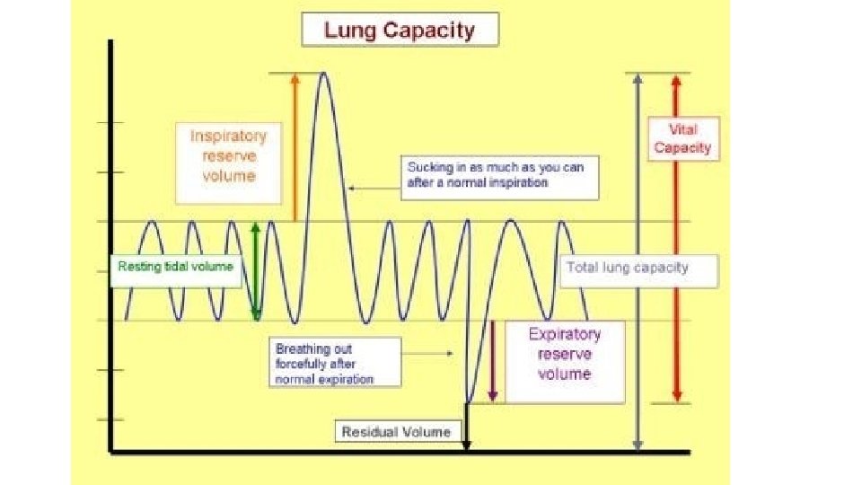
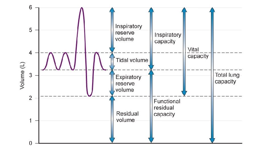
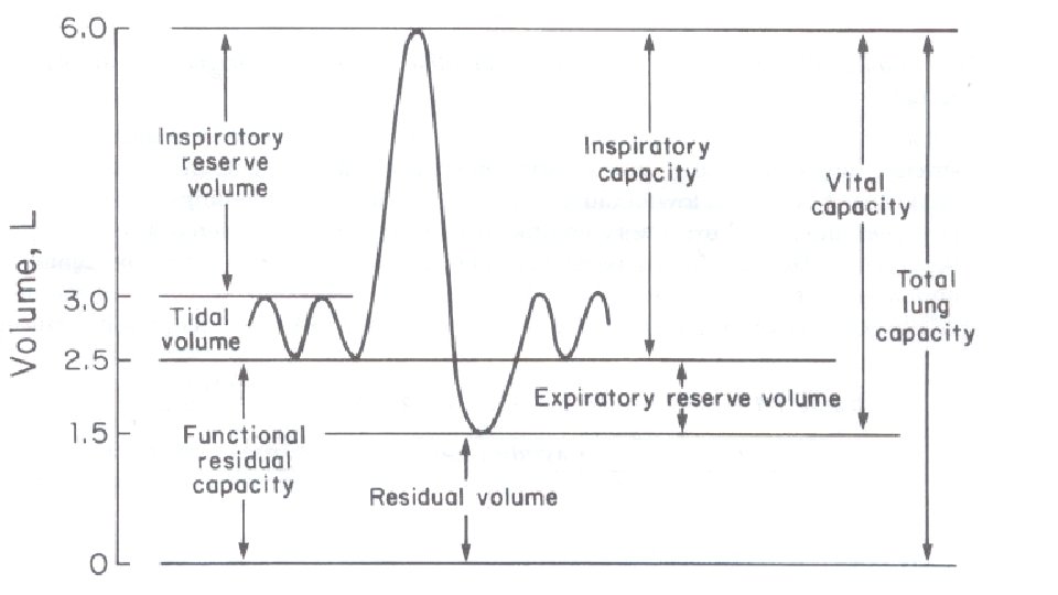
C. Pulmonary capacities 1. Tidal volume (TV): This is the amount of air passing into and out of the lungs during each cycle of breathing. It is about 500 m. L at rest. 2. Inspiratory reserve volume (IRV): This is the volume of air that can be inhaled into the lungs during normal inspiration. It is about 1500 ml. 3. Expiratory reserve volume (ERV): This is the total volume of air which can be expelled from the lungs forcefully during normal expiration. It is about 1100 ml. 4. Inspiratory capacity (IC): This is the amount of air that can be inspired with maximum effort. It consists of the tidal volume (500 ml) plus the inspiratory reserve volume. IC = TV + IRV. It is about 2000 ml 5. Functional residual capacity (FRC): This is the amount of air remaining in the lungs after normal expiration. It is equal to ERV+ RV= 1100+1200= 2300 ml. The functional residual volume also prevents collapse of the alveoli on expiration. 6. Residual volume (RV): This is the volume of air remaining in the lungs after forceful expiration. It is about 1200 ml. 7. Vital capacity (VC): This is the maximum volume of air which can be expired after forceful inspiration in single breath. VC= TV+IRV+ERV= 500+1100. VC of athletes is more than normal person. 8. Total lung capacity (TLC): This is the maximum amount of air the lungs can hold after forceful inspiration. It is normally about 5000 -6000 ml in adult. LC = VC + RV. 9. Dead space: The lungs and the air passages are never empty. Out of 500 ml of air inspired during normal respiration, 350 ml are exchanged across the walls of the alveolar ducts. About 150 ml of air always remains in the respiratory passage called as dead space.
D. Factors affecting respiration 1. Altitude 2. Surface area of the alveoli – direct relationship
3. Minute volume – direct relationship 4. Condition of respiratory passageways
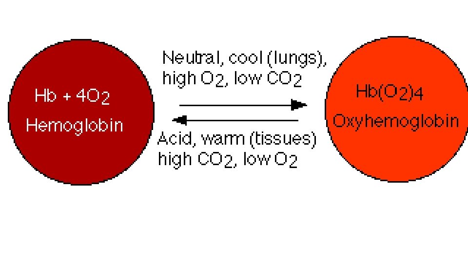
E. Transport of gases 1. Oxygen a. Combines with the heme portion of hemoglobin to form oxyhemoglobin b. The amount combining depends on temperature, p. H …
c. CO poisoning occurs because CO bonds with hemoglobin just as O 2, but more strongly.
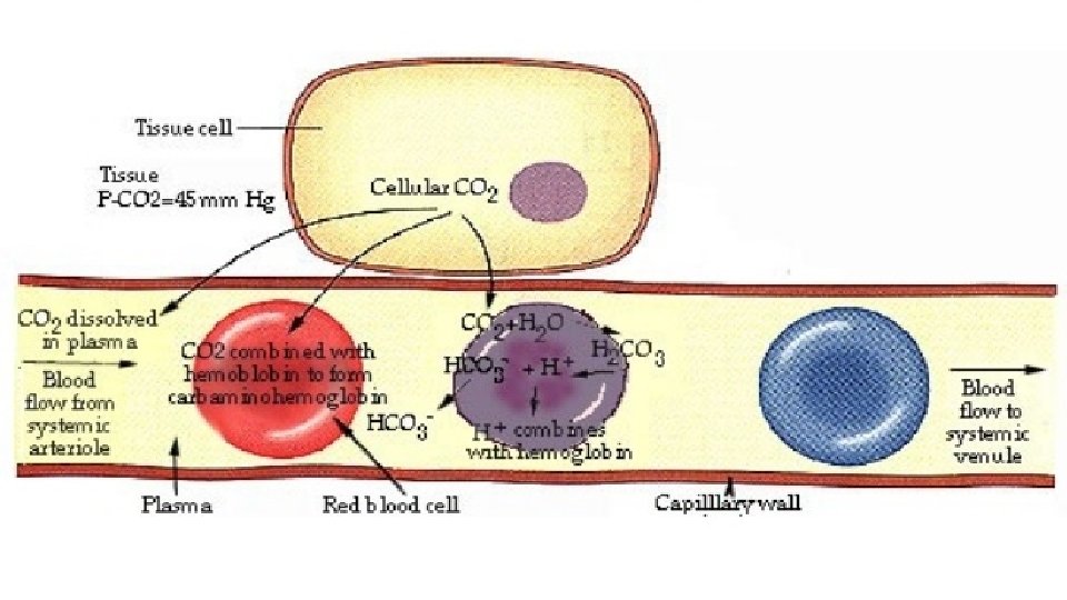
2. CO 2 a. Is carried in blood dissolved, as carbaminohemoglobin, or as bicarbonate ions. The bicarbonate ions combine with water to form carbonic acid.
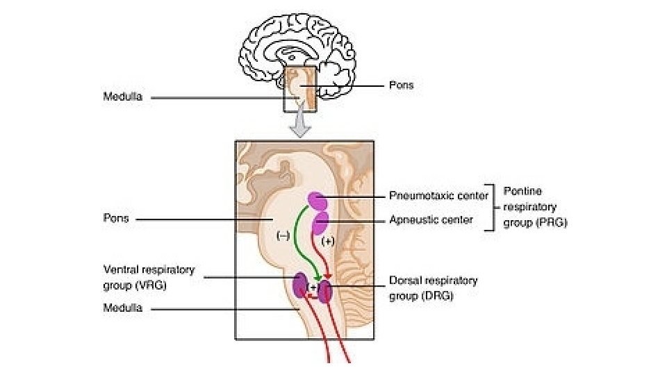
F. Control of Respiration 1. Nervous control a. Respiratory center in the brain stem
* Medullary rhythmicity area in medulla oblongata to controls basic rhythm * Pneumotaxic area in pons to coordinate in expiration by inhibiting inspiration * Apneustic area in pons to coordinate in expiration by stimulating inspiration
2. Body demands a. Cerebral cortex you can hold your breath on purpose
https: //www. youtube. com/watch? v=ASWd. De. No 54 o
b. Inflation reflex stretch receptors in the bronchi and lungs prevent overinflation
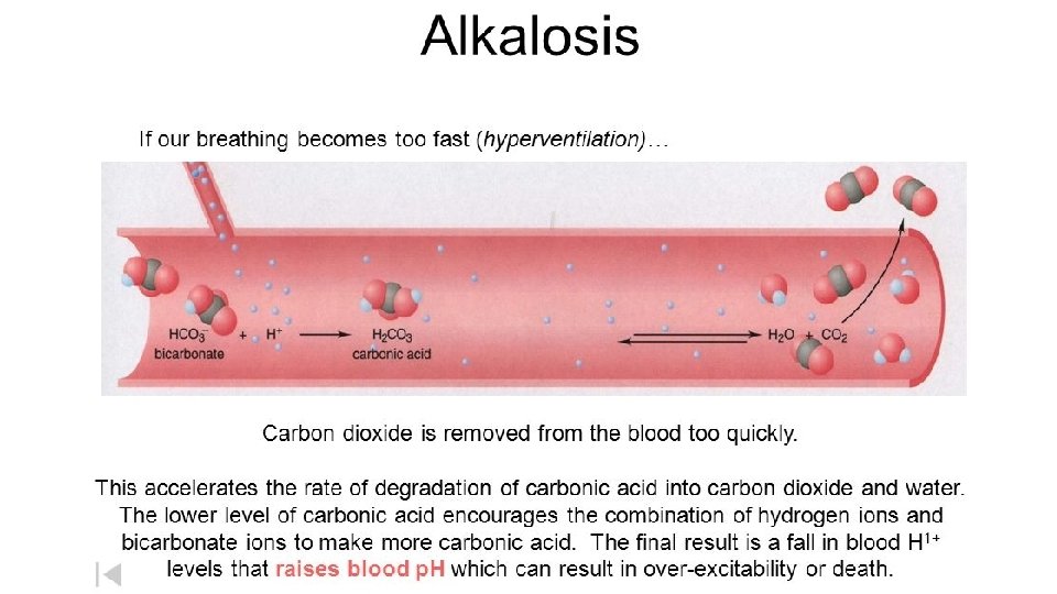
c. Chemicals CO 2 - the H+ ion that it creates, changes blood p. H. Hypercapnia, an increase in CO 2, increases the rate of breathing (hyperventilation), to rid the body of the CO 2. Hypoventilation is a slow rate of respiration.
3. Other factors – pain, emotions, temperature, weight, alcohol, drugs