The Reflex Arc Reflexes are an automatic and


 Sensory receptors that terminate where tendons joint to muscle")


- Slides: 16
The Reflex Arc Reflexes are an automatic and rapid response to a particular stimulation If the command centre for the reflex is located in the brain – cerebral reflex If the command centre for the reflex is located in the spinal cord – spinal reflex
Autonomic and Somatic Reflexes Autonomic reflexes are mediated by the autonomic division of the nervous system Involve activation of smooth and cardiac muscles as well as glands Regulate bodily functions such as digestion, elimination, blood pressure, salivation, and sweating Somatic Reflexes involve stimulation of skeletal muscles
The Reflex Arc The name given to the pathway along which the initial stimulus and the corresponding response message travel
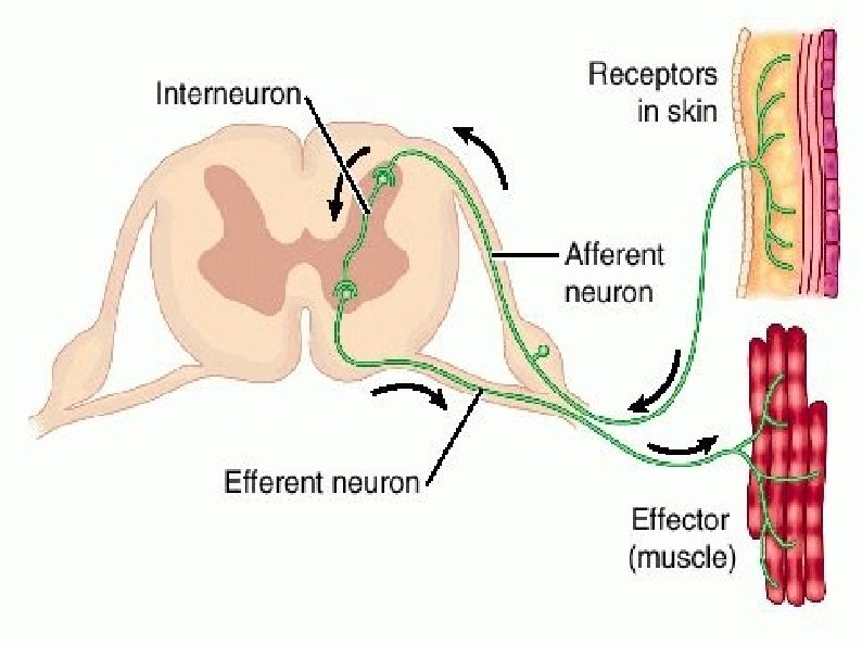
There are 5 parts to a reflex arc: 1. Receptor: receives the initial stimulus i. e. A loud noise, pinprick to the skin) 2. Sensory nerve: carries the impulse to the spinal column or brain (aka afferent nerve) 3. Intermediate nerve fibre: interprets the signal and issues and appropriate response
Cont. . . 4. Motor nerve: carries the response message from the spinal cord to the muscle or organ (aka efferent nerve) 5. Effector organ: carries out the response (i. e. Skeletal muscle – moving your hand away from danger)
Proprioceptors and the Control of Movement Proprioceptors – specialized receptors found in tendons, muscles, and joints Provide sensory information about the state of muscle contraction, the position of limbs, and body posture and balance This feedback is provided primarily by afferent (sensory) input from two sensory receptors: tendon organs and muscle spindles
Golgi Tendon Organs (Tension Reflex) Sensory receptors that terminate where tendons joint to muscle fibres Since they are aligned with muscle, any stretching of the muscle also stretches the GTO The job of GTO is to detect increased tension exerted on the tendon
GTO cont. . . When a change in tension is detected an impulse is sent along afferent (sensory) neurons to the CNS The efferent (motor) neurons transmit an impulse causing the muscle to relax This prevents injury
Muscle Spindles Lie parallel to the muscle fibre Send constant signals to the spinal cord Help maintain muscle tension Unlike GTO, they are sensitive to changes in muscle length rather than tension
Muscle Spindles cont. . . Contains two afferent and one efferent nerve fibres Two sensory nerves explains high level of sensitivity as well as the critical role they play in regulating muscle contraction Responds to changes in length by sending a message to the spinal cord Resulting contraction allows the muscle to maintain proper muscle tension or tone (i. e. Erect posture)
The Stretch Reflex Simplest spinal reflex Depends only on the single connection between primary afferent fibres and motor neurons of the same muscle i. e. Knee-jerk test
1. The receptor muscle senses the action of the hammer against the patella ligament through the muscle spindle's sensory neuron 2. The message is transmitted along the afferent (sensory) nerve axon to the spinal cord 3. The afferent neuron synapses with the efferent pathway (motor neuron) of the same muscle 4. An impulse is transmitted along the efferent pathway (motor neuron) to the muscle 5. The motor units contract (knee-jerk to accomodate additional stretch)
Reciprocal Inhibition During a reflex, the opposing muscle group is simultaneously stimulated In a knee-jerk reflex the quadriceps contract to extend the knee while the hamstrings are inhibited in a slightly delayed response (they do not flex)
Polysynaptic Reflexes With other spinal reflexes, one or more interneurons lie between the primary sensory fibres and the motor neurons The more interactions involved, the more complex and slow the reflex
The Withdrawal Reflex The withdrawal of a body part from a painful stimulus Reflex action involves transferring the impulse from a sensory neuron to a motor neuron through a connecting interneuron in the spinal cord Extremely rapid, brain doesn't have time to interpret information