The presence of crystals is called crytalluria The


- Slides: 49
The presence of crystals is called crytalluria. The type of crystal that forms, depends on the p. H of the urine. Other factors include urine concentration, temperature, and the solubility of the elements.
Refrigeration of samples can either increase or decrease the number of crystals in the sample. The materials that make up crystals are less soluble at lower temperatures. Sometimes crystals dissolve when a refrigerated sample is warmed to room temperature.
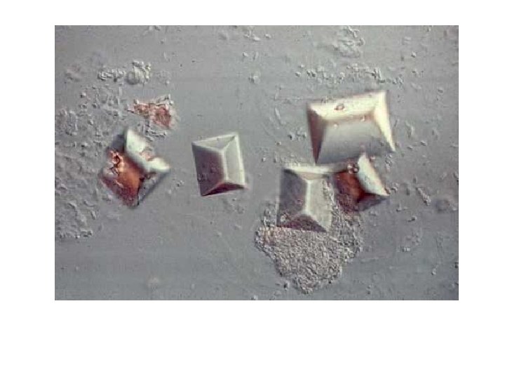
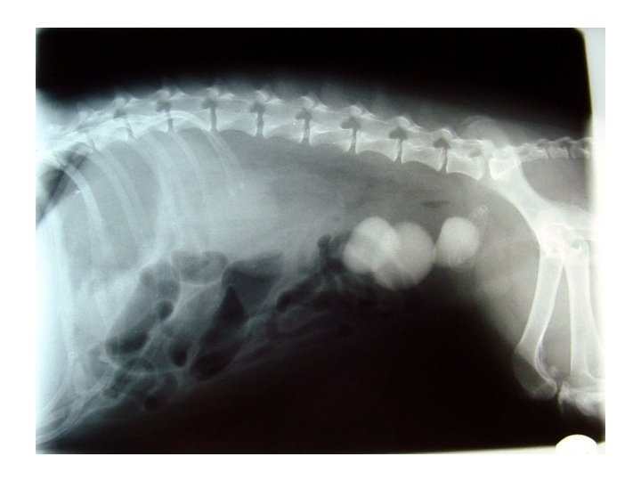
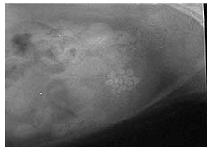
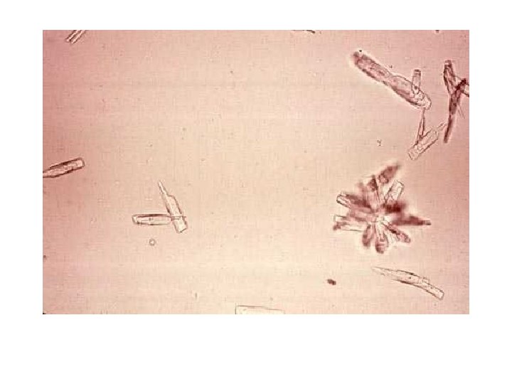
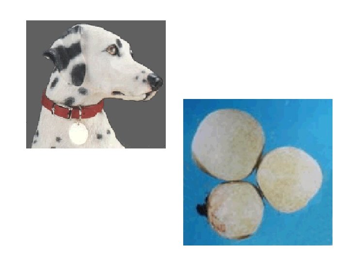
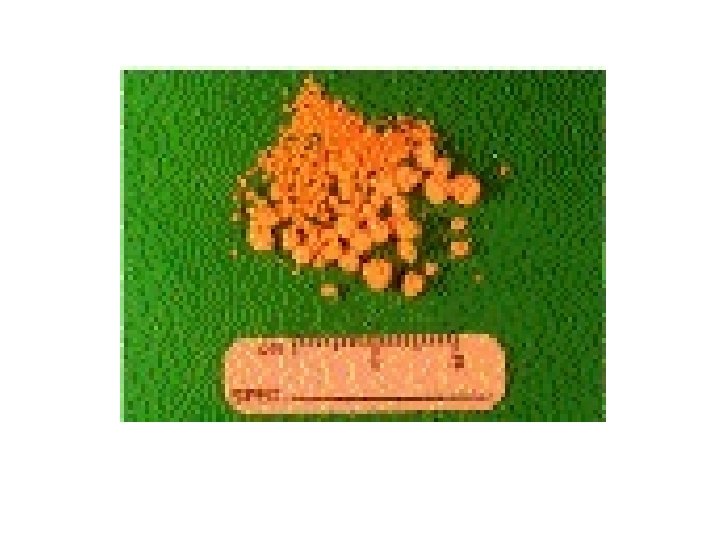
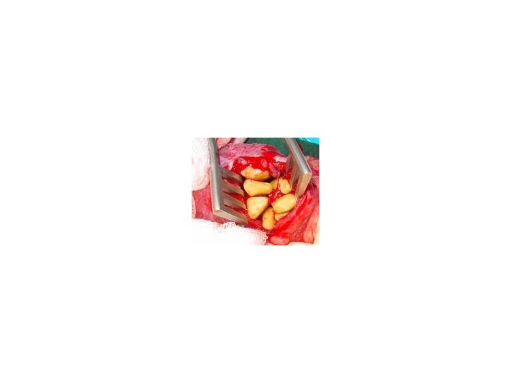
Struvite crystals are also referred to as triple phosphate crystals. They are found in alkaline to slightly acidic urine. Struvite crystals are prisms with tapering sides and are described as resembling a coffin. These are the most common type of crystal that you will identify.
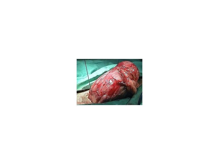
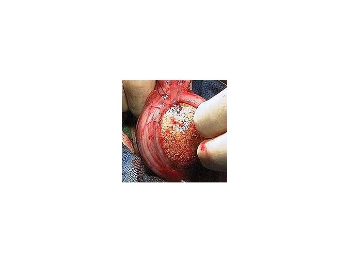
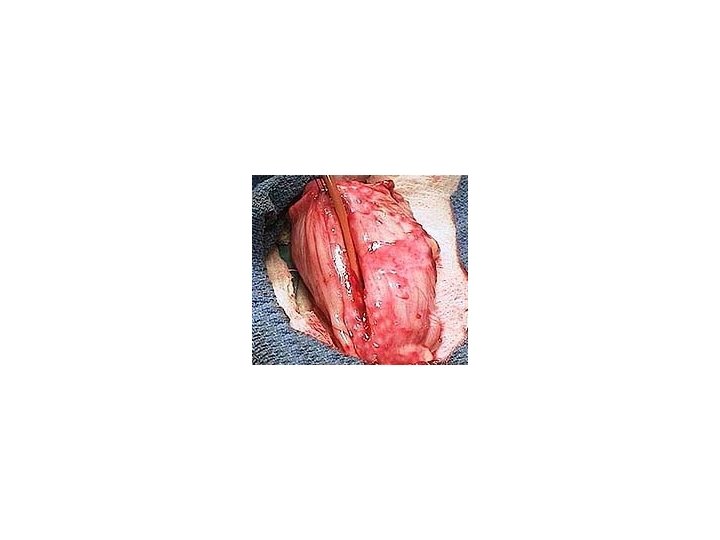
How a stone is formed: Urea is a component excreted in the urine. If infected with bacteria, the bacteria digests the urea and breaks it down into ammonia. Ammonia is toxic to the cells of the bladder wall causing inflammation. Proteins are released to form a matrix which the crystals use to form a stone.
The general rule in dogs is : No infection No stone
85% of canines with struvite stone occurrence occurs in females. Predisposed breeds include beagle, schnauzer and cocker spaniels. The average for stone occurrence is 2 ½ years.
Magnesium ammonium phosphate
Magnesium ammonium phosphate
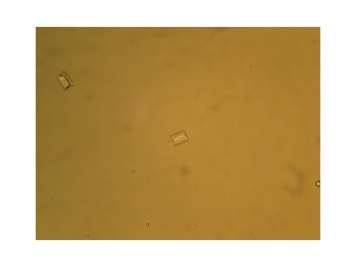
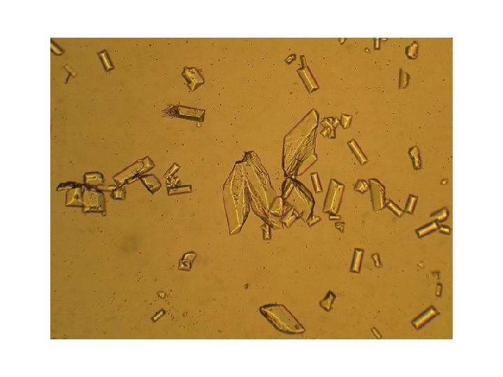
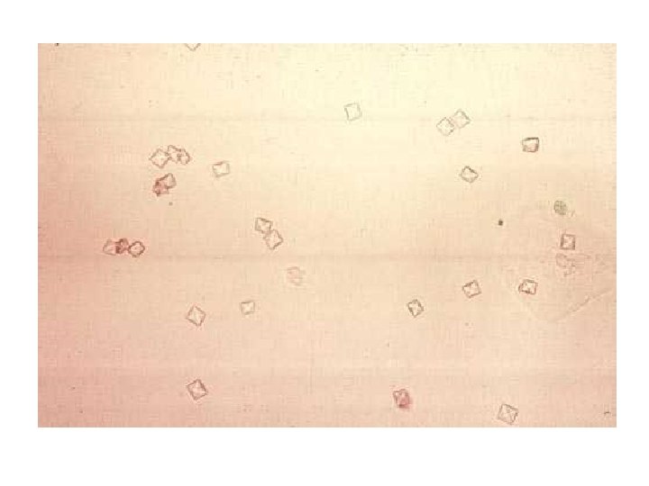
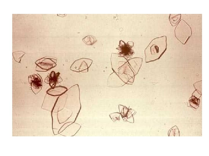
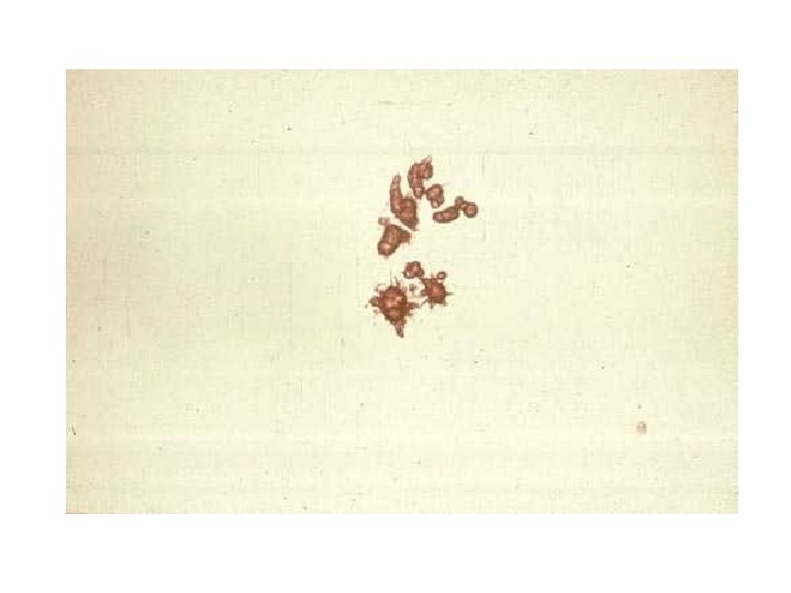
Calcium oxalate dihydrate crystals appear as small squares containing an X. Calcium oxalate monohydrate may be shaped like a dumb bell or they may be elongated and pointed at each end, resembling a picket fence slat.
Calcium oxalate crystals are formed in acidic and neutral urine and may be seen in small numbers normally in dogs. The urine of animals poisoned with ethylene glycol often contains large numbers of the monohydrate crystals.
Large numbers of calcium oxalate crystals seen in the urine may indicate a predisposition to oxalate urolithiasis.
73% of canines affected by calcium oxalate crystals and stones are male. Breeds predisposed include, schauzer, lhasa, yorkie, poodle, shih tzu, bichon. The average is 5 – 12 years
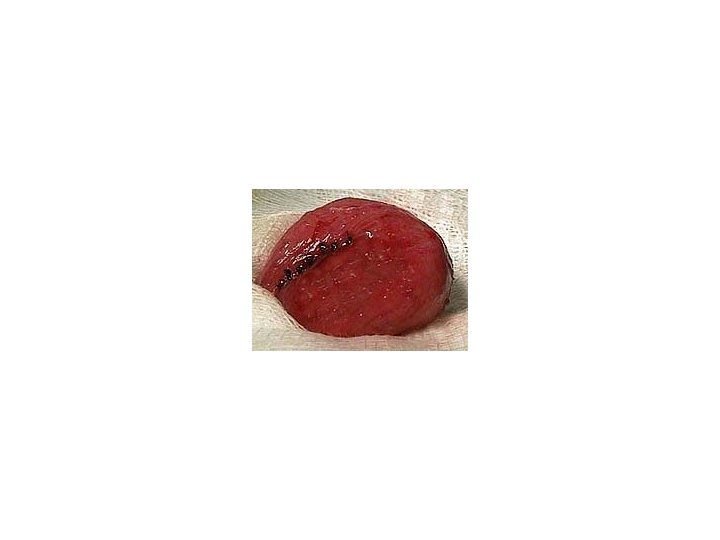
Calcium oxalate stones can not be dissolved by diet and must be surgically removed. 50% of animals who undergo cystotomy sx will redevelop stones within 3 years.
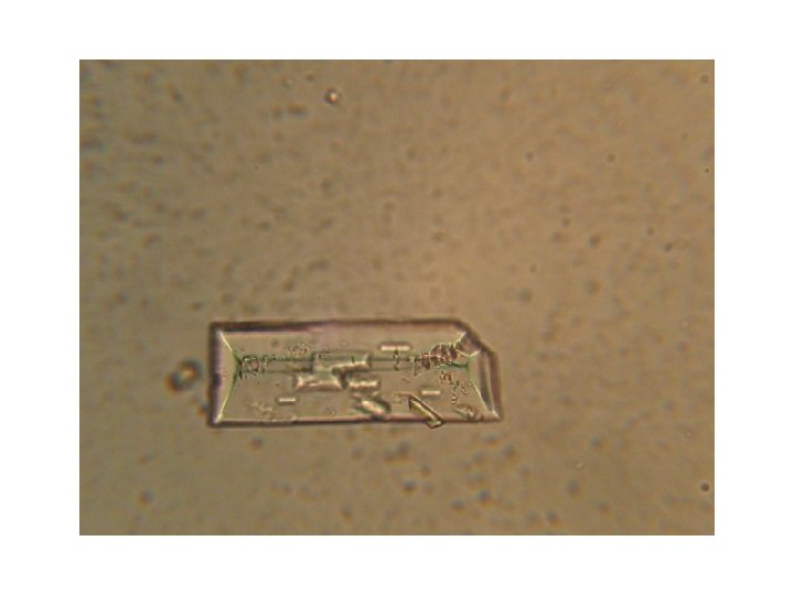
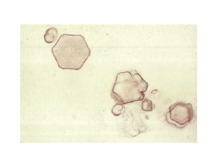
Calcium phosphate crystals assume various forms including the rosette and pointed finger forms. They appear most often in alkaline urine.
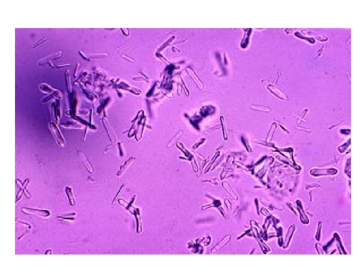
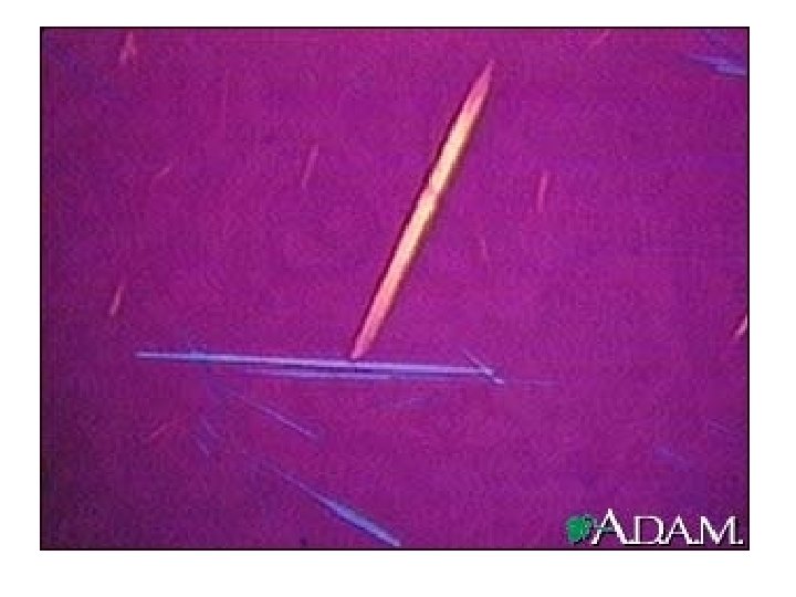
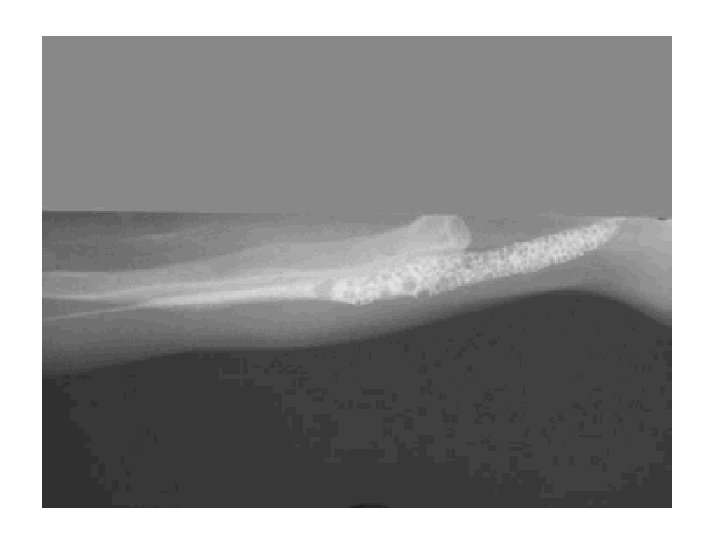
Uric acid crystals appear yellow to yellow brown. The most common shapes seen are diamond, rhomboid or rosettes. These crystals are very uncommon in small animals but are seen in dalmations.
• We eat purines when we eat meat and drink them when we drink coffee and our livers convert them ultimately into something called “Allantoin” which is readily soluble in water and easy for us to unload in that waste/water mixture known as urine. • Dalmatians just cannot seem to convert uric acid to allantoin; the process described above never gets past the uric acid stage. Dalmatian liver cells simply cannot absorb uric acid which is where the conversion to allantoin ought to take place. Dalmatians must excrete uric acid in their urine and the problem is that the stuff just is not that water soluble. Being unable to convert uric acid to allantoin is the main predisposing factor to uric acid stone formation and accounts for why 80% of uric acid bladder stones come from Dalmatians.
Ammonium urate crystals are seen in slightly acidic, neutral and alkaline urine. The shape of these crystals is called the “thorn apple” These crystals are not common but are frequently observed in dogs with portal vascular anomalies. Common breeds are Dalmations and English Bulldogs
Cystine crystalluria is not a normal phenomenon. These crystals may develop in animals with metabolic disorders
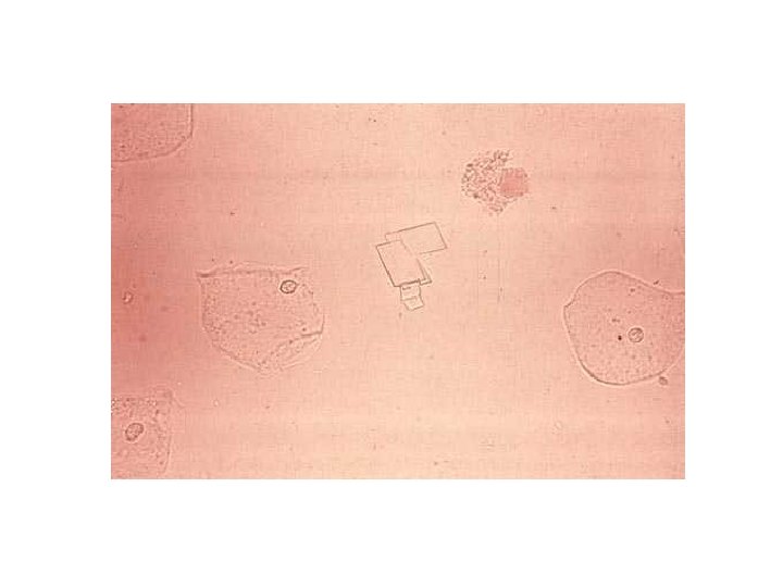
Cholesterol crystals are seen in the center of this field with squamous epithelial cells on either side. Cholesterol crystals are found in acid or neutral urine. They appear as regular or irregular transparent plates. They may occur singly or in large numbers. Usually one or more corners are cut off or notched, justifying their description as "stair step crystals". They are not commonly seen and are always considered pathological. They can be found in various renal diseases.
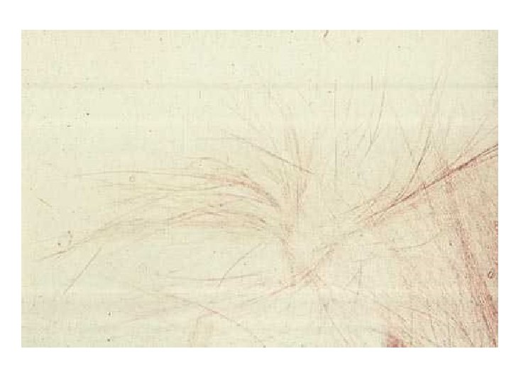
Tyrosine crystals are not normally found in urine. They are products of protein metabolism and appear in urine of animals with tissue degeneration or necrosis (acute liver disease) They are present only when urine is acid. They are colorless to yellowish brown, needle shaped crystals and have a fine silky appearance. The needles may be single or arranged in sheaves or rosettes. Tyrosine crystals usually appear in urinary sediment together with leucine crystals
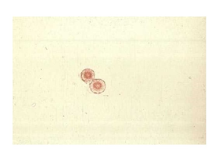
Leucine crystals are not normally found in urine They appear in urine in association with tyrosine and are manifestations of the same clinical conditions. When found, leucine crystals are in acid urine in the form of spheroids with concentric striations. They are dense, highly refractive and appear as yellowish brown bodies.
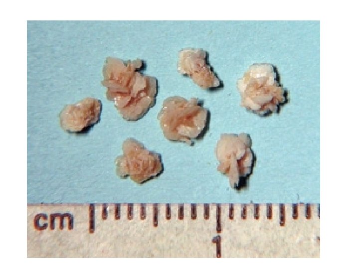
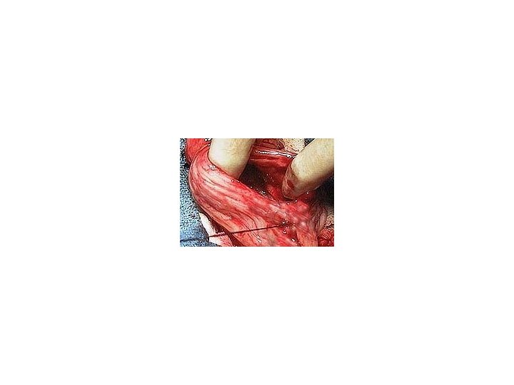
Cystotomy :