The Pleural cavities Pleural membranes Each lung occupies

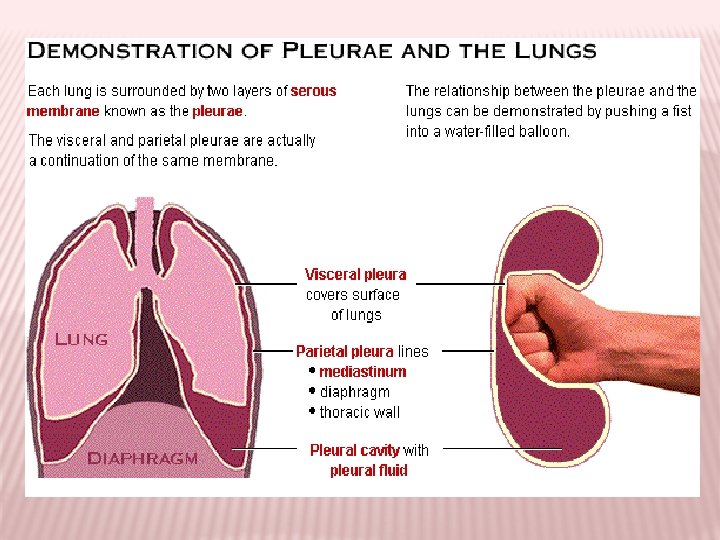
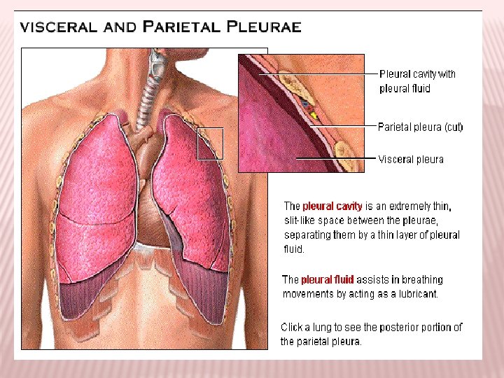
The Pleural cavities & Pleural membranes: Each lung occupies a single pleural cavity which is formed by a serous membrane called the pleura. • the parietal pleura = covers the inner surface of the thoracic wall & extends over the diaphragm & mediastinum. • the visceral pleura = covers the outer surface of the lungs. A small amount of pleural fluid is secreted by both pleurae, the fluid gives a moist, slippery coating that provides lubrication.

Pulmonary Ventilation: Pulmonary ventilation is the physical movement of air into & out of the respiratory tract, while the alveolar ventilation is movement into & out of the alveoli. *Boyle’s Law : there is an inverse relationship between the pressure & volume of a gas In a closed container. (gas pressure is inversely proportional to volume). • decrease volume of a gas → pressure rise. • Increase volume of a gas → pressure falls. P=1/V

Pressure & airflow to lungs: -The air flow from an area of higher pressure to an area of lower pressure. Inhalation & exhalation involves changes in the volume of the lungs, these changes create pressure gradients that move air into or out of the respiratory tract. So movement of the chest wall or diaphragm have a direct effect on the volume of the lungs). -the diaphragm contracts & moves inferiorly this movement increase the volume of the thoracic cavity & when the diaphragm relaxes the volume of thoracic cavity decreases. -superior movement of the rib cage increase the depth & width of the thoracic cavity, inferior movement reduces the volume of the thoracic cavity.
— they are, in fact, about a")
Compliance : -The lungs are very distensible (stretchable)— they are, in fact, about a hundred times more distensible than a toy balloon. Another term for distensibility is compliance, which here refers to the ease with which the lungs can expand under pressure. Factors affecting compliance: • the connective tissue structure of the lungs. (loss of tissue resulting from alveolar damage → ↑es compliance. • the level of surfactant production. Inadequate surfactant lead to collapse of alveoli → reduce compliance. • the mobility of the thoracic cage: any skeletal disorder that affect the articulations of the ribs will → reduce compliance. * if compliance is reduced an individual may become exhausted trying to breath.

Pressure changes during Inhalation & Exhalation: The atmospheric pressure = 760 mm. Hg. The intrapulmonary pressure (or intra alveolar pressure) is the pressure inside the respiratory tract, at the alveoli. -at quiet breathing the pressure difference between the atmospheric & intrapulmonary pressure is small. (on inhalation, the lungs expand & the intrapulmonary pressure drops to about 759 mm. Hg (or − 1 mm. Hg)). (on exhalation, the lungs recoil & the intrapulmonary pressure rise to 761 mm. Hg (or +1 mm. Hg). -at heavy breathing the pressure gradient increase about -30 mm. Hg during inspiration.

The Intra Plural Pressure : - is the pressure measured in the space between the parietal & visceral pleurae. (the intrapleural pressure is about -4 mm. Hg & reaches -18 mm. Hg during a powerful inspiration. ) ** The pressure is below atmospheric pressure due to the relationship between the lungs & body wall. The lungs are highly elastic tend to recoil but they cannot recoil significantly because of the fluid bond between the parietal & visceral pleurae. So the intrapleural pressure remains below atmospheric pressure throughout normal inhalation & exhalation

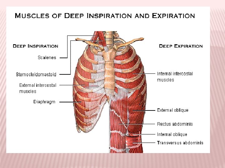
Respiration Muscles : 1 - Inhalation : - is an active process involving contraction of one or more of these muscles : a- diaphragm : ↑es volume of the thoracic cavity , it’s responsible for 75% of air movement in quiet breathing. b- external intercostals : assist by elevating the ribs about 25% of the volume of air in the lungs. c- accessory muscles : sternocleido mastoid , serratus anterior , pectoralis minor & scalenes. These assist the external intercostals in elevating the ribs.

2 - Exhalation : - exhalation may be passive or active, if it’s active it may involve one or more of the following muscles : a- internal intercostals & transverses thoracis : depress the ribs & reduce the width & depth of the thoracic cavity. b- abdominal muscles : - external & internal obliques , transversus abdomens & the rectus abdominis. These assist the internal intercost in expiration by compressing the abdomen & forcing the diaphragm upward.
 inhalation involves muscular")
Modes of Breathing : a- Quiet breathing : (or eupnea ) inhalation involves muscular contractions, but exhalation is a passive process. - diaphragm breathing ( or deep breathing ) : this provides the necessary changes in thoracic volume. Contraction causes inhalation & exhalation occurs when diaphragm relaxes. - costal breathing : ( or shallow breathing ) : the thoracic volume changes because the rib cage changes shape. Inhalation occurs when external intercostals contract & exhalation occurs passively when these muscles relax.
 involves active inspiratory & expiratory movements.")
b- Forced breathing : ( or hyperpnea ) involves active inspiratory & expiratory movements. In forced breathing the accessory muscles assist with inspiration & expiration contraction of internal intercostals & abdominal muscles. Respiratory Rate : is number of breaths per minute. Adult = 12 - 18 breaths / min. Children = 18 – 20 breaths / min. Respiratory Minute Volume = amount of air moved each minute. VE ( respiratory minute volume ) = f (respiratory rate) × VT (tidal volume) = 12 × 500 ml /min. = 6000 ml / min. = 6 lit / min.
- Slides: 13