The Plasma Membrane Fluid Dynamics and Cell Transportation

The Plasma Membrane Fluid Dynamics and Cell Transportation

The Plasma Membrane • The plasma membrane is the boundary that separates the living cell from its nonliving surroundings • The plasma membrane exhibits selective permeability, meaning some substances can go through, others cannot.

Fluid Mosaic Model • The plasma membrane is primarily made of phospholipids. • Phospholipids contain both hydrophobic (waterhating) and hydrophilic (water-loving) regions • The fluid mosaic model states that a membrane is constantly moving with a mixture of proteins embedded in it

LE 7 -2 WATER Hydrophilic head Hydrophobic tail WATER

LE 7 -3 Hydrophilic region of protein Phospholipid bilayer Hydrophobic region of protein
 Saturated hydrocarbon")
LE 7 -5 b Fluid Unsaturated hydrocarbon tails with kinks Viscous (Thick) Saturated hydrocarbon tails

• Cholesterol helps to maintain the homeostasis of membranes, keeping them fluid. o At warm temperatures, cholesterol keeps phospholipids from moving around too much. o At cool temperatures, it maintains fluidity by preventing tight packing

LE 7 -5 c Cholesterol within the animal cell membrane

Membrane Proteins and Their Functions • Proteins determine most of the membrane’s specific functions • Peripheral proteins are on the surface of the membrane, either outside or inside the cell. • Integral proteins go all the way through the phospholipid bilayer and contact both sides.
 Glycoprotein Carbohydrate Glycolipid EXTRACELLULAR SIDE OF")
LE 7 -7 Fibers of extracellular matrix (ECM) Glycoprotein Carbohydrate Glycolipid EXTRACELLULAR SIDE OF MEMBRANE Cholesterol Microfilaments of cytoskeleton Peripheral proteins Integral protein CYTOPLASMIC SIDE OF MEMBRANE

Membrane Proteins and Their Functions • Six major functions of membrane proteins: 1. 2. 3. 4. 5. 6. Transport substances across the cell membrane Serve as enzymes for reactions Transmit signals across the cell membrane Cell-cell recognition Joining of two cells together Attachment to the cytoskeleton

Membrane Carbohydrates • Cells can recognize each other by binding to carbohydrates on the plasma membrane. • These carbohydrates vary among species, individuals, and even cell types in an individual. o This is how the immune system recognizes “self” and “foreign” cells. o Example: Blood type (A, B, AB, O) is determined by markers on your red blood cell membranes.

Permeability of the Lipid Bilayer • The cell membrane is selectively permeable. • Nonpolar (hydrophobic) molecules can dissolve in the lipid bilayer and pass through the membrane rapidly o Examples: Oxygen, carbon dioxide, hormones made of lipids • Large polar (hydrophilic) molecules, cannot cross the membrane as easily. o Examples: Glucose, sucrose, proteins

Transport Proteins • Transport proteins are needed to allow passage of polar substances across the membrane • Channel proteins, act like a tunnel that ions or other molecules can use to enter the cell. o Example: Aquaporins facilitate the passage of water. • Carrier proteins, bind to molecules and change shape to shuttle them across the membrane o A transport protein is specific for one substance only.

Passive Transport • Diffusion is the movement of molecules areas of greater concentration to areas of lower concentration. • This is considered passive transport because no energy is required.
 WATER Net diffusion Diffusion")
LE 7 -11 a Molecules of dye Membrane (cross section) WATER Net diffusion Diffusion of one solute Net diffusion Equilibrium

LE 7 -11 b Net diffusion Diffusion of two solutes Net diffusion Equilibrium

Osmosis and Water Balance • Osmosis is the diffusion of water across a selectively permeable membrane • The direction of osmosis is determined only by a difference in the concentration of solutes. o Solutes are substances dissolved in water, like sugar or salts. • Water diffuses across a membrane from the region of lower solute concentration to the region of higher solute concentration
 Higher concentration of sugar H 2")
LE 7 -12 Lower concentration of solute (sugar) Higher concentration of sugar H 2 O sugar molecules cannot pass through pores, but water molecules can Osmosis Same concentration of sugar

Water Balance of Cells Without Walls • These are three types of solutions that cells can be placed in: • Isotonic solution: concentration of solutes (sugars, salts) is the same as that inside the cell o cell does not change • Hypertonic solution: concentration of solutes is greater outside the cell o cell loses water and shrivels up • Hypotonic solution: concentration of solutes is lower than that inside the cell o cell gains water and bursts

Effect of Tonicity on Animal and Plant Cells • Can you predict what will happen when a red blood cell is exposed to different types of solutions?

LE 7 -13 Isotonic solution Distilled water Saltwater Animal cell H 2 O Shriveled Lysed Plant cell H 2 O Turgid (normal) H 2 O Flaccid H 2 O Plasmolyzed

LE 7 -13 Isotonic solution Distilled water Salt water Animal cell H 2 O Turgid (normal) H 2 O Flaccid H 2 O Shriveled Normal Lysed Plant cell H 2 O Plasmolyzed

LE 7 -13 Isotonic solution Distilled water Saltwater Animal cell H 2 O Turgid (normal) H 2 O Flaccid H 2 O Shriveled Normal Lysed Plant cell H 2 O Plasmolyzed

LE 7 -13 Distilled water Isotonic solution Salt water Animal cell H 2 O Turgid (normal) H 2 O Flaccid H 2 O Shriveled Normal Lysed Plant cell H 2 O Plasmolyzed

• Animal cells cannot survive in a hypertonic or hypotonic environment because their cell membranes are too thin. • Some organisms have adaptations to allow them to survive in these environments. o Example: The protist Paramecium lives in a hypotonic environment. • It has vacuole that can absorb excess water and pump it back out of the cell.

LE 7 -14 Filling vacuole Contracting vacuole 50 µm

Water Balance of Cells with Walls • Plant cells have cell walls. • Cell walls are thicker then plasma membranes and do not burst. • This allows plants to survive in different environments.

LE 7 -13 Saltwater solution Isotonic solution Distilled water Animal cell H 2 O Turgid (normal) H 2 O Flaccid H 2 O Shriveled Normal Lysed Plant cell H 2 O Plasmolyzed

LE 7 -13 Hypotonic solution Isotonic solution Hypertonic solution Animal cell H 2 O Turgid (normal) H 2 O Flaccid H 2 O Shriveled Normal Lysed Plant cell H 2 O Plasmolyzed

LE 7 -13 Hypotonic solution Isotonic solution Hypertonic solution Animal cell H 2 O Turgid (normal) H 2 O Flaccid H 2 O Shriveled Normal Lysed Plant cell H 2 O Plasmolyzed

LE 7 -13 Hypotonic solution Isotonic solution Hypertonic solution Animal cell H 2 O Turgid (normal) H 2 O Flaccid H 2 O Shriveled Normal Lysed Plant cell H 2 O Plasmolyzed

Facilitated Diffusion • In facilitated diffusion, transport proteins aid movement of molecules across the plasma membrane o This increases the rate of transport. o This is considered passive transport because no energy is used.

• Channel proteins provide corridors that allow a specific molecule or ion to cross the membrane EXTRACELLULAR FLUID Channel protein Solute CYTOPLASM

• Carrier proteins undergo a subtle change in shape that translocates the solute-binding site across the membrane Carrier protein Solute

Active Transport • Active transport moves substances against their concentration gradient • Active transport requires energy, usually in the form of ATP o ATP is the smallest, most basic energy-containing molecule that cells use. • Active transport is performed by specific proteins embedded in the membranes

Neurons
![LE 7 -16 EXTRACELLULAR [Na+] high FLUID [K+] low Na+ Na+ CYTOPLASM [Na+] low](http://slidetodoc.com/presentation_image_h2/99369add0c26a286cd7f4ccaa541b1f2/image-38.jpg "LE 7 -16 EXTRACELLULAR [Na+] high FLUID [K+] low Na+ Na+ CYTOPLASM [Na+] low")
LE 7 -16 EXTRACELLULAR [Na+] high FLUID [K+] low Na+ Na+ CYTOPLASM [Na+] low [K+] high Na+ bonds to the sodium-potassium pump P ATP P ADP ATP is broken down into ADP + P, generating energy. The phosphorus causes the protein to change shape, releasing the sodium to the Outside of the cell. K+ K+ P Potassium from outside the cell bonds to the protein pump, causing the phosphorus to be removed. K+ The potassium is released to the inside of the cell and the protein returns to its original shape. The protein is now ready to repeat the process.
![LE 7 -16 EXTRACELLULAR [Na+] high FLUID [K+] low Na+ Na+ CYTOPLASM [Na+] low](http://slidetodoc.com/presentation_image_h2/99369add0c26a286cd7f4ccaa541b1f2/image-39.jpg "LE 7 -16 EXTRACELLULAR [Na+] high FLUID [K+] low Na+ Na+ CYTOPLASM [Na+] low")
LE 7 -16 EXTRACELLULAR [Na+] high FLUID [K+] low Na+ Na+ CYTOPLASM [Na+] low [K+] high Na+ bonds to the sodium-potassium pump P ATP P ADP ATP is broken down into ADP + P, generating energy. The phosphorus causes the protein to change shape, releasing the sodium to the Outside of the cell. K+ K+ P Potassium from outside the cell bonds to the protein pump, causing the phosphorus to be removed. K+ The potassium is released to the inside of the cell and the protein returns to its original shape. The protein is now ready to repeat the process.
![LE 7 -16 EXTRACELLULAR [Na+] high FLUID [K+] low Na+ Na+ CYTOPLASM [Na+] low](http://slidetodoc.com/presentation_image_h2/99369add0c26a286cd7f4ccaa541b1f2/image-40.jpg "LE 7 -16 EXTRACELLULAR [Na+] high FLUID [K+] low Na+ Na+ CYTOPLASM [Na+] low")
LE 7 -16 EXTRACELLULAR [Na+] high FLUID [K+] low Na+ Na+ CYTOPLASM [Na+] low [K+] high Na+ bonds to the sodium-potassium pump P ATP P ADP ATP is broken down into ADP + P, generating energy. The phosphorus causes the protein to change shape, releasing the sodium to the Outside of the cell. K+ K+ P Potassium from outside the cell bonds to the protein pump, causing the phosphorus to be removed. K+ The potassium is released to the inside of the cell and the protein returns to its original shape. The protein is now ready to repeat the process.
![LE 7 -16 EXTRACELLULAR [Na+] high FLUID [K+] low Na+ Na+ CYTOPLASM [Na+] low](http://slidetodoc.com/presentation_image_h2/99369add0c26a286cd7f4ccaa541b1f2/image-41.jpg "LE 7 -16 EXTRACELLULAR [Na+] high FLUID [K+] low Na+ Na+ CYTOPLASM [Na+] low")
LE 7 -16 EXTRACELLULAR [Na+] high FLUID [K+] low Na+ Na+ CYTOPLASM [Na+] low [K+] high Na+ bonds to the sodium-potassium pump P ATP P ADP ATP is broken down into ADP + P, generating energy. The phosphorus causes the protein to change shape, releasing the sodium to the Outside of the cell. K+ K+ P Potassium from outside the cell bonds to the protein pump, causing the phosphorus to be removed. K+ The potassium is released to the inside of the cell and the protein returns to its original shape. The protein is now ready to repeat the process.
![LE 7 -16 EXTRACELLULAR [Na+] high FLUID [K+] low Na+ Na+ CYTOPLASM [Na+] low](http://slidetodoc.com/presentation_image_h2/99369add0c26a286cd7f4ccaa541b1f2/image-42.jpg "LE 7 -16 EXTRACELLULAR [Na+] high FLUID [K+] low Na+ Na+ CYTOPLASM [Na+] low")
LE 7 -16 EXTRACELLULAR [Na+] high FLUID [K+] low Na+ Na+ CYTOPLASM [Na+] low [K+] high Na+ bonds to the sodium-potassium pump P ATP P ADP ATP is broken down into ADP + P, generating energy. The phosphorus causes the protein to change shape, releasing the sodium to the Outside of the cell. K+ K+ P Potassium from outside the cell bonds to the protein pump, causing the phosphorus to be removed. K+ The potassium is released to the inside of the cell and the protein returns to its original shape. The protein is now ready to repeat the process.
![LE 7 -16 EXTRACELLULAR [Na+] high FLUID [K+] low Na+ Na+ CYTOPLASM [Na+] low](http://slidetodoc.com/presentation_image_h2/99369add0c26a286cd7f4ccaa541b1f2/image-43.jpg "LE 7 -16 EXTRACELLULAR [Na+] high FLUID [K+] low Na+ Na+ CYTOPLASM [Na+] low")
LE 7 -16 EXTRACELLULAR [Na+] high FLUID [K+] low Na+ Na+ CYTOPLASM [Na+] low [K+] high Na+ bonds to the sodium-potassium pump P ATP P ADP ATP is broken down into ADP + P, generating energy. The phosphorus causes the protein to change shape, releasing the sodium to the Outside of the cell. K+ K+ P Potassium from outside the cell bonds to the protein pump, causing the phosphorus to be removed. K+ The potassium is released to the inside of the cell and the protein returns to its original shape. The protein is now ready to repeat the process.

Cotransport • Cotransport occurs when active transport of a solute indirectly drives transport of another solute.

LE 7 -19 – + H+ ATP H+ – + Proton pump H+ H+ – H+ + H+ Sucrose-H+ cotransporter Diffusion of H+ H+ – – + + Sucrose

Exocytosis • In exocytosis, transport vesicles from the Golgi fuse with the cell membrane and release their contents to the outside.

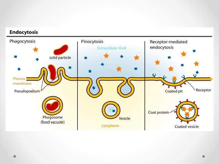
Endocytosis • In endocytosis, the cell takes in large molecules by forming vesicles from the plasma membrane • Endocytosis is a reversal of exocytosis. • Three types of endocytosis: o Phagocytosis (“cellular eating”): Cell engulfs particle in a vacuole o Pinocytosis (“cellular drinking”): Cell creates vesicle around fluid o Receptor-mediated: Particles or fluid must bond to protein receptors first, then are engulfed.

Sodium – Potassium Pumps The sodium potassium pump works with only Sodium and Potassium. It moves 3 Sodium ions out of a cell, and allows 2 potassium ions into the cell. Because there is already usually more sodium outside the cell, and the pump continues to move more sodium outside the cell, the sodium potassium pump works against the concentration gradient. Meaning the rich get richer and therefore is active transport, because equilibrium is not the goal
- Slides: 49